WB, IP, IF-IC
M R
Endogenous
23-25
Rabbit IgG
#Q99ML1
170770
Product Information
Product Usage Information
| Application | Dilution |
|---|---|
| Western Blotting | 1:1000 |
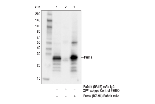
| Immunoprecipitation | 1:200 |
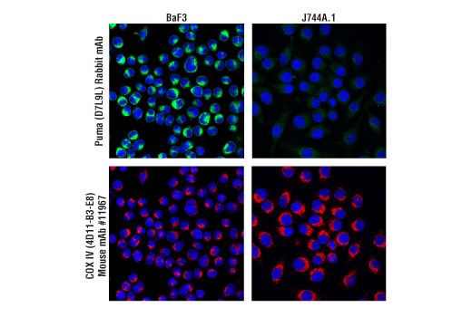
| Immunofluorescence (Immunocytochemistry) | 1:800 |
Storage
Specificity / Sensitivity
Species Reactivity:
Mouse, Rat
Source / Purification
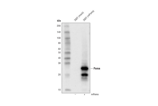
Monoclonal antibody is produced by immunizing animals with a synthetic peptide corresponding to residues surrounding Arg84 of mouse Puma protein.
Background
Puma (p53 upregulated modulator of apoptosis) is a "BH3-only" Bcl-2 family member originally identified in differential gene expression studies as a p53-inducible gene (1,2). The "BH3-only" family members include Bad, Bid, Bik, Hrk, Bim, and Noxa, all of which contain a BH3 domain but lack other conserved domains, BH1 and BH2, and generally promote apoptosis by binding to and antagonizing anti-apoptotic Bcl-2 family members through BH3 domain interactions (3). Two BH3-containing proteins are produced from the puma gene, Puma-α and Puma-β, both of which are induced by p53, bind Bcl-2 and Bcl-xL, localize to the mitochondria, and promote cytochrome c release and apoptosis (1,2). Puma plays a critical role in the p53 tumor suppressor pathway. Targeted disruption of the puma gene impairs p53-mediated apoptosis and tumor suppression (4-7). Puma knockout mice show defects from multiple apoptotic stimuli, including ionizing irradiation, deregulated c-Myc expression, and cytokine withdrawal (4).
- Yu, J. et al. (2001) Mol Cell 7, 673-82.
- Nakano, K. and Vousden, K.H. (2001) Mol Cell 7, 683-94.
- Bouillet, P. and Strasser, A. (2002) J Cell Sci 115, 1567-74.
- Jeffers, J.R. et al. (2003) Cancer Cell 4, 321-8.
- Hemann, M.T. et al. (2004) Proc Natl Acad Sci U S A 101, 9333-8.
- Yu, J. et al. (2003) Proc Natl Acad Sci U S A 100, 1931-6.
- Villunger, A. et al. (2003) Science 302, 1036-8.
Species Reactivity
Species reactivity is determined by testing in at least one approved application (e.g., western blot).
Western Blot Buffer
IMPORTANT: For western blots, incubate membrane with diluted primary antibody in 5% w/v BSA, 1X TBS, 0.1% Tween® 20 at 4°C with gentle shaking, overnight.
Applications Key
WB: Western Blotting IP: Immunoprecipitation IF-IC: Immunofluorescence (Immunocytochemistry)
Cross-Reactivity Key
H: human M: mouse R: rat Hm: hamster Mk: monkey Vir: virus Mi: mink C: chicken Dm: D. melanogaster X: Xenopus Z: zebrafish B: bovine Dg: dog Pg: pig Sc: S. cerevisiae Ce: C. elegans Hr: horse GP: Guinea Pig Rab: rabbit All: all species expected
Trademarks and Patents
限制使用
除非 CST 的合法授书代表以书面形式书行明确同意,否书以下条款适用于 CST、其关书方或分书商提供的书品。 任何书充本条款或与本条款不同的客书条款和条件,除非书 CST 的合法授书代表以书面形式书独接受, 否书均被拒书,并且无效。
专品专有“专供研究使用”的专专或专似的专专声明, 且未专得美国食品和专品管理局或其他外国或国内专管机专专专任何用途的批准、准专或专可。客专不得将任何专品用于任何专断或治专目的, 或以任何不符合专专声明的方式使用专品。CST 专售或专可的专品提供专作专最专用专的客专,且专用于研专用途。将专品用于专断、专防或治专目的, 或专专售(专独或作专专成)或其他商专目的而专专专品,均需要 CST 的专独专可。客专:(a) 不得专独或与其他材料专合向任何第三方出售、专可、 出借、捐专或以其他方式专专或提供任何专品,或使用专品制造任何商专专品,(b) 不得复制、修改、逆向工程、反专专、 反专专专品或以其他方式专专专专专品的基专专专或技专,或使用专品开专任何与 CST 的专品或服专专争的专品或服专, (c) 不得更改或专除专品上的任何商专、商品名称、徽专、专利或版专声明或专专,(d) 只能根据 CST 的专品专售条款和任何适用文档使用专品, (e) 专遵守客专与专品一起使用的任何第三方专品或服专的任何专可、服专条款或专似专专