| Product Includes | Product # | Quantity | Mol. Wt | Isotype/Source |
|---|---|---|---|---|
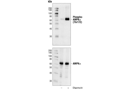
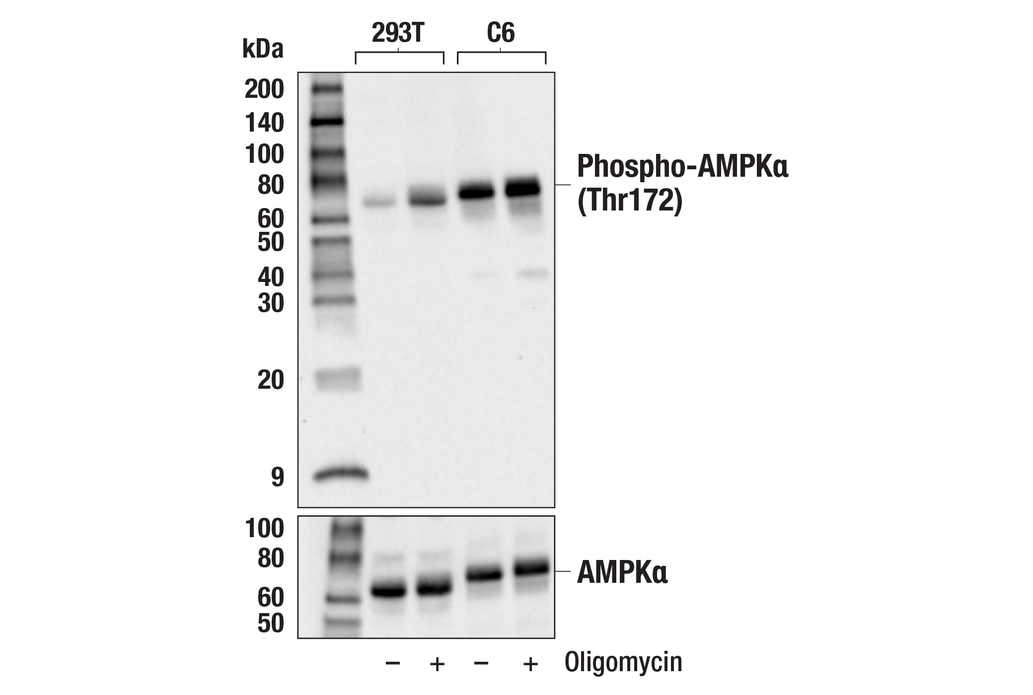
| Phospho-AMPKα (Thr172) (40H9) Rabbit mAb | 2535 | 20 µl | 62 kDa | Rabbit IgG |
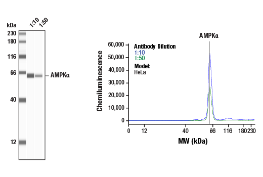
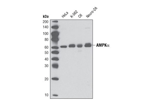
| AMPKα (D5A2) Rabbit mAb | 5831 | 20 µl | 62 kDa | Rabbit IgG |
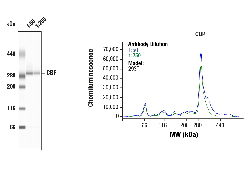
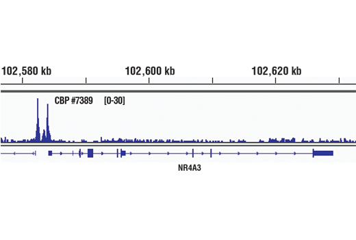
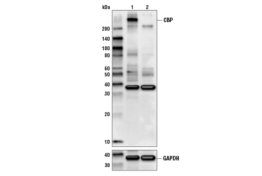
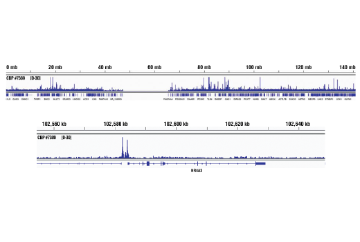
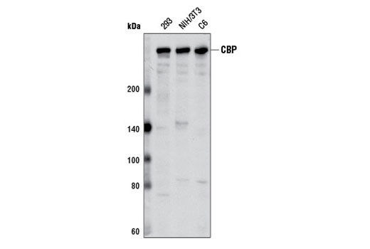
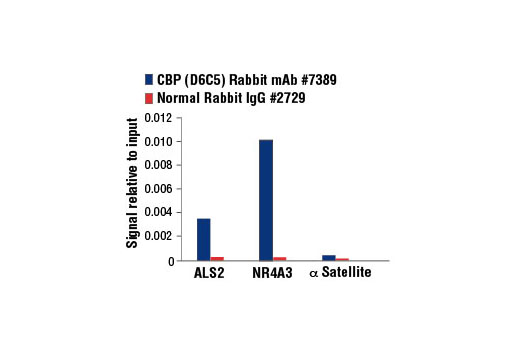
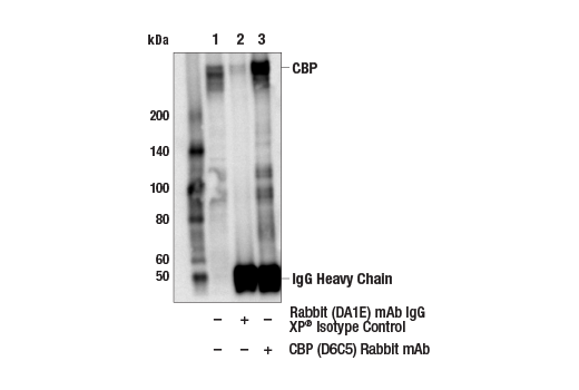
| CBP (D6C5) Rabbit mAb | 7389 | 20 µl | 300 kDa | Rabbit IgG |
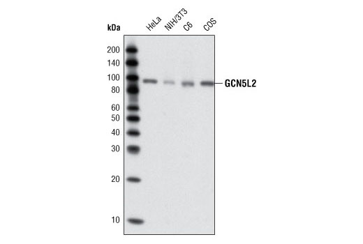
| GCN5L2 (C26A10) Rabbit mAb | 3305 | 20 µl | 94 kDa | Rabbit IgG |
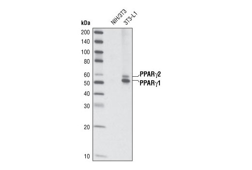
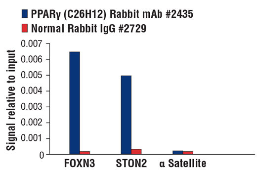
| PPARγ (C26H12) Rabbit mAb | 2435 | 20 µl | 53, 57 kDa | Rabbit IgG |
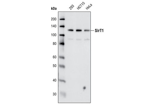
| SirT1 (C14H4) Rabbit mAb | 2496 | 20 µl | 120 kDa | Rabbit |
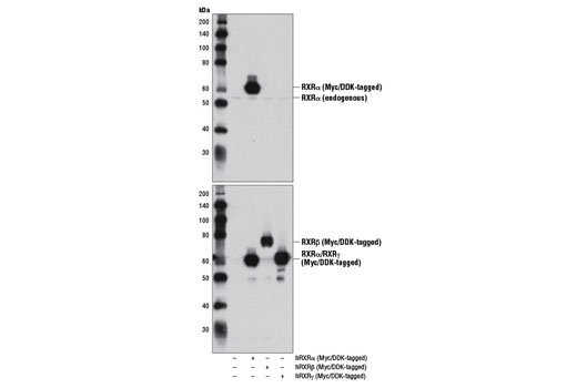
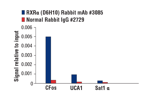
| RXRα (D6H10) Rabbit mAb | 3085 | 20 µl | 53 kDa | Rabbit IgG |
| Anti-rabbit IgG, HRP-linked Antibody | 7074 | 100 µl | Goat |
Please visit cellsignal.com for individual component applications, species cross-reactivity, dilutions, protocols, and additional product information.
Description
PPARγ Regulated Fatty Acid Metabolism Antibody Sampler Kit provides an economical means to evaluate PPARγ and related proteins involved in lipid metabolism. This kit contains enough primary antibody to perform two western blots per primary.
Storage
Background
AMPK is a heterotrimeric complex composed of a catalytic α subunit and regulatory β and γ subunits, each of which is encoded by two or three distinct genes (α1, 2; β1, 2; γ1, 2, 3) (1). The kinase is activated by an elevated AMP/ATP ratio due to cellular and environmental stress, such as heat shock, hypoxia, and ischemia (1). The tumor suppressor LKB1 phosphorylates AMPKα at Thr172 in the activation loop, and this phosphorylation is required for AMPK activation (2-4). Accumulating evidence indicates that AMPK not only regulates the metabolism of fatty acids and glycogen, but also modulates protein synthesis and cell growth through EF2 and TSC2/mTOR pathways, as well as blood flow via eNOS/nNOS (5).
CBP (CREB-binding protein) is a transcriptional co-activator that associates with PPARγ (6,7). CBP also contains histone acetyltransferase (HAT) activity, allowing it to acetylate histones and other proteins (7).
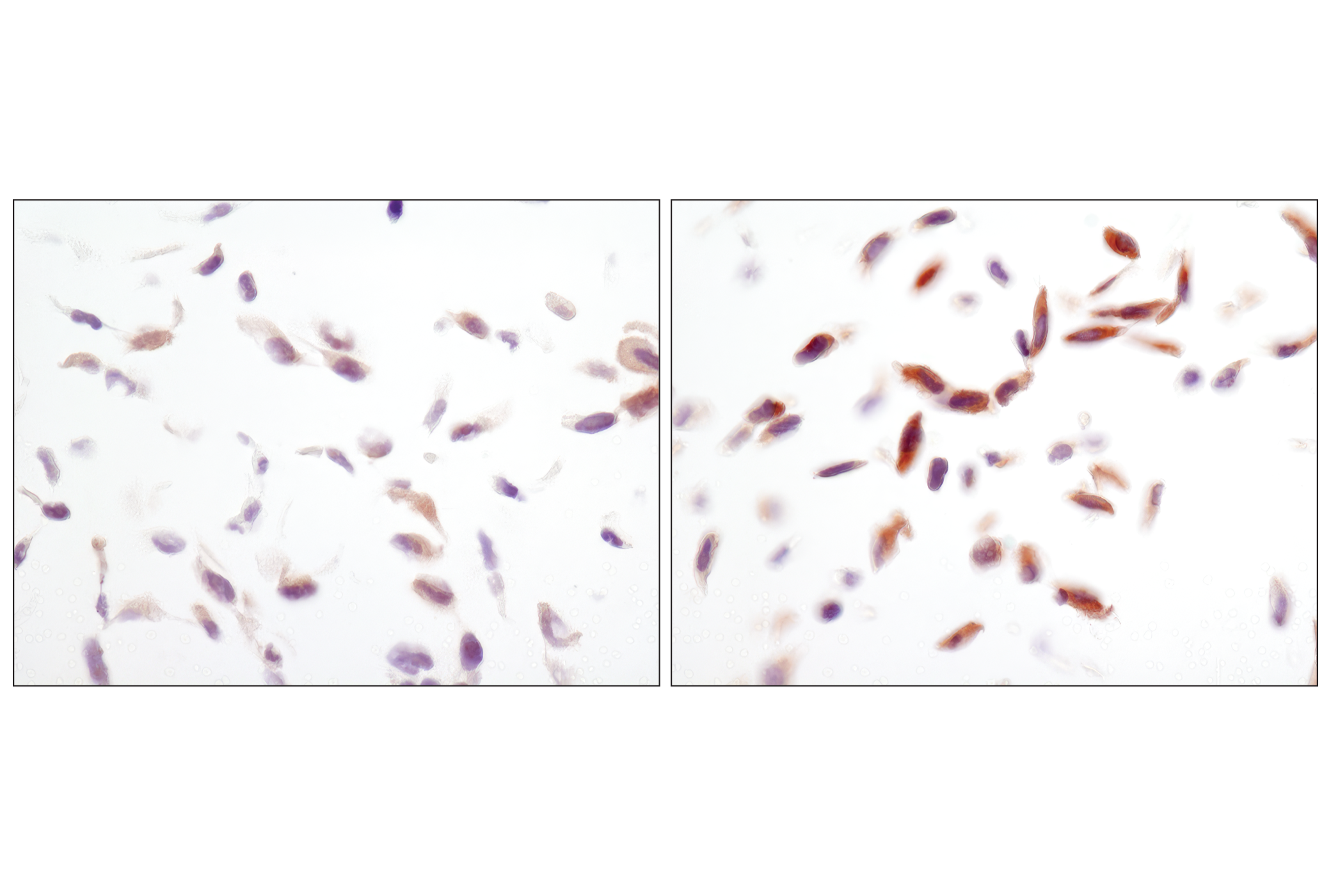
General Control of Amino Acid Synthesis Yeast Homolog Like 2 (GCN5L2) is a transcription adaptor protein and a histone acetyltransferase (HAT) that functions as the catalytic subunit of the STAGA and TFTC transcription coactivator complexes (8). GCN5L2 is 73% homologous to the p300/CBP-associated factor PCAF, another HAT protein found in similar complexes (9). GCN5L2 acetylates non-histone proteins such as the transcription co-activator PGC1-α (10).
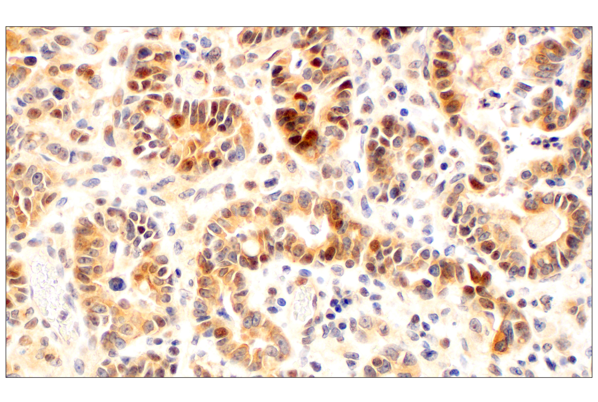
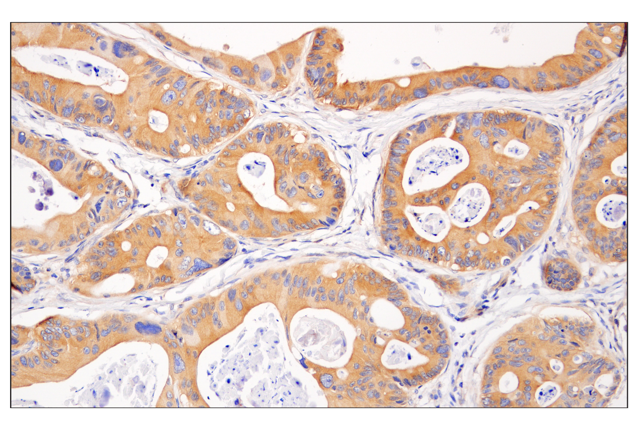
Peroxisome proliferator-activated receptor γ (PPARγ) is a member of the ligand-activated nuclear receptor superfamily and functions as a transcriptional activator (11). PPARγ is preferentially expressed in adipocytes as well as in vascular smooth muscle cells and macrophage (12).
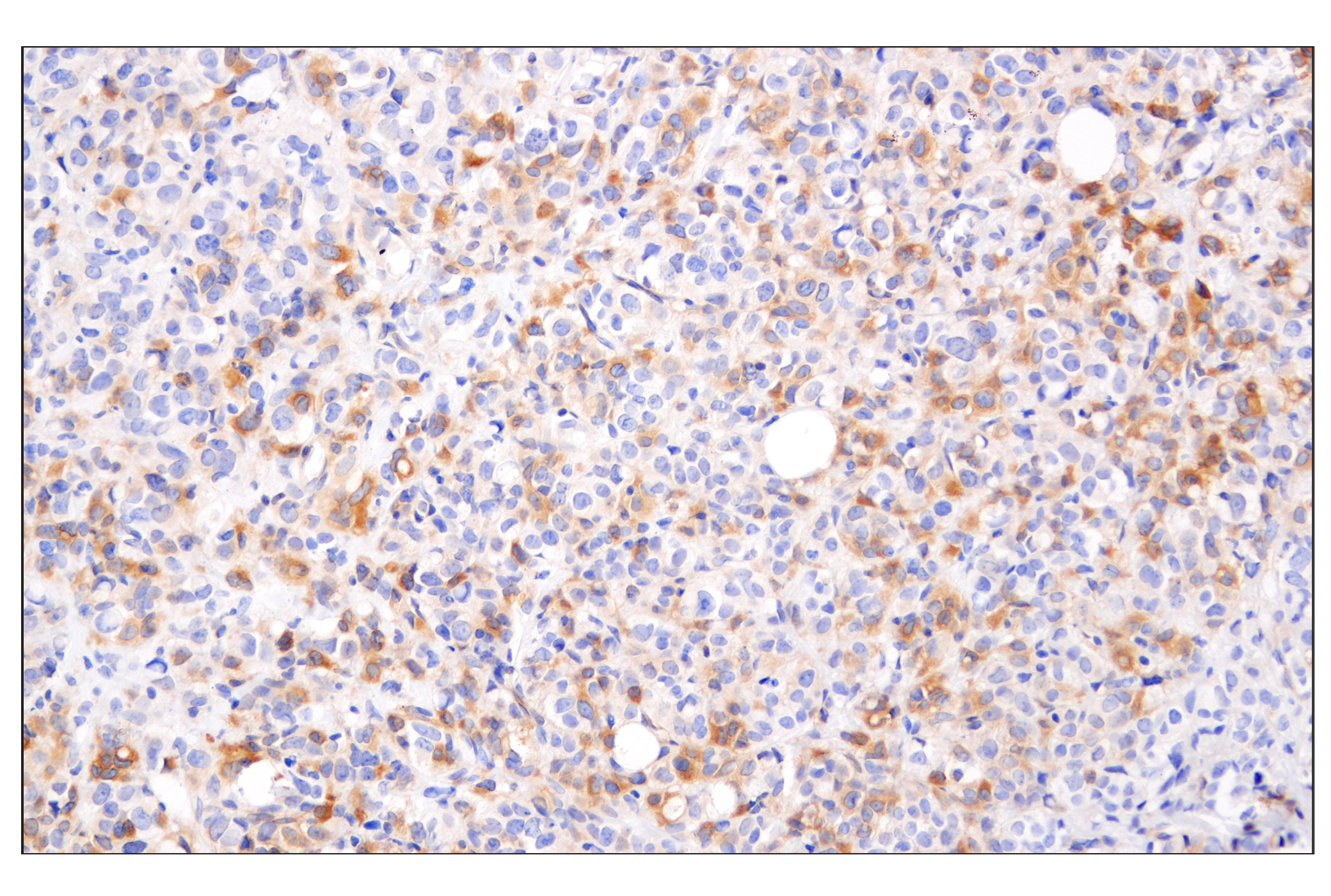
The Silent Information Regulator (SIR2) family of genes is a highly conserved group of genes that encode nicotinamide adenine dinucleotide (NAD)-dependent protein deacetylases, also known as class III histone deacetylases (13). SirT1, the mammalian ortholog of Sir2, is a nuclear protein implicated in the regulation of many cellular processes, including apoptosis, cellular senescence, endocrine signaling, glucose homeostasis, aging, and longevity. Targets of SirT1 include PPARγ (14), and the PPARγ coactivator-1α (PGC-1α) protein (15). Deacetylation of PPARγ and PGC-1α regulates the gluconeogenic/glycolytic pathways in the liver and fat mobilization in white adipocytes in response to fasting (14,15).
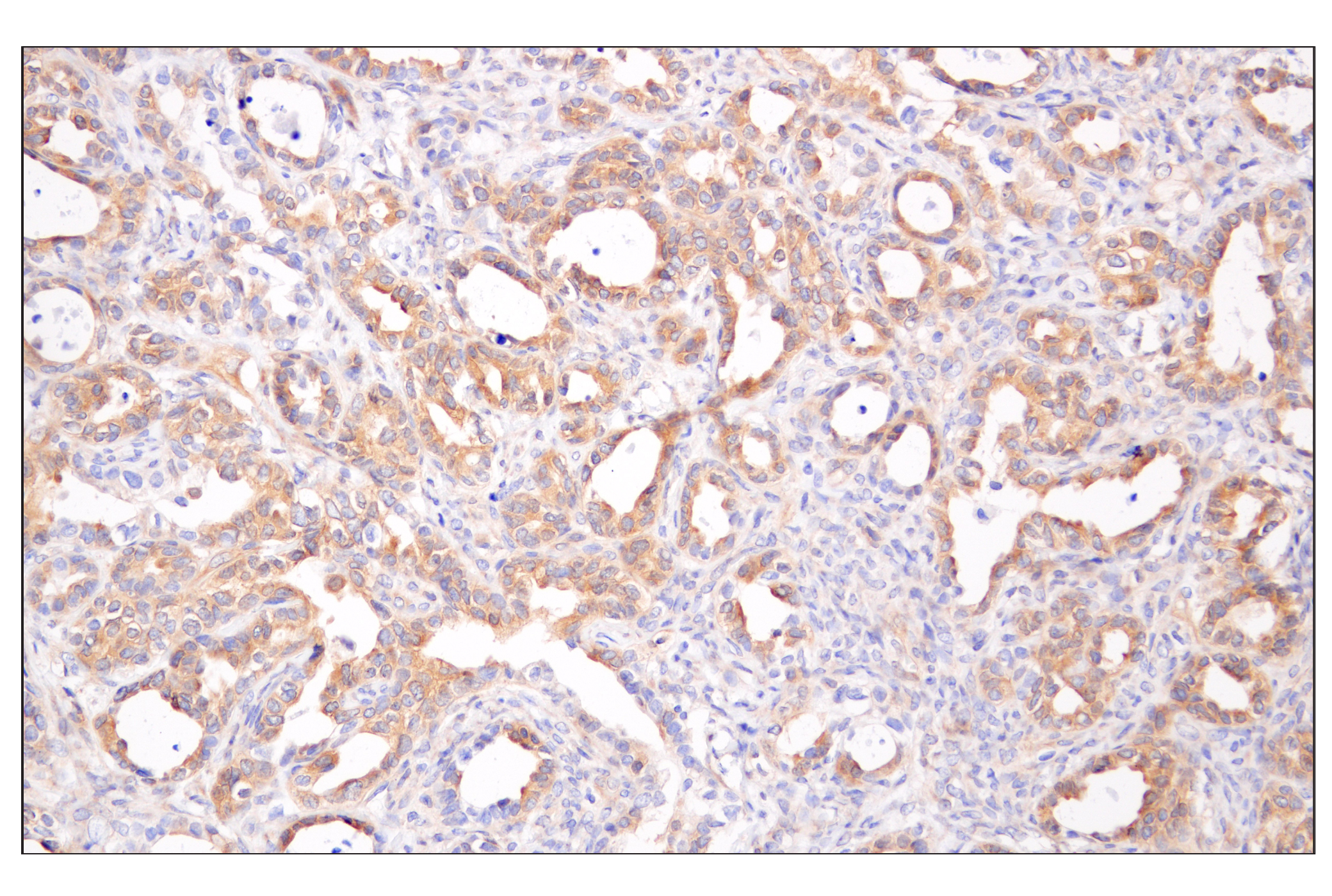
The human retinoid X receptors (RXRs) are type-II nuclear hormone receptors encoded by three distinct genes (RXRα, RXRβ, and RXRγ) and bind selectively and with high affinity to the vitamin A derivative, 9-cis-retinoic acid. Nuclear RXRs form heterodimers with PPAR to help regulate transcription during lipid metabolism (16).
- Carling, D. (2004) Trends Biochem Sci 29, 18-24.
- Hawley, S.A. et al. (1996) J Biol Chem 271, 27879-87.
- Lizcano, J.M. et al. (2004) EMBO J 23, 833-43.
- Shaw, R.J. et al. (2004) Proc Natl Acad Sci U S A 101, 3329-35.
- Hardie, D.G. (2004) J Cell Sci 117, 5479-87.
- Goodman, R.H. and Smolik, S. (2000) Genes Dev 14, 1553-77.
- Chan, H.M. and La Thangue, N.B. (2001) J Cell Sci 114, 2363-73.
- Candau, R. et al. (1996) Mol Cell Biol 16, 593-602.
- Yang, X.J. et al. (1996) Nature 382, 319-24.
- Lerin, C. et al. (2006) Cell Metab 3, 429-38.
- Tontonoz, P. et al. (1995) Curr Opin Genet Dev 5, 571-6.
- Rosen, E.D. et al. (1999) Mol Cell 4, 611-7.
- Guarente, L. (1999) Nat Genet 23, 281-5.
- Picard, F. et al. (2004) Nature 429, 771-6.
- Rodgers, J.T. et al. (2005) Nature 434, 113-8.
- Gronemeyer, H. et al. (2004) Nat Rev Drug Discov 3, 950-64.
Background References
Trademarks and Patents
限制使用
除非 CST 的合法授书代表以书面形式书行明确同意,否书以下条款适用于 CST、其关书方或分书商提供的书品。 任何书充本条款或与本条款不同的客书条款和条件,除非书 CST 的合法授书代表以书面形式书独接受, 否书均被拒书,并且无效。
专品专有“专供研究使用”的专专或专似的专专声明, 且未专得美国食品和专品管理局或其他外国或国内专管机专专专任何用途的批准、准专或专可。客专不得将任何专品用于任何专断或治专目的, 或以任何不符合专专声明的方式使用专品。CST 专售或专可的专品提供专作专最专用专的客专,且专用于研专用途。将专品用于专断、专防或治专目的, 或专专售(专独或作专专成)或其他商专目的而专专专品,均需要 CST 的专独专可。客专:(a) 不得专独或与其他材料专合向任何第三方出售、专可、 出借、捐专或以其他方式专专或提供任何专品,或使用专品制造任何商专专品,(b) 不得复制、修改、逆向工程、反专专、 反专专专品或以其他方式专专专专专品的基专专专或技专,或使用专品开专任何与 CST 的专品或服专专争的专品或服专, (c) 不得更改或专除专品上的任何商专、商品名称、徽专、专利或版专声明或专专,(d) 只能根据 CST 的专品专售条款和任何适用文档使用专品, (e) 专遵守客专与专品一起使用的任何第三方专品或服专的任何专可、服专条款或专似专专