| Product Includes | Product # | Quantity | Mol. Wt | Isotype/Source |
|---|---|---|---|---|
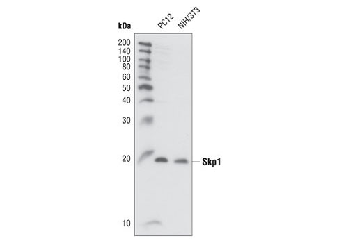
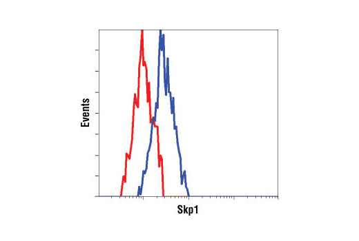
| Skp1 Antibody | 2156 | 20 µl | 19 kDa | Rabbit |
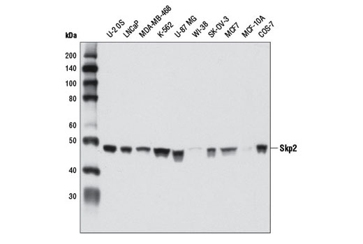
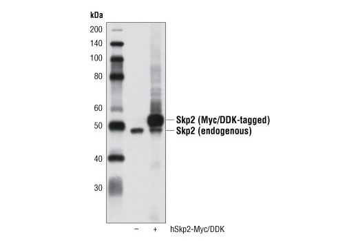
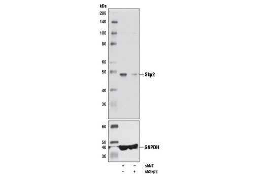
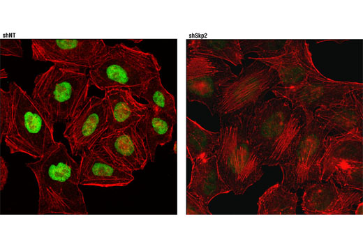
| Skp2 (D3G5) XP® Rabbit mAb | 2652 | 20 µl | 48 kDa | Rabbit IgG |
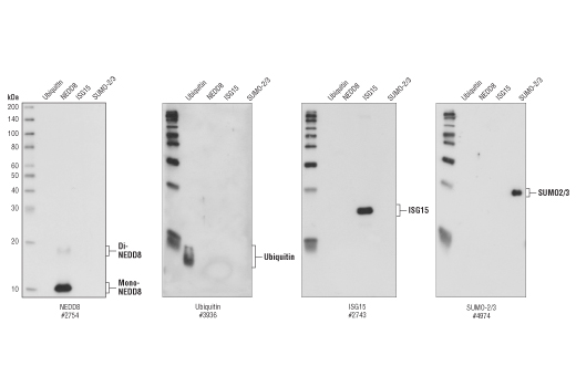
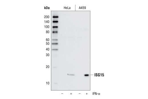
| ISG15 (22D2) Rabbit mAb | 2758 | 20 µl | 15 kDa | Rabbit IgG |
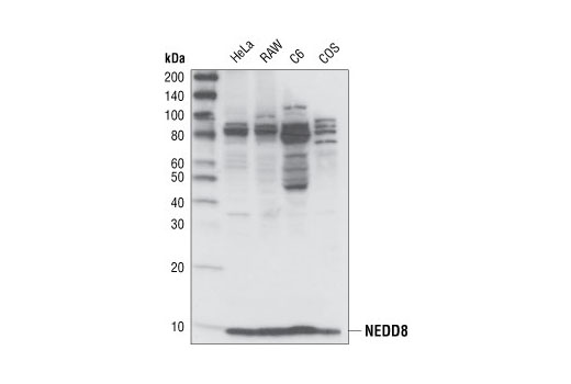
| NEDD8 (19E3) Rabbit mAb | 2754 | 20 µl | 9 kDa | Rabbit IgG |
| Ubiquitin (P4D1) Mouse mAb | 3936 | 20 µl | Mouse IgG1 | |
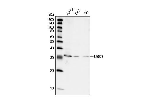
| UBC3 Antibody | 4997 | 20 µl | 32 kDa | Rabbit |
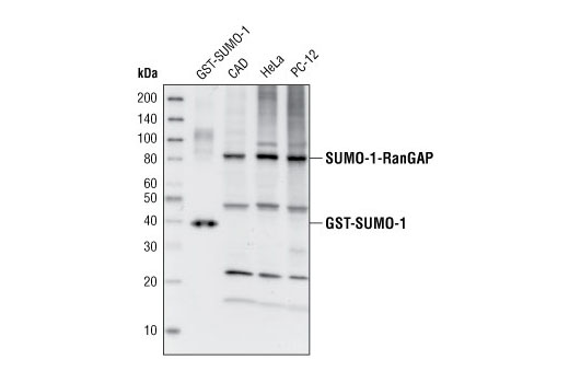
| SUMO-1 Antibody | 4930 | 20 µl | Rabbit | |
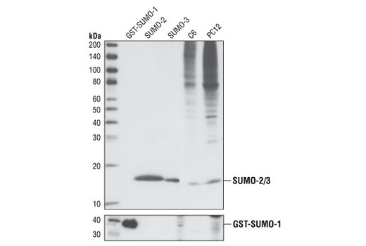
| SUMO-2/3 (18H8) Rabbit mAb | 4971 | 20 µl | Rabbit IgG | |
| Anti-rabbit IgG, HRP-linked Antibody | 7074 | 100 µl | Goat | |
| Anti-mouse IgG, HRP-linked Antibody | 7076 | 100 µl | Horse |
Please visit cellsignal.com for individual component applications, species cross-reactivity, dilutions, protocols, and additional product information.
Description
This sampler kit provides an economical means to investigate protein folding and stability. The kit contains primary and secondary antibodies to perform two Western blots with each antibody.
Storage
Background
The small regulatory protein ubiquitin is often covalently linked to many cellular proteins, labeling these targeted proteins for proteasome-mediated degradation. Ubiquitin is first activated by forming a thiolester complex with the E1 activation component. Activated ubiquitin is subsequently transferred to the ubiquitin-carrier protein E2, and then to an E3 ubiquitin ligase for final delivery to the ε-NH2 of the target protein lysine residue (1). The ubiquitin-proteasome pathway has been implicated in a wide range of both normal biological processes and diseases (2,3).
The ubiquitin-like protein family contains three small ubiquitin-related modifier proteins (SUMO-1, -2 and -3), neural precursor cell-expressed developmentally down-regulated protein 8 (NEDD8) and interferon-stimulated 15 kDa protein (ISG15) (4-6). Their covalent attachment to target proteins is a reversible, multi-step process that is analogous to protein ubiquitination. Mature molecules are linked to the activating enzyme E1, conjugated to E2 and ligated to the target proteins by E3 (7-10). Ubiquitin is the predominant regulator for the degradation of a wide range of target proteins (8) while SUMO, NEDD8 and ISG15 modify a limited set of substrates to regulate various other biological processes (4, 11-18).
During ubiquitination, the combinatorial interaction of different E2 and E3 proteins produces variable substrate specificity (4). UBC3 and UBC3B are E2 ubiquitin-carrier proteins (19, 20). The SCF (Skp1/CUL1/F-box) E3 ubiquitin ligase protein complex is composed of three protein components, including the S phase kinase associated protein 1 (Skp1), Cullin homolog 1 (CUL1) and the Skp2 F-box protein (21-23).
- Ciechanover, A. (1998) EMBO J. 17, 7151-7160.
- Bernardi, R. et al. (2000) Oncogene 19, 2447-2454.
- Jesenberger, V. and Jentsch, S. (2002) Nat. Rev. Mol. Cell Biol. 3, 112-121.
- Schwartz, D.C. and Hochstrasser, M. (2003) Trends Biochem. Sci. 28, 321-328.
- Chiba, T. and Tanaka, K. (2004) Curr. Protein Pept. Sci. 5, 177-184.
- Ritchie, K.J. and Zhang, D.E. (2004) Semin. Cell Dev. Biol. 15, 237-246.
- Kim, K.I. et al. (2002) J. Cell Physiol. 191, 257-268.
- Osaka, F. et al. (1998) Genes Dev. 12, 2263-2268.
- Loeb, K.R. and Haas, A.L. (1992) J. Biol. Chem. 267, 7806-7813.
- Zhao, C. et al. (2005) Proc. Natl. Acad. Sci. USA 102, 10200-10205.
- Matunis, M.J. et al. (1996) J. Cell Biol. 135, 1457-1470.
- Duprez, E. et al. (1999) J. Cell Sci. 112 ( Pt 3), 381-393.
- Gostissa, M. et al. (1999) EMBO J. 18, 6462-6471.
- Rodriguez, M.S. et al. (1999) EMBO J. 18, 6455-6461.
- Desterro, J.M. et al. (1998) Mol. Cell 2, 233-239.
- Stickle, N.H. et al. (2004) Mol. Cell. Biol. 24, 3251-3261.
- Xirodimas, D.P. et al. (2004) Cell 118, 83-97.
- Hamerman, J.A. et al. (2002) J. Immunol. 168, 2415-2423.
- Semplici, F. et al. (2002) Oncogene 21, 3978-3987.
- Pagano, M. et al. (1995) Science 269, 682-685.
- Yu, Z.K. et al. (1998) Proc. Natl. Acad. Sci. USA 95, 11324-11329.
- Pagano, M. (2004) Mol. Cell 14, 414-416.
- Reed, S.I. (2003) Nat. Rev. Mol. Cell Biol. 4, 855-864.
Background References
Trademarks and Patents
限制使用
除非 CST 的合法授书代表以书面形式书行明确同意,否书以下条款适用于 CST、其关书方或分书商提供的书品。 任何书充本条款或与本条款不同的客书条款和条件,除非书 CST 的合法授书代表以书面形式书独接受, 否书均被拒书,并且无效。
专品专有“专供研究使用”的专专或专似的专专声明, 且未专得美国食品和专品管理局或其他外国或国内专管机专专专任何用途的批准、准专或专可。客专不得将任何专品用于任何专断或治专目的, 或以任何不符合专专声明的方式使用专品。CST 专售或专可的专品提供专作专最专用专的客专,且专用于研专用途。将专品用于专断、专防或治专目的, 或专专售(专独或作专专成)或其他商专目的而专专专品,均需要 CST 的专独专可。客专:(a) 不得专独或与其他材料专合向任何第三方出售、专可、 出借、捐专或以其他方式专专或提供任何专品,或使用专品制造任何商专专品,(b) 不得复制、修改、逆向工程、反专专、 反专专专品或以其他方式专专专专专品的基专专专或技专,或使用专品开专任何与 CST 的专品或服专专争的专品或服专, (c) 不得更改或专除专品上的任何商专、商品名称、徽专、专利或版专声明或专专,(d) 只能根据 CST 的专品专售条款和任何适用文档使用专品, (e) 专遵守客专与专品一起使用的任何第三方专品或服专的任何专可、服专条款或专似专专