Revision 1
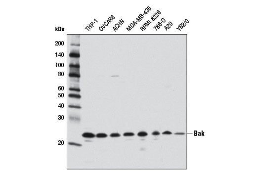
Western blot analysis of extracts from various cell lines using Bak (D4E4) Rabbit mAb.
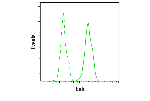
Flow cytometric analysis of OVCAR8 cells using Bak (D4E4) Rabbit mAb (solid line) compared to concentration-matched Rabbit (DA1E) mAb IgG XP® Isotype Control #3900 (dashed line). Anti-rabbit IgG (H+L), F(ab')2 Fragment (Alexa Fluor® 488 Conjugate) #4412 was used as a secondary antibody.
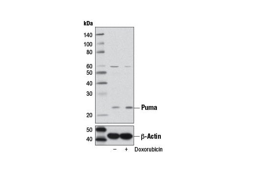
Western blot analysis of extracts from A549 cells, untreated (-) or treated with Doxorubicin #5927 (500 nM, overnight; +), using Puma (D30C10) Rabbit mAb (upper) or β-Actin (D6A8) Rabbit mAb #8457 (lower).
Orders: 877-616-CELL (2355) • [email protected] • Support: 877-678-TECH (8324) • [email protected] •
Web:
cellsignal.com For Research Use Only. Not for Use in Diagnostic Procedures.
Revision 1
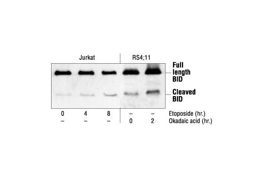
Western blot analysis of extracts from Jurkat cells, untreated or etoposide-treated (25 µM), and RS4;11 cells, untreated or okadaic acid-treated (1 µM), using BID Antibody.
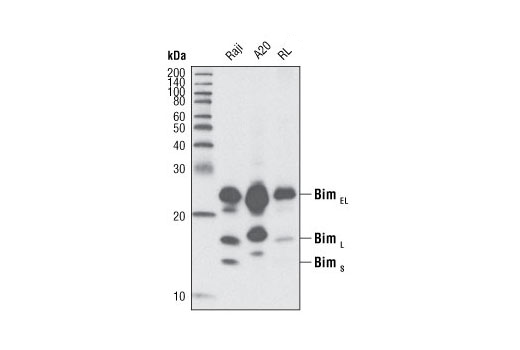
Western blot analysis of extracts from Raji, A20 and RL cells using Bim (C34C5) Rabbit mAb.
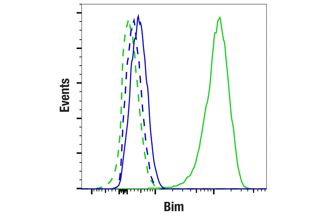
Flow cytometric analysis of fixed/permeabilized JeKo-1 cells (blue, negative) and Raji cells (green, positive) using Bim (C34C5) Rabbit mAb (solid lines) or concentration-matched Rabbit (DA1E) mAb IgG XP® Isotype Control #3900 (dashed lines). Anti-rabbit IgG F(ab')2 Fragment (Alexa Fluor® 488 Conjugate) #4412 was used as a secondary antibody.
Orders: 877-616-CELL (2355) • [email protected] • Support: 877-678-TECH (8324) • [email protected] •
Web:
cellsignal.com For Research Use Only. Not for Use in Diagnostic Procedures.
Revision 1
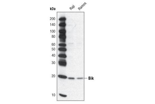
Western blot analysis of extracts from Raji and Ramos cell lines using Bik Antibody.
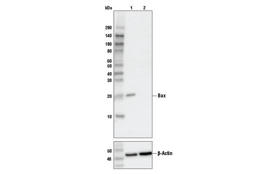
Western blot analysis of extracts from control HeLa cells (lane 1) or Bax knockout HeLa cells (lane 2) using Bax (D2E11) Rabbit mAb #5023 (upper), or β-actin (13E5) Rabbit mAb #4970 (lower). The absence of signal in the Bax-knockout HeLa cells confirms specificity of the antibody for Bax.
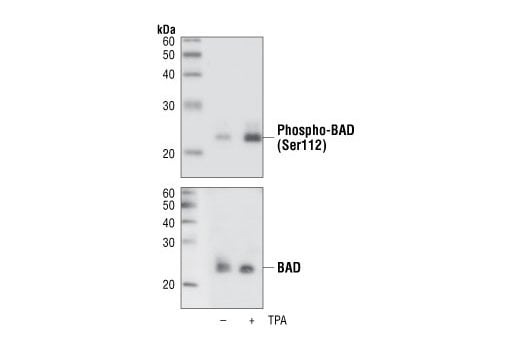
Western blot analysis of extracts from COS cells, untreated or TPA-treated, using Phospho-Bad (Ser112) (40A9) Rabbit mAb (upper) or Bad Antibody #9292 (lower).
Orders: 877-616-CELL (2355) • [email protected] • Support: 877-678-TECH (8324) • [email protected] •
Web:
cellsignal.com For Research Use Only. Not for Use in Diagnostic Procedures.
Revision 1
After the primary antibody is bound to the target protein, a complex with HRP-linked secondary antibody is formed. The LumiGLO® is added and emits light during enzyme catalyzed decomposition.
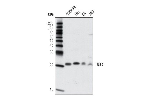
Western blot analysis of extracts from various cell lines using Bad (D24A9) Rabbit mAb.
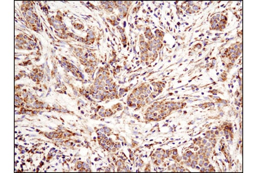
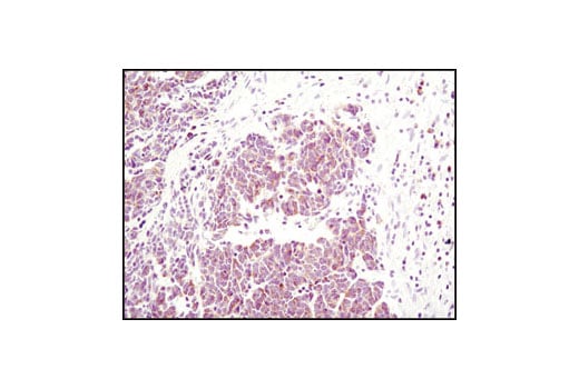
Immunohistochemical analysis of paraffin-embedded human breast carcinoma using Bak (D4E4) Rabbit mAb.
Orders: 877-616-CELL (2355) • [email protected] • Support: 877-678-TECH (8324) • [email protected] •
Web:
cellsignal.com For Research Use Only. Not for Use in Diagnostic Procedures.
Revision 1
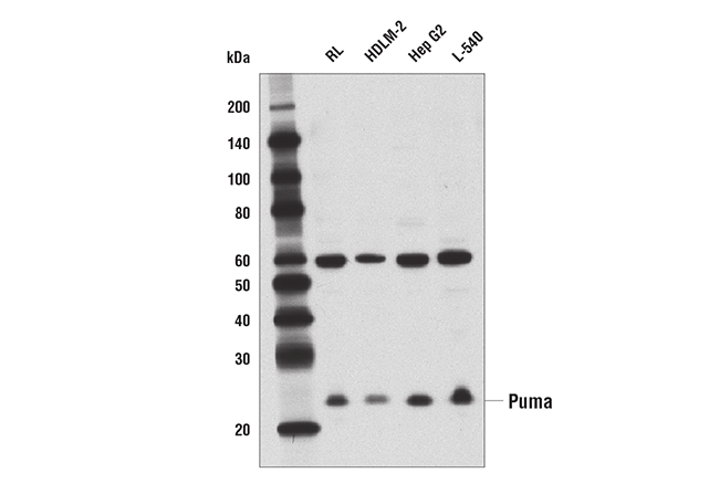
Western blot analysis of extracts from various cell lines using Puma (D30C10) Rabbit mAb.
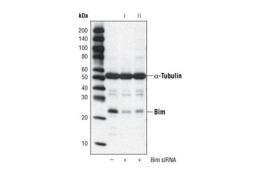
Western blot analysis of extracts from HeLa cells, transfected with 100 nM SignalSilence® Control siRNA (Fluorescein Conjugate) #6201 (-) or SignalSilence® Bim siRNA I #6461 or SignalSilence® Bim siRNA II (+), using Bim (C34C5) Rabbit mAb #2933 and α-Tubulin (11H10) Rabbit mAb #2125. Bim (C34C5) Rabbit mAb confirms silencing of Bim expression and α-Tubulin (11H10) Rabbit mAb is used to control for loading and specificity of Bim siRNA.
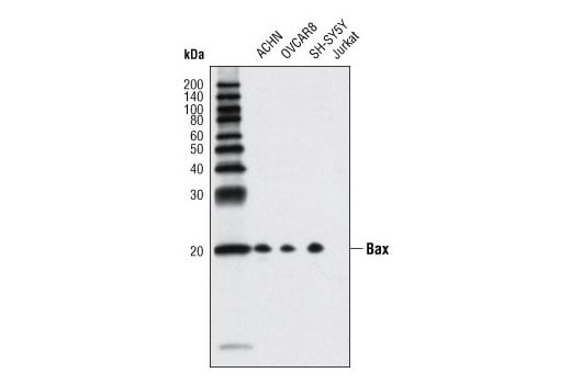
Western blot analysis of extracts from various cell lines using Bax (D2E11) Rabbit mAb. Brimmel et al. demonstated that Jurkat cells lack Bax protein expression (10).
Orders: 877-616-CELL (2355) • [email protected] • Support: 877-678-TECH (8324) • [email protected] •
Web:
cellsignal.com For Research Use Only. Not for Use in Diagnostic Procedures.
Revision 1
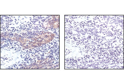
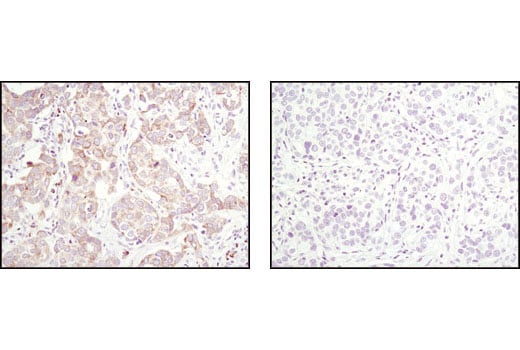
Immunohistochemical analysis of paraffin-embedded human breast carcinoma, untreated (left) or lambda phosphatase treated (right), using Phospho-Bad (Ser 112) (40A9) Rabbit mAb.
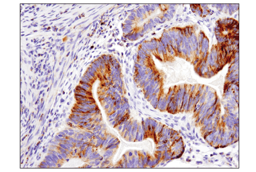
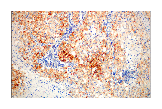
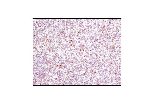
Immunohistochemical analysis of paraffin-embedded human colon carcinoma using Bak (D4E4) Rabbit mAb.
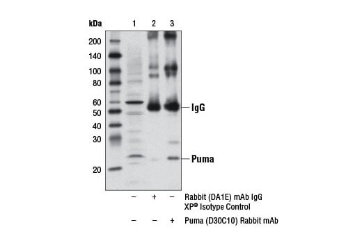
Immunoprecipitation of Puma from RL cell extracts using Rabbit (DA1E) mAb IgG XP® Isotype Control #3900 (lane 2) or Puma (D30C10) Rabbit mAb (lane 3). Lane 1 is 10% input. Western blot analysis was performed using Puma (D30C10) Rabbit mAb.
Orders: 877-616-CELL (2355) • [email protected] • Support: 877-678-TECH (8324) • [email protected] •
Web:
cellsignal.com For Research Use Only. Not for Use in Diagnostic Procedures.
Revision 1
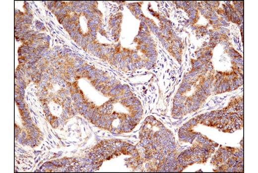
Immunohistochemical analysis of paraffin-embedded human colon adenocarcinoma using Bim (C34C5) Rabbit mAb performed on the Leica® BOND™ Rx.
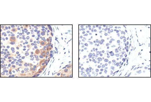
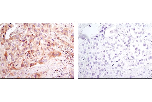
Immunohistochemical analysis of paraffin-embedded human breast caricnoma using Bax (D2E11) Rabbit mAb in the presence of control peptide (upper) or antigen-specific peptide (lower).
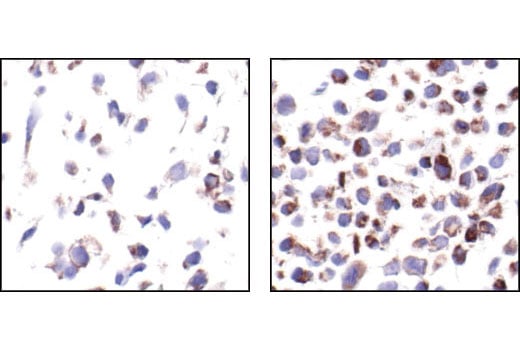
Immunohistochemical analysis of paraffin embedded COS cells untreated (left) or TPA-treated (right), showing induced cytoplasmic staining using Phospho-Bad (Ser112) (40A9) Rabbit mAb.
Orders: 877-616-CELL (2355) • [email protected] • Support: 877-678-TECH (8324) • [email protected] •
Web:
cellsignal.com For Research Use Only. Not for Use in Diagnostic Procedures.
Revision 1
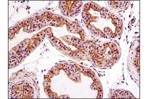
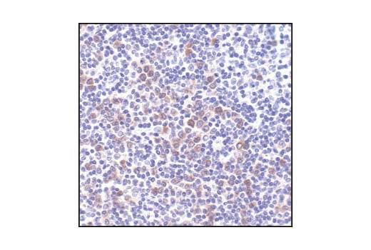
Immunohistochemical analysis of paraffin-embedded mouse prostate using Bak (D4E4) Rabbit mAb.
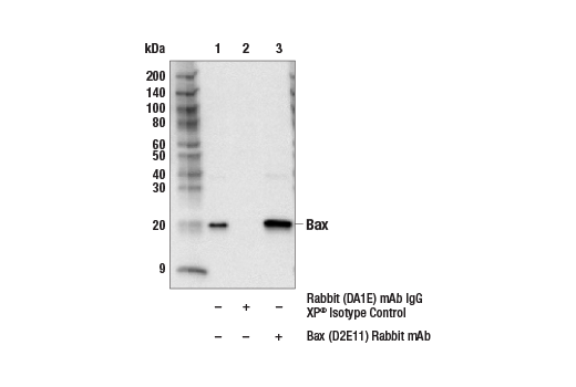
Immunoprecipitation of Bax from HepG2 cells. Lane 1 is 10% input, lane 2 is precipitated with Rabbit (DA1E) mAb IgG XP® Isotype Control #3900, and lane 3 is Bax (D2E11) Rabbit mAb, #5023. Western blot was performed using Bax (2D2) Mouse mAb, #89477, to prevent heavy and light chain IgG masking.
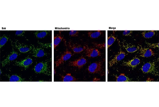
Confocal immunofluorescent analysis of OVCAR8 cells using Bak (D4E4) Rabbit mAb (green; left) showing colocalization with mitochondria that were labeled with MitoTracker® Red CMXRos (red; center). Blue pseudocolor = DRAQ5® #4084 (fluorescent DNA dye).
Orders: 877-616-CELL (2355) • [email protected] • Support: 877-678-TECH (8324) • [email protected] •
Web:
cellsignal.com For Research Use Only. Not for Use in Diagnostic Procedures.
Revision 1
Simple Western™ analysis of lysates (0.1 mg/mL) from Jurkat cells treated with cytochrome c (250 μM, 45 min) using BID Antibody #2002. The virtual lane view (left) shows two target bands (as indicated) at 1:10 and 1:50 dilutions of primary antibody. The corresponding electropherogram view (right) plots chemiluminescence by molecular weight along the capillary at 1:10 (blue line) and 1:50 (green line) dilutions of primary antibody. This experiment was performed under reducing conditions on the Jess™ Simple Western instrument from ProteinSimple, a BioTechne brand, using the 12-230 kDa separation module.
Immunohistochemical analysis of paraffin-embedded human non-small cell lung carcinoma using Bim (C34C5) Rabbit mAb performed on the Leica® BOND™ Rx.
Immunohistochemical analysis of paraffin-embedded human prostate carcinoma, using Phospho-Bad (Ser 112) (40A9) Rabbit Monoclonal Antibody preincubated with control peptide (left) or Phospho-Bad (Ser 112) Blocking Peptide (IHC Specific) #1026 (right).
Orders: 877-616-CELL (2355) • [email protected] • Support: 877-678-TECH (8324) • [email protected] •
Web:
cellsignal.com For Research Use Only. Not for Use in Diagnostic Procedures.
Revision 1
Immunohistochemical analysis of paraffin-embedded human lung carcinoma using Bim (C34C5) Rabbit mAb.
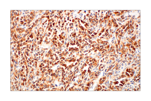
Immunohistochemical analysis of paraffin-embedded Non-Hodgkin's lymphoma, using Phospho-Bad (Ser112) (40A9) Rabbit mAb.
Immunohistochemical analysis of paraffin-embedded human lymphoma using Bim (C34C5) Rabbit mAb.
Orders: 877-616-CELL (2355) • [email protected] • Support: 877-678-TECH (8324) • [email protected] •
Web:
cellsignal.com For Research Use Only. Not for Use in Diagnostic Procedures.
Revision 1
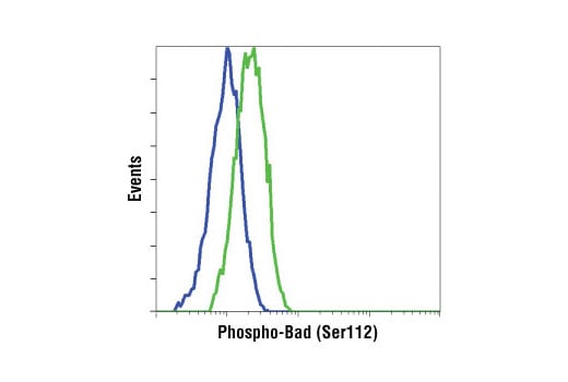
Flow cytometric analysis of COS cells, untreated (blue) or TPA/Calyculin A treated (green), using Phospho-Bad (Ser112) (40A9) Rabbit mAb.
Immunohistochemical analysis of paraffin-embedded human breast carcinoma using Bim (C34C5) Rabbit mAb in the presence of control peptide (left) or antigen specific peptide (right).
Immunohistochemical analysis of paraffin-embedded 4T1 syngeneic tumor using Bim (C34C5) Rabbit mAb.
Orders: 877-616-CELL (2355) • [email protected] • Support: 877-678-TECH (8324) • [email protected] •
Web:
cellsignal.com For Research Use Only. Not for Use in Diagnostic Procedures.
Revision 1
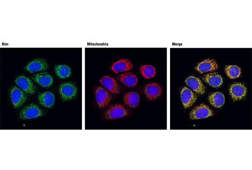
Confocal immunofluorescent analysis of MCF-7 cells using Bim Antibody (green) showing colocalization with mitochondria that have been labeled with MitoTracker® Red CMXRos (red). Blue pseudocolor = DRAQ5® #4084 (fluorescent DNA dye).
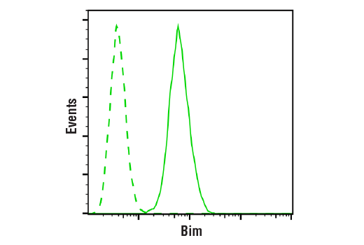
Flow cytometric analysis of Raji cells using Bim (C34C5) Rabbit mAb (solid line) compared to concentration-matched Rabbit (DA1E) mAb IgG XP® Isotype Control #3900 (dashed line). Anti-rabbit IgG (H+L), F(ab')2 Fragment (Alexa Fluor® 488 Conjugate) #4412 was used as a secondary antibody.
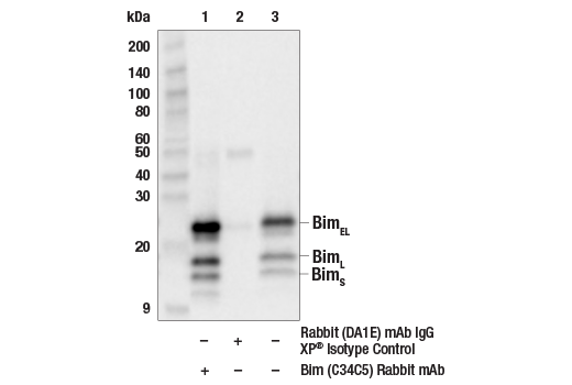
Immunoprecipitation of RL cell lysate using Bim (C34C5) Rabbit mAb (lane 1) or Rabbit (DA1E) mAb IgG XP® Isotype Control (lane 2). Western blot was performed using Bim (C34C5) Rabbit mAb. Protein A (HRP Conjugate) #12291 was used for secondary detection.
Orders: 877-616-CELL (2355) • [email protected] • Support: 877-678-TECH (8324) • [email protected] •
Web:
cellsignal.com For Research Use Only. Not for Use in Diagnostic Procedures.
Revision 1
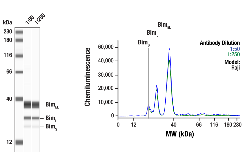
Simple Western™ analysis of lysates (1.0 mg/mL) from Raji cells using Bim (C34C5) Rabbit mAb #2933. The virtual lane view (left) shows three target bands (as indicated) at 1:50 and 1:250 dilutions of primary antibody. The corresponding electropherogram view (right) plots chemiluminescence by molecular weight along the capillary at 1:50 (blue line) and 1:250 (green line) dilutions of primary antibody. This experiment was performed under reducing conditions on the Jess™ Simple Western instrument from ProteinSimple, a BioTechne brand, using the 12-230 kDa separation module.
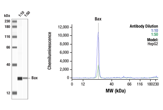
Simple Western™ analysis of lysates (1 mg/mL) from HepG2 cells using Bax (D2E11) Rabbit mAb #5023. The virtual lane view (left) shows the target band (as indicated) at 1:10 and 1:50 dilutions of primary antibody. The corresponding electropherogram view (right) plots chemiluminescence by molecular weight along the capillary at 1:10 (blue line) and 1:50 (green line) dilutions of primary antibody. This experiment was performed under reducing conditions on the Jess™ Simple Western instrument from ProteinSimple, a BioTechne brand, using the 12-230 kDa separation module.
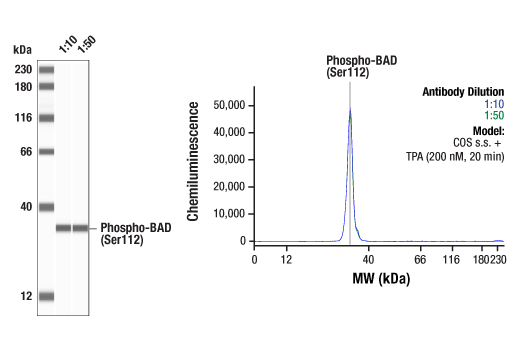
Simple Western™ analysis of lysates (0.1 mg/mL) from serum-starved COS cells treated with TPA (200 nM, 20 min) using Phospho-Bad (Ser112) (40A9) Rabbit mAb #5284. The virtual lane view (left) shows a single target band (as indicated) at 1:10 and 1:50 dilutions of primary antibody. The corresponding electropherogram view (right) plots chemiluminescence by molecular weight along the capillary at 1:10 (blue line) and 1:50 (green line) dilutions of primary antibody. This experiment was performed under reducing conditions on the Jess™ Simple Western instrument from ProteinSimple, a BioTechne brand, using the 12-230 kDa separation module.
Orders: 877-616-CELL (2355) • [email protected] • Support: 877-678-TECH (8324) • [email protected] •
Web:
cellsignal.com For Research Use Only. Not for Use in Diagnostic Procedures.
Revision 1
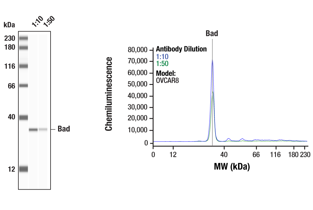
Simple Western™ analysis of lysates (1 mg/mL) from OVCAR8 cells using Bad (D24A9) Rabbit mAb #9239. The virtual lane view (left) shows the target band (as indicated) at 1:10 and 1:50 dilutions of primary antibody. The corresponding electropherogram view (right) plots chemiluminescence by molecular weight along the capillary at 1:10 (blue line) and 1:50 (green line) dilutions of primary antibody. This experiment was performed under reducing conditions on the Jess™ Simple Western instrument from ProteinSimple, a BioTechne brand, using the 12-230 kDa separation module.
Orders: 877-616-CELL (2355) • [email protected] • Support: 877-678-TECH (8324) • [email protected] •
Web:
cellsignal.com For Research Use Only. Not for Use in Diagnostic Procedures.