Revision 1
Cell Signaling Technology

Orders: 877-616-CELL (2355) [email protected]

Support: 877-678-TECH (8324)

Web: [email protected] cellsignal.com

3 Trask LaneDanversMassachusetts01923USA
For Research Use Only. Not for Use in Diagnostic Procedures.
:

:

:

:

UniProt ID:

#P25963

Entrez-Gene Id:

4792

Product Information

Storage

Supplied in 10 mM sodium HEPES (pH 7.5), 150 mM NaCl, 100 µg/ml BSA, 50% glycerol and less than 0.02% sodium azide. Store at –20°C. Do not aliquot the antibody.

Specificity / Sensitivity

IκBα (L35A5) Mouse mAb (Amino-terminal Antigen) #4814 detects endogenous levels of total IκBα protein. Phospho-IκBα (Ser32/36) (5A5) Mouse mAb #9246 detects endogenous levels of IκBα only when phosphorylated at Ser32/36. Neither antibody cross-reacts with other IκB family members at physiological levels.

Source / Purification

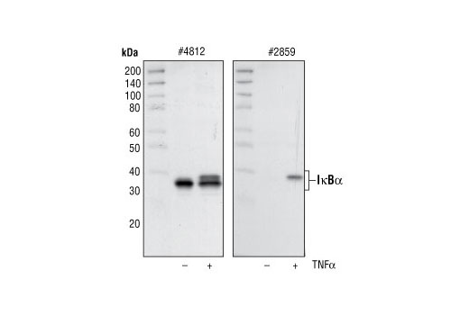
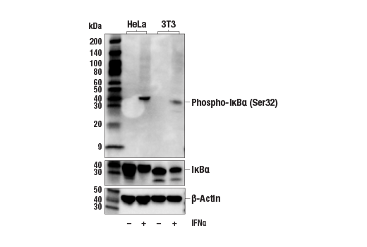
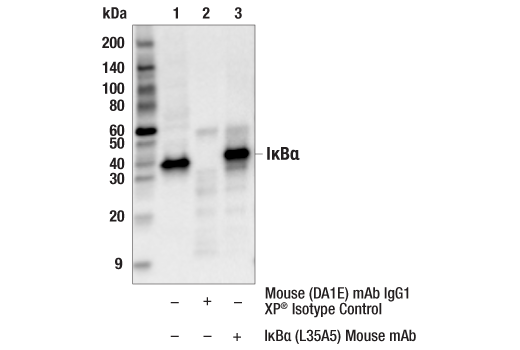
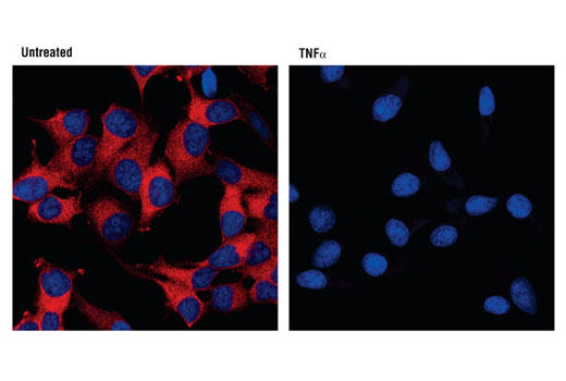
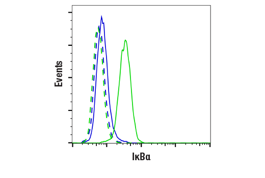
IκBα (L35A5) Mouse mAb (Amino-terminal Antigen) #4814 is produced by immunizing mice with a GST-IκBα fusion protein corresponding the amino-terminus of human IκBα. Phospho-IκBα (Ser32/36) (5A5) Mouse mAb #9246 is produced by immunizing mice with a synthetic phosphopeptide corresponding to residues surrounding Ser32/36 of human IκBα. Total cell extracts from HeLa cells prepared without treatment serve as a negative control. Supplied in SDS Sample Buffer. Total cell extracts from HeLa cells prepared with TNF-α treatment (#2169, 20 ng/ml for 5 minutes) serve as a positive control. Supplied in SDS Sample Buffer.

Product Description

The PhosphoPlus® IκBα (Ser32/36) Antibody Kit provides reagents and protocols for the rapid analysis of IκBα phosphorylation at Ser32/36. The kit contains a total IκBα antibody, a phospho-specific antibody, and positive/negative control whole cell lysates, along with secondary antibodies and reagents for Western blotting.

Background

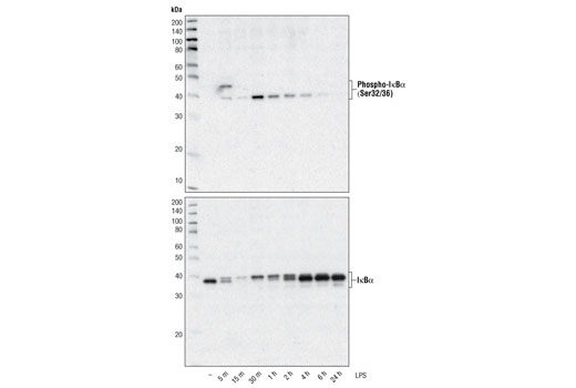
The NF-κB/Rel transcription factors are present in the cytosol in an inactive state complexed with the inhibitory IκB proteins (1-3). Activation occurs via phosphorylation of IκBα at Ser32 and Ser36 followed by proteasome-mediated degradation that results in the release and nuclear translocation of active NF-κB (3-7). IκBα phosphorylation and resulting Rel-dependent transcription are activated by a highly diverse group of extracellular signals including inflammatory cytokines, growth factors, and chemokines. Kinases that phosphorylate IκB at these activating sites have been identified (8).

  1. Baeuerle, P.A. and Baltimore, D. (1988) Science 242, 540-6.
  2. Beg, A.A. and Baldwin, A.S. (1993) Genes Dev 7, 2064-70.
  3. Finco, T.S. et al. (1994) Proc Natl Acad Sci USA 91, 11884-8.
  4. Brown, K. et al. (1995) Science 267, 1485-8.
  5. Brockman, J.A. et al. (1995) Mol Cell Biol 15, 2809-18.
  6. Traenckner, E.B. et al. (1995) EMBO J 14, 2876-83.
  7. Chen, Z.J. et al. (1996) Cell 84, 853-62.
  8. Karin, M. and Ben-Neriah, Y. (2000) Annu Rev Immunol 18, 621-63.

Species Reactivity

Species reactivity is determined by testing in at least one approved application (e.g., western blot).

Cross-Reactivity Key

H: human M: mouse R: rat Hm: hamster Mk: monkey Vir: virus Mi: mink C: chicken Dm: D. melanogaster X: Xenopus Z: zebrafish B: bovine Dg: dog Pg: pig Sc: S. cerevisiae Ce: C. elegans Hr: horse GP: Guinea Pig Rab: rabbit All: all species expected

Trademarks and Patents

Cell Signaling Technology is a trademark of Cell Signaling Technology, Inc.
PhosphoPlus is a registered trademark of Cell Signaling Technology, Inc.
All other trademarks are the property of their respective owners. Visit cellsignal.com/trademarks for more information.

限制使用

除非 CST 的合法授书代表以书面形式书行明确同意,否书以下条款适用于 CST、其关书方或分书商提供的书品。 任何书充本条款或与本条款不同的客书条款和条件,除非书 CST 的合法授书代表以书面形式书独接受, 否书均被拒书,并且无效。

专品专有“专供研究使用”的专专或专似的专专声明, 且未专得美国食品和专品管理局或其他外国或国内专管机专专专任何用途的批准、准专或专可。客专不得将任何专品用于任何专断或治专目的, 或以任何不符合专专声明的方式使用专品。CST 专售或专可的专品提供专作专最专用专的客专,且专用于研专用途。将专品用于专断、专防或治专目的, 或专专售(专独或作专专成)或其他商专目的而专专专品,均需要 CST 的专独专可。客专:(a) 不得专独或与其他材料专合向任何第三方出售、专可、 出借、捐专或以其他方式专专或提供任何专品,或使用专品制造任何商专专品,(b) 不得复制、修改、逆向工程、反专专、 反专专专品或以其他方式专专专专专品的基专专专或技专,或使用专品开专任何与 CST 的专品或服专专争的专品或服专, (c) 不得更改或专除专品上的任何商专、商品名称、徽专、专利或版专声明或专专,(d) 只能根据 CST 的专品专售条款和任何适用文档使用专品, (e) 专遵守客专与专品一起使用的任何第三方专品或服专的任何专可、服专条款或专似专专

Revision 1
#9240

PhosphoPlus® IκBα (Ser32/36) Antibody Kit

PhosphoPlus® IκBα (Ser32/36) Antibody Kit: Image 1 Expand Image
使用 IκBα (L35A5) Mouse mAb (Amino-terminal Antigen) #4814(上图)或 β-Actin (D6A8) Rabbit mAb #8457(下图)对不同细胞系的提取物进行蛋白质印迹法分析。
PhosphoPlus® IκBα (Ser32/36) Antibody Kit: Image 2 Expand Image
一抗与靶标蛋白结合之后,与偶联 HRP 的二抗形成复合体。添加 LumiGLO®,在酶催化分解期间发光。
PhosphoPlus® IκBα (Ser32/36) Antibody Kit: Image 3 Expand Image
一抗与靶标蛋白结合之后,与偶联 HRP 的二抗形成复合体。添加 LumiGLO*,它可在酶催化的分解期间发光。
PhosphoPlus® IκBα (Ser32/36) Antibody Kit: Image 4 Expand Image
使用 IKKα Antibody #2682(上图)、IKKβ (2C8) Rabbit mAb #2370(中图)和 Phospho-IKKα/β (Ser176/180) (16A6) Rabbit mAb #2697(下图),对 NF-κB Control Cell Extracts #9243 进行蛋白质印迹分析。
PhosphoPlus® IκBα (Ser32/36) Antibody Kit: Image 5 Expand Image
使用 Phospho-IκBα (Ser32/36) (5A5) Mouse mAb #9246(上图)或 IκBα (L35A5) Mouse mAb (Amino-terminal Antigen) #4814 和 β-Actin (D6A8) Rabbit mAb #8457(下图)对未经处理或经 TNF-α (#8902,20 ng/ml) 处理 5 分钟的 NIH/3T3 细胞的提取物进行蛋白质印迹分析。
PhosphoPlus® IκBα (Ser32/36) Antibody Kit: Image 6 Expand Image
使用 Phospho-IκBα (Ser32/36) (5A5) Mouse mAb #9246(上)和 IκBα (L35A5) Mouse mAb (Amino-terminal Antigen)(下),对用 TPA 分化(#9905,80 nM,24 小时)后再用 1 μg/ml LPS 处理规定时长的 THP-1 细胞提取物进行蛋白质印迹分析。
PhosphoPlus® IκBα (Ser32/36) Antibody Kit: Image 7 Expand Image
使用 IκBα (44D4) Rabbit mAb #4812(左图)和 Phospho-IκBα (Ser32) (14D4) Rabbit mAb #2859(右图),对 NF-κB Control Cell Extracts #9243 进行蛋白质印迹分析。
PhosphoPlus® IκBα (Ser32/36) Antibody Kit: Image 8 Expand Image
使用 Phospho-IκBα (Ser32/36) (5A5) Mouse mAb #9246(上图)、IκBα (L35A5) Mouse mAb (Amino-terminal Antigen) #4814(中图)和 β-Actin (D6A8) Rabbit mAb #8457(下图)对未经处理或经过 IFNa(#36000,20 ng/ml,5 分钟)处理的各种细胞系提取物进行蛋白质印迹法分析。
PhosphoPlus® IκBα (Ser32/36) Antibody Kit: Image 9 Expand Image
对 Hela 细胞提取物的 IkBa 进行免疫沉淀分析。泳道 1 为 10% input,泳道 2 用 Mouse (G3A1) mAb IgG1 Isotype Control #5415 进行沉淀,泳道 3 为 IκBα (L35A5) Mouse mAb (Amino-terminal Antigen)#4814。使用 IκBα Antibody #9242 进行蛋白质印迹。
PhosphoPlus® IκBα (Ser32/36) Antibody Kit: Image 10 Expand Image
使用 Phospho-IκBα (Ser32/36) (5A5) Mouse mAb(上)和 IκBα (L35A5) Mouse mAb (Amino-terminal Antigen) #4814(下),对经 TPA(#9905,80 nM 24 小时)分化后在指定时间经 1 μg/ml LPS 处理的 THP-1 细胞的提取物进行蛋白质印迹分析。
PhosphoPlus® IκBα (Ser32/36) Antibody Kit: Image 11 Expand Image
使用 IκBα (L35A5) Mouse mAb (Amino-terminal Antigen) 对石蜡包埋的人平滑肌瘤进行免疫组织化学分析。
PhosphoPlus® IκBα (Ser32/36) Antibody Kit: Image 12 Expand Image
使用 IκBα (L35A5) Mouse mAb (Amino-terminal Antigen) 对石蜡包埋的人肺癌进行免疫组织化学分析。
PhosphoPlus® IκBα (Ser32/36) Antibody Kit: Image 13 Expand Image
使用 IκBα (L35A5) Mouse mAb (Amino-terminal Antigen) 对石蜡包埋的人肾腺癌进行免疫组织化学分析。
PhosphoPlus® IκBα (Ser32/36) Antibody Kit: Image 14 Expand Image
使用 IκBα (L35A5) Mouse mAb (Amino-terminal Antigen)(红色),对未经处理(左)或经 TNF-α 处理(#8902,10 ng/ml,15 分钟,右)的 HeLa 细胞进行共聚焦免疫荧光分析。蓝色伪彩 = DRAQ5® #4084(DNA 荧光染料)。
PhosphoPlus® IκBα (Ser32/36) Antibody Kit: Image 15 Expand Image
使用IκBα (L35A5) Mouse mAb (Amino-terminal Antigen)(实线)或浓度匹配的 Mouse (G3A1) mAb IgG1 Isotype Control #5415(虚线),对经过 TNF-α(#8902,10 ng/ml,5 分钟;蓝色,阴性)处理或未经处理(绿色,阳性)的 NIH/3T3 细胞进行流式细胞分析。Anti-mouse IgG (H+L),F(ab')2 Fragment (Alexa Fluor® 488 Conjugate) #4408 作为二抗。