WB, IHC-P, IF-F, IF-IC, FC-FP
H M R Mk
Endogenous
46, 48
Rabbit IgG
#Q92597
10397
Product Information
Product Usage Information
| Application | Dilution |
|---|---|
| Western Blotting | 1:1000 |
| Immunohistochemistry (Paraffin) | 1:250 - 1:1000 |
| Immunofluorescence (Frozen) | 1:50 - 1:100 |
| Immunofluorescence (Immunocytochemistry) | 1:200 - 1:800 |
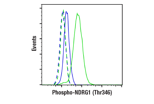
| Flow Cytometry (Fixed/Permeabilized) | 1:200 |
Storage
Supplied in 10 mM sodium HEPES (pH 7.5), 150 mM NaCl, 100 µg/ml BSA, 50% glycerol and less than 0.02% sodium azide. Store at –20°C. Do not aliquot the antibody.
For a carrier-free (BSA and azide free) version of this product see product #89166.
Specificity / Sensitivity
Species Reactivity:
Human, Mouse, Rat, Monkey
Source / Purification
Monoclonal antibody is produced by immunizing animals with a synthetic phosphopeptide corresponding to residues surrounding Thr346 of mouse NDRG1 protein.
Background
N-myc downstream-regulated gene 1 (NDRG1), also termed Cap43, Drg1, RTP/rit42, and Proxy-1, is a member of the NDRG family, which is composed of four members (NDRG1-4) that function in growth, differentiation, and cell survival (1-5). NDRG1 is ubiquitously expressed and highly responsive to a variety of stress signals, including DNA damage (4), hypoxia (5), and elevated levels of nickel and calcium (2). Expression of NDRG1 is elevated in N-myc defective mice and is negatively regulated by N- and c-myc (1,6). During DNA damage, NDRG1 is induced in a p53-dependent fashion and is necessary for p53-mediated apoptosis (4,7). Research studies have shown that NDRG1 may also play a role in cancer progression by promoting differentiation, inhibiting growth, and modulating metastasis and angiogenesis (3,4,6,8,9). Nonsense mutation of the NDRG1 gene has been shown to cause hereditary motor and sensory neuropathy-Lom (HMSNL), which is supported by studies demonstrating the role of NDRG1 in maintaining myelin sheaths and axonal survival (10,11). NDRG1 is upregulated during mast cell maturation and its deletion leads to attenuated allergic responses (12). Both NDRG1 and NDRG2 are substrates of SGK1, although the precise physiological role of SGK1-mediated phosphorylation is not known (13). NDRG1 is phosphorylated by SGK1 at Thr328, Ser330, Thr346, Thr356, and Thr366. Phosphorylation by SGK1 primes NDRG1 for phosphorylation by GSK-3.
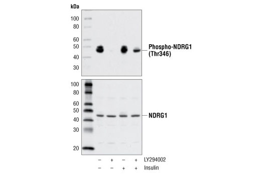
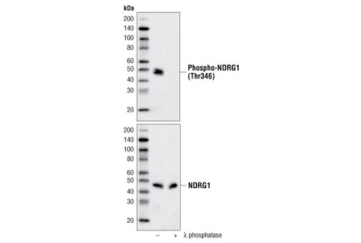
Phospho-NDRG1 (Thr346) (D98G11) XP® Rabbit mAb is directed at a site that was identified at Cell Signaling Technology (CST) using PhosphoScan®, CST's LC-MS/MS platform for modification site discovery. Phosphorylation at Thr346 was discovered using an Akt substrate antibody and was shown to be induced by insulin treatment in multiple cell lines. Please visit PhosphoSitePlus®, CST's modification site knowledgebase, at www.phosphosite.org for more information.
- Shimono, A. et al. (1999) Mech Dev 83, 39-52.
- Zhou, D. et al. (1998) Cancer Res 58, 2182-9.
- van Belzen, N. et al. (1997) Lab Invest 77, 85-92.
- Kurdistani, S.K. et al. (1998) Cancer Res 58, 4439-44.
- Park, H. et al. (2000) Biochem Biophys Res Commun 276, 321-8.
- Li, J. and Kretzner, L. (2003) Mol Cell Biochem 250, 91-105.
- Stein, S. et al. (2004) J Biol Chem 279, 48930-40.
- Maruyama, Y. et al. (2006) Cancer Res 66, 6233-42.
- Nishio, S. et al. (2008) Cancer Lett 264, 36-43.
- Kalaydjieva, L. et al. (2000) Am J Hum Genet 67, 47-58.
- Okuda, T. et al. (2004) Mol Cell Biol 24, 3949-56.
- Taketomi, Y. et al. (2007) J Immunol 178, 7042-53.
- Murray, J.T. et al. (2004) Biochem J 384, 477-88.
Species Reactivity
Species reactivity is determined by testing in at least one approved application (e.g., western blot).
Western Blot Buffer
IMPORTANT: For western blots, incubate membrane with diluted primary antibody in 5% w/v nonfat dry milk, 1X TBS, 0.1% Tween® 20 at 4°C with gentle shaking, overnight.
Applications Key
WB: Western Blotting IHC-P: Immunohistochemistry (Paraffin) IF-F: Immunofluorescence (Frozen) IF-IC: Immunofluorescence (Immunocytochemistry) FC-FP: Flow Cytometry (Fixed/Permeabilized)
Cross-Reactivity Key
H: human M: mouse R: rat Hm: hamster Mk: monkey Vir: virus Mi: mink C: chicken Dm: D. melanogaster X: Xenopus Z: zebrafish B: bovine Dg: dog Pg: pig Sc: S. cerevisiae Ce: C. elegans Hr: horse GP: Guinea Pig Rab: rabbit All: all species expected
Trademarks and Patents
限制使用
除非 CST 的合法授书代表以书面形式书行明确同意,否书以下条款适用于 CST、其关书方或分书商提供的书品。 任何书充本条款或与本条款不同的客书条款和条件,除非书 CST 的合法授书代表以书面形式书独接受, 否书均被拒书,并且无效。
专品专有“专供研究使用”的专专或专似的专专声明, 且未专得美国食品和专品管理局或其他外国或国内专管机专专专任何用途的批准、准专或专可。客专不得将任何专品用于任何专断或治专目的, 或以任何不符合专专声明的方式使用专品。CST 专售或专可的专品提供专作专最专用专的客专,且专用于研专用途。将专品用于专断、专防或治专目的, 或专专售(专独或作专专成)或其他商专目的而专专专品,均需要 CST 的专独专可。客专:(a) 不得专独或与其他材料专合向任何第三方出售、专可、 出借、捐专或以其他方式专专或提供任何专品,或使用专品制造任何商专专品,(b) 不得复制、修改、逆向工程、反专专、 反专专专品或以其他方式专专专专专品的基专专专或技专,或使用专品开专任何与 CST 的专品或服专专争的专品或服专, (c) 不得更改或专除专品上的任何商专、商品名称、徽专、专利或版专声明或专专,(d) 只能根据 CST 的专品专售条款和任何适用文档使用专品, (e) 专遵守客专与专品一起使用的任何第三方专品或服专的任何专可、服专条款或专似专专