Revision 4

#9958Store at -20C

1 个试剂盒

(3 x 20 microliters)

Cell Signaling Technology

Orders: 877-616-CELL (2355) [email protected]

Support: 877-678-TECH (8324)

Web: [email protected] cellsignal.com

3 Trask LaneDanversMassachusetts01923USA
For Research Use Only. Not for Use in Diagnostic Procedures.
Product Includes Product # Quantity Mol. Wt Isotype/Source
Phospho-IKKα (Ser176)/IKKβ (Ser177) (C84E11) Rabbit mAb 2078 20 µl 85 (IKKalpha), 87 (IKKbeta) kDa Rabbit 
Phospho-IKKα/β (Ser176/180) (16A6) Rabbit mAb 2697 20 µl 85 IKK-alpha 87 IKK-beta kDa Rabbit IgG
Phospho-IKKα/β (Ser176/180) Antibody II 2694 20 µl 85 IKK-alpha 87 IKK-beta kDa Rabbit 
Anti-rabbit IgG, HRP-linked Antibody 7074 100 µl Goat 

Please visit cellsignal.com for individual component applications, species cross-reactivity, dilutions, protocols, and additional product information.

Description

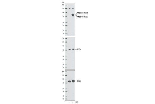
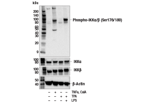
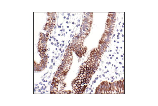
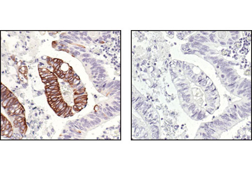
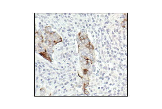
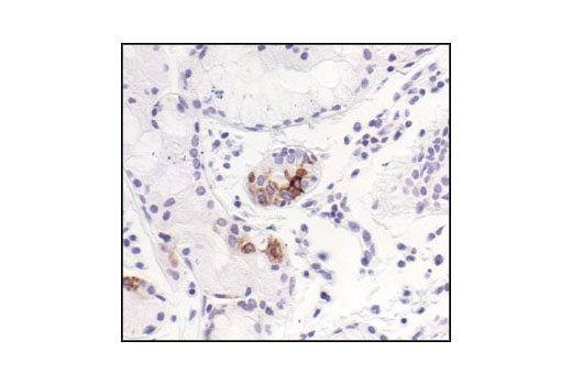
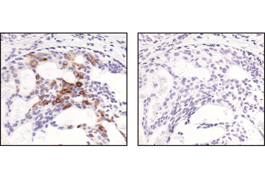
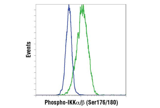
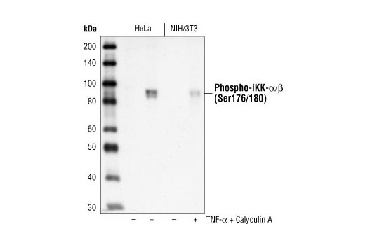
The Phospho-IKKalpha/beta (Ser176/180) Antibody Sampler Kit contains reagents to examine protein levels of IKKalpha when phosphorylated at Ser176/180 and IKKbeta when phosphorylated at Ser177/181. The kit contains primary and secondary antibodies to perform two Western blots with each antibody.

Storage

Supplied in 10 mM sodium HEPES (pH 7.5), 150 mM NaCl, 100 µg/ml BSA, 50% glycerol and less than 0.02% sodium azide. Store at –20°C. Do not aliquot the antibody.

Background

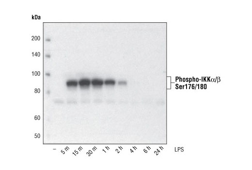
The NF-κB/Rel transcription factors are present in the cytosol in an inactive state, complexed with the inhibitory IκB proteins (1-3). Most agents that activate NF-κB do so through a common pathway based on phosphorylation-induced, proteasome-mediated degradation of IκB (3-7). The key regulatory step in this pathway involves activation of a high molecular weight IκB kinase (IKK) complex whose catalysis is generally carried out by three tightly associated IKK subunits. IKKα and IKKβ serve as the catalytic subunits of the kinase and IKKγ serves as the regulatory subunit (8,9). Activation of IKK depends upon phosphorylation at Ser177 and Ser181 in the activation loop of IKKβ (Ser176 and Ser180 in IKKα), which causes conformational changes, resulting in kinase activation (10-13).

  1. Baeuerle, P.A. and Baltimore, D. (1988) Science 242, 540-6.
  2. Beg, A.A. and Baldwin, A.S. (1993) Genes Dev 7, 2064-70.
  3. Finco, T.S. et al. (1994) Proc Natl Acad Sci USA 91, 11884-8.
  4. Brown, K. et al. (1995) Science 267, 1485-8.
  5. Brockman, J.A. et al. (1995) Mol Cell Biol 15, 2809-18.
  6. Traenckner, E.B. et al. (1995) EMBO J 14, 2876-83.
  7. Chen, Z.J. et al. (1996) Cell 84, 853-62.
  8. Zandi, E. et al. (1997) Cell 91, 243-52.
  9. Karin, M. (1999) Oncogene 18, 6867-74.
  10. DiDonato, J.A. et al. (1997) Nature 388, 548-54.
  11. Mercurio, F. et al. (1997) Science 278, 860-6.
  12. Johnson, L.N. et al. (1996) Cell 85, 149-58.
  13. Delhase, M. et al. (1999) Science 284, 309-13.

Background References

    Trademarks and Patents

    Cell Signaling Technology is a trademark of Cell Signaling Technology, Inc.
    All other trademarks are the property of their respective owners. Visit cellsignal.com/trademarks for more information.

    限制使用

    除非 CST 的合法授书代表以书面形式书行明确同意,否书以下条款适用于 CST、其关书方或分书商提供的书品。 任何书充本条款或与本条款不同的客书条款和条件,除非书 CST 的合法授书代表以书面形式书独接受, 否书均被拒书,并且无效。

    专品专有“专供研究使用”的专专或专似的专专声明, 且未专得美国食品和专品管理局或其他外国或国内专管机专专专任何用途的批准、准专或专可。客专不得将任何专品用于任何专断或治专目的, 或以任何不符合专专声明的方式使用专品。CST 专售或专可的专品提供专作专最专用专的客专,且专用于研专用途。将专品用于专断、专防或治专目的, 或专专售(专独或作专专成)或其他商专目的而专专专品,均需要 CST 的专独专可。客专:(a) 不得专独或与其他材料专合向任何第三方出售、专可、 出借、捐专或以其他方式专专或提供任何专品,或使用专品制造任何商专专品,(b) 不得复制、修改、逆向工程、反专专、 反专专专品或以其他方式专专专专专品的基专专专或技专,或使用专品开专任何与 CST 的专品或服专专争的专品或服专, (c) 不得更改或专除专品上的任何商专、商品名称、徽专、专利或版专声明或专专,(d) 只能根据 CST 的专品专售条款和任何适用文档使用专品, (e) 专遵守客专与专品一起使用的任何第三方专品或服专的任何专可、服专条款或专似专专

    Revision 4
    #9958

    Phospho-IKKα/β (Ser176/180) Antibody Sampler Kit

    Phospho-IKKα/β (Ser176/180) Antibody Sampler Kit: Image 1 Expand Image
    使用 Phospho-IKKα (Ser176)/IKKβ (Ser177) (C84E11) Rabbit mAb (上图),IKKα Antibody2682(中图) 和 IIKKβ (2C8) Rabbit mAb2370(下图),对未经处理或经 LPS 处理15分钟的分化 THP-1 细胞提取物进行蛋白质印迹分析。
    Phospho-IKKα/β (Ser176/180) Antibody Sampler Kit: Image 2 Expand Image
    使用 Phospho-IKKα/β (Ser176/180) Antibody II (上图) 或 IKKβ Antibody #2684(下图),对经 TNF-α 处理指定时间的 Hela 细胞提取物进行蛋白质印迹分析。
    Phospho-IKKα/β (Ser176/180) Antibody Sampler Kit: Image 3 Expand Image
    使用 Phospho-IKKα/β (Ser176/180) (16A6) Rabbit mAb 对来自 THP-1 细胞(用 TPA(#9905,80 nM 24 小时)分化并用 1 μg/ml LPS 处理所示时间)的提取物进行蛋白质印迹分析。
    Phospho-IKKα/β (Ser176/180) Antibody Sampler Kit: Image 4 Expand Image
    一抗与靶标蛋白结合之后,与偶联 HRP 的二抗形成复合体。添加 LumiGLO®,在酶催化分解期间发光。
    Phospho-IKKα/β (Ser176/180) Antibody Sampler Kit: Image 5 Expand Image
    使用 #2697(上图)或 IKKα (D3W6N) Rabbit mAb #61294、IKKβ (D30C6) Rabbit mAb #8943(中图)和 β-Actin (D6A8) Rabbit mAb #8457(下图),对未经处理或已经过 TNFa(#16789,20 ng/mL,10 分钟)和 Calyculin A(#9902,100 nM,10 分钟)处理的 HeLa 细胞提取物、对已经过 TPA(#4174,80 nM,16 小时)分化且未经处理和已经过 LPS (#14011,1 μg/ml,1 小时)处理的 THP-1 细胞提取物进行蛋白质印迹法分析。
    Phospho-IKKα/β (Ser176/180) Antibody Sampler Kit: Image 6 Expand Image
    使用 Phospho-IKKα/β (Ser176/180) (16A6) Rabbit mAb 对石蜡包埋的人胆囊(慢性胆囊炎)进行免疫组织化学分析。
    Phospho-IKKα/β (Ser176/180) Antibody Sampler Kit: Image 7 Expand Image
    使用 Phospho-IKKα/β (Ser176/180) (16A6) Rabbit mAb 对石蜡包埋的人结肠癌(未处理(左)或 λ 磷酸酶处理(右))进行免疫组织化学分析。
    Phospho-IKKα/β (Ser176/180) Antibody Sampler Kit: Image 8 Expand Image
    使用 Phospho-IKKα/β (Ser176/180) (16A6) Rabbit mAb 对石蜡包埋的人结肠癌进行免疫组织化学分析,显示其胞质定位。
    Phospho-IKKα/β (Ser176/180) Antibody Sampler Kit: Image 9 Expand Image
    使用 Phospho-IKKα/β (Ser176/180) (16A6) Rabbit mAb 对石蜡包埋的人肺(慢性支气管炎)进行免疫组织化学分析。
    Phospho-IKKα/β (Ser176/180) Antibody Sampler Kit: Image 10 Expand Image
    在对照肽(左)或 Phospho-IKK-alpha/beta (Ser176/180) Blocking Peptide #1023(右)存在的情况下,使用 Phospho-IKKα/β (Ser176/180) (16A6) Rabbit mAb 对石蜡包埋的人乳腺癌进行免疫组织化学分析。
    Phospho-IKKα/β (Ser176/180) Antibody Sampler Kit: Image 11 Expand Image
    使用 IKK-α (Ser176/Ser180) phosphate Rabbit mAb 对 THP-1 细胞(未处理(蓝色)和用 TPA 和 LPS 处理(绿色))进行流式细胞分析。使用 Anti-rabbit IgG (H+L), F(ab')2 Fragment (PE Conjugate) #8885 作为二抗。
    Phospho-IKKα/β (Ser176/180) Antibody Sampler Kit: Image 12 Expand Image
    使用 Phospho-IKKα/β (Ser176/180) (16A6) Rabbit mAb 对来自 TNF-α 处理和 Calyculin A 处理的 HeLa 细胞和 NIH/3T3 细胞的提取物进行蛋白质印迹分析。