WB, IP
H M
Endogenous
140, 124, 120
Rabbit
#P56524, #Q9UQL6, #Q8WUI4
9759, 10014, 51564
Product Information
Product Usage Information
| Application | Dilution |
|---|---|
| Western Blotting | 1:1000 |
| Immunoprecipitation | 1:25 |
Storage
Specificity / Sensitivity
Species Reactivity:
Human, Mouse
Source / Purification
Polyclonal antibodies are produced by immunizing animals with a synthetic peptide corresponding to human HDAC7 protein phosphorylated on Ser486. Antibodies are purified by protein A and peptide affinity chromatography.
Background
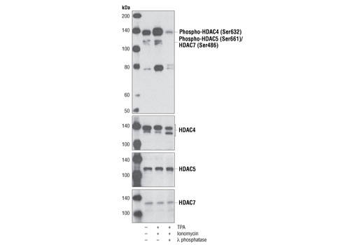
Acetylation of the histone tail causes chromatin to adopt an "open" conformation, allowing increased accessibility of transcription factors to DNA. The identification of histone acetyltransferases (HATs) and their large multiprotein complexes has yielded important insights into how these enzymes regulate transcription (1,2). HAT complexes interact with sequence-specific activator proteins to target specific genes. In addition to histones, HATs can acetylate nonhistone proteins, suggesting multiple roles for these enzymes (3). In contrast, histone deacetylation promotes a "closed" chromatin conformation and typically leads to repression of gene activity (4). Mammalian histone deacetylases can be divided into three classes on the basis of their similarity to various yeast deacetylases (5). Class I proteins (HDACs 1, 2, 3, and 8) are related to the yeast Rpd3-like proteins, those in class II (HDACs 4, 5, 6, 7, 9, and 10) are related to yeast Hda1-like proteins, and class III proteins are related to the yeast protein Sir2. Inhibitors of HDAC activity are now being explored as potential therapeutic cancer agents (6,7).
Histone deacetylases (HDACs) interact with an increasing number of transcription factors, including myocyte enhancer factor 2 (MEF2), to negatively regulate gene expression. HDACs are regulated in part by shuttling between the nucleus and cytoplasm, where export to the cytoplasm facilitates gene activation by removing HDACs from their target genes (8,9). The cytoplasmic export is facilitated by 14-3-3 proteins, which bind to specific phospho-serine residues on the HDAC proteins (8,9). These phospho-serine 14-3-3 binding modules are highly conserved between HDAC proteins, allowing for their collective regulation in response to specific cell stimuli. For example, the highly conserved HDAC 4 Ser632, HDAC 5 Ser498 and HDAC 7 Ser486 residues are all phosphorylated by CAMK and PKD kinases in response to multiple cell stimuli, including VEGF-induced angiogenesis in endothelial cells, B cell and T cell activation, and differentiation of myoblasts into muscle fiber (10-14).
- Marmorstein, R. (2001) Cell Mol Life Sci 58, 693-703.
- Gregory, P.D. et al. (2001) Exp Cell Res 265, 195-202.
- Liu, Y. et al. (2000) Mol Cell Biol 20, 5540-53.
- Cress, W.D. and Seto, E. (2000) J Cell Physiol 184, 1-16.
- Gray, S.G. and Ekström, T.J. (2001) Exp Cell Res 262, 75-83.
- Thiagalingam, S. et al. (2003) Ann. N.Y. Acad. Sci. 983, 84-100.
- Vigushin, D.M. and Coombes, R.C. (2004) Curr Cancer Drug Targets 4, 205-18.
- Grozinger, C.M. and Schreiber, S.L. (2000) Proc Natl Acad Sci USA 97, 7835-40.
- Wang, A.H. et al. (2000) Mol Cell Biol 20, 6904-12.
- Ha, C.H. et al. (2008) J Biol Chem 283, 14590-9.
- Wang, S. et al. (2008) Proc Natl Acad Sci U S A 105, 7738-43.
- Matthews, S.A. et al. (2006) Mol Cell Biol 26, 1569-77.
- Parra, M. et al. (2005) J Biol Chem 280, 13762-70.
- McKinsey, T.A. et al. (2000) Nature 408, 106-11.
Species Reactivity
Species reactivity is determined by testing in at least one approved application (e.g., western blot).
Western Blot Buffer
IMPORTANT: For western blots, incubate membrane with diluted primary antibody in 5% w/v BSA, 1X TBS, 0.1% Tween® 20 at 4°C with gentle shaking, overnight.
Applications Key
WB: Western Blotting IP: Immunoprecipitation
Cross-Reactivity Key
H: human M: mouse R: rat Hm: hamster Mk: monkey Vir: virus Mi: mink C: chicken Dm: D. melanogaster X: Xenopus Z: zebrafish B: bovine Dg: dog Pg: pig Sc: S. cerevisiae Ce: C. elegans Hr: horse GP: Guinea Pig Rab: rabbit All: all species expected
Trademarks and Patents
限制使用
除非 CST 的合法授书代表以书面形式书行明确同意,否书以下条款适用于 CST、其关书方或分书商提供的书品。 任何书充本条款或与本条款不同的客书条款和条件,除非书 CST 的合法授书代表以书面形式书独接受, 否书均被拒书,并且无效。
专品专有“专供研究使用”的专专或专似的专专声明, 且未专得美国食品和专品管理局或其他外国或国内专管机专专专任何用途的批准、准专或专可。客专不得将任何专品用于任何专断或治专目的, 或以任何不符合专专声明的方式使用专品。CST 专售或专可的专品提供专作专最专用专的客专,且专用于研专用途。将专品用于专断、专防或治专目的, 或专专售(专独或作专专成)或其他商专目的而专专专品,均需要 CST 的专独专可。客专:(a) 不得专独或与其他材料专合向任何第三方出售、专可、 出借、捐专或以其他方式专专或提供任何专品,或使用专品制造任何商专专品,(b) 不得复制、修改、逆向工程、反专专、 反专专专品或以其他方式专专专专专品的基专专专或技专,或使用专品开专任何与 CST 的专品或服专专争的专品或服专, (c) 不得更改或专除专品上的任何商专、商品名称、徽专、专利或版专声明或专专,(d) 只能根据 CST 的专品专售条款和任何适用文档使用专品, (e) 专遵守客专与专品一起使用的任何第三方专品或服专的任何专可、服专条款或专似专专