IHC-Bond, IHC-P
H
Endogenous
Mouse IgG2a
#Q15116
5133
Product Information
Product Usage Information
| Application | Dilution |
|---|---|
| IHC Leica Bond | 1:100 |
| Immunohistochemistry (Paraffin) | 1:200 |
Storage
Supplied in 10 mM sodium HEPES (pH 7.5), 150 mM NaCl, 100 µg/ml BSA, 50% glycerol and less than 0.02% sodium azide. Store at –20°C. Do not aliquot the antibody.
For a carrier-free (BSA and Azide) version of this product see product #60847.
Specificity / Sensitivity
Species Reactivity:
Human
Source / Purification
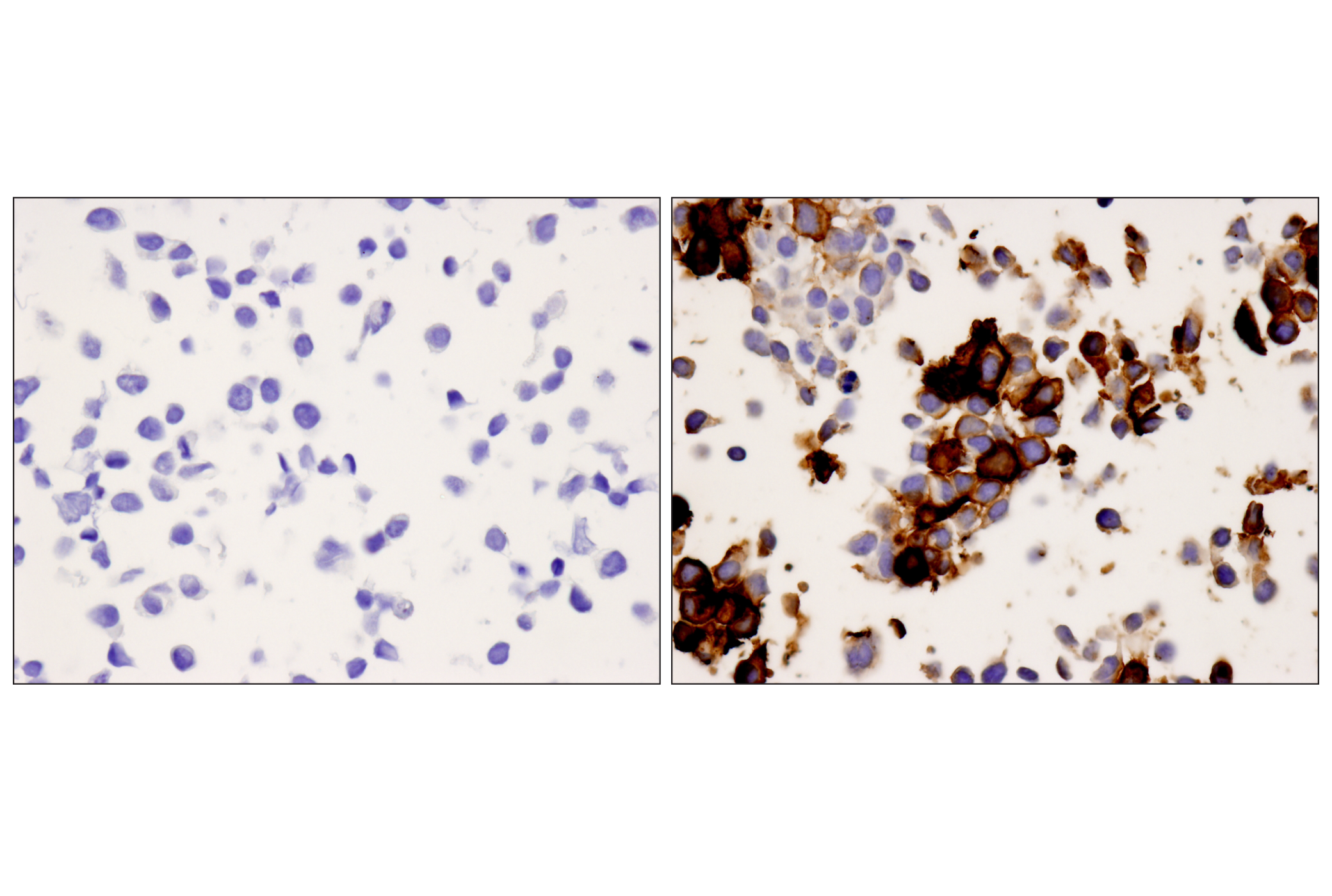
Monoclonal antibody is produced by immunizing animals with recombinant protein specific to the amino terminus of human PD-1 protein.
Background
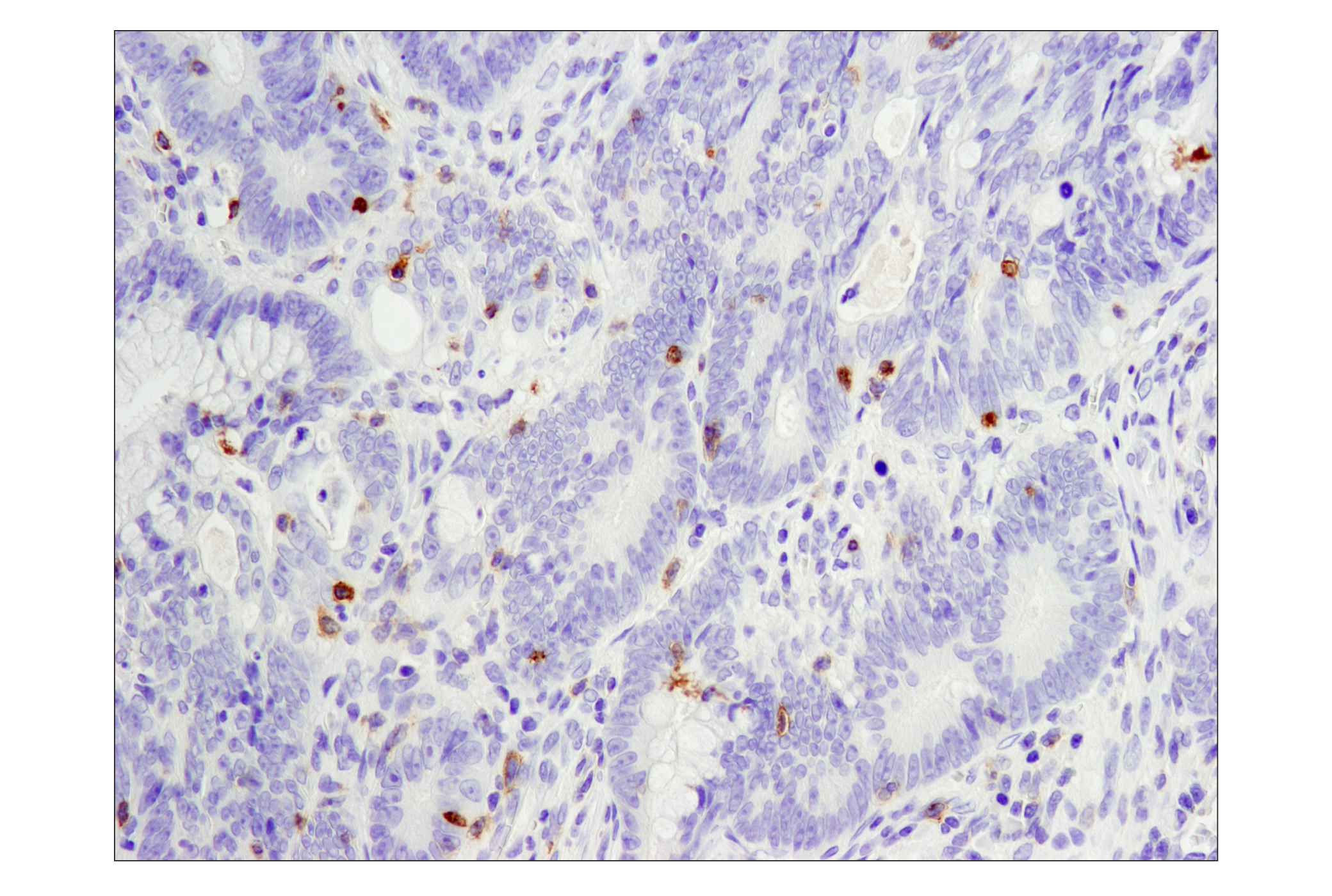
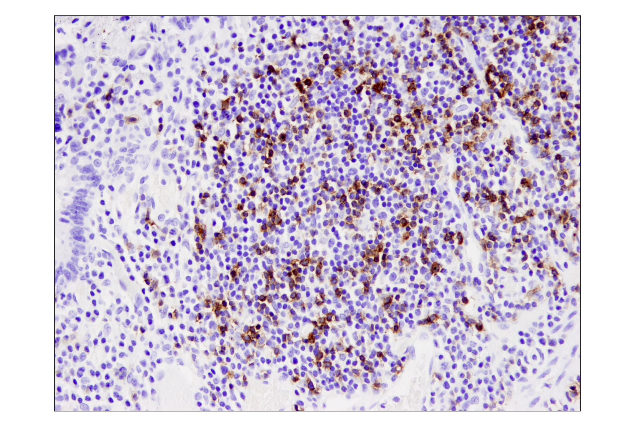
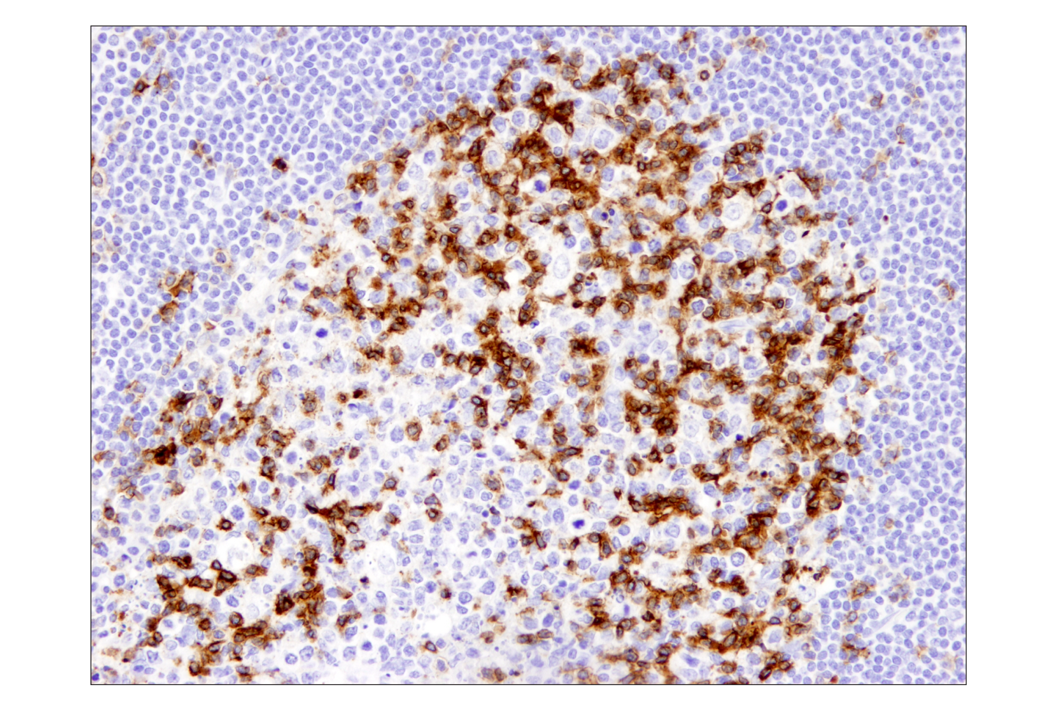
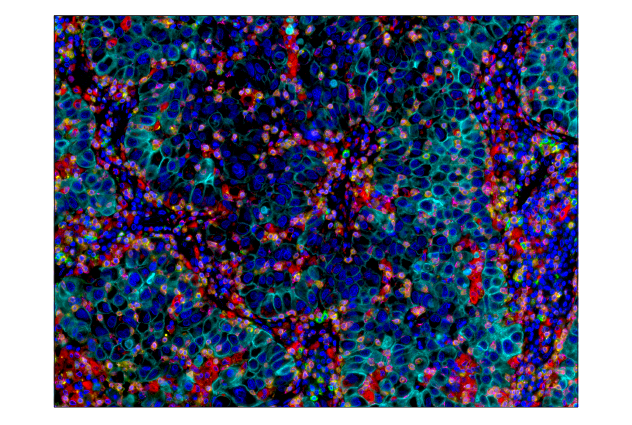
The programmed cell death 1 protein (PD-1, PDCD1, CD279) is a member of the CD28 family of immunoreceptors that regulate T cell activation and immune responses (1-3). The PD-1 protein contains an extracellular Ig V domain, a transmembrane domain, and a cytoplasmic tail that includes an immunoreceptor tyrosine-based inhibitory motif (ITIM) and an immunoreceptor tyrosine-based switch motif (ITSM). PD-1 is activated by the cell surface ligands PD-L1 and PD-L2 (4). Upon activation, PD-1 ITIM and ITSM phosphorylation leads to the recruitment of the protein tyrosine phosphatases SHP-1 and SHP-2, which suppress TCR signaling (5-7). In addition to activated T cells, PD-1 is expressed in activated B cells and monocytes, although its function in these cell types has not been fully characterized (8). The PD-1 pathway plays an important role in immune tolerance (3); however, research studies show that cancer cells often adopt this pathway to escape immune surveillance (9). Consequently, blockade of PD-1 and its ligands is proving to be a sound strategy for neoplastic intervention (10).
- Ishida, Y. et al. (1992) EMBO J 11, 3887-95.
- Shinohara, T. et al. (1994) Genomics 23, 704-6.
- Nishimura, H. et al. (1999) Immunity 11, 141-51.
- Freeman, G.J. et al. (2000) J Exp Med 192, 1027-34.
- Yokosuka, T. et al. (2012) J Exp Med 209, 1201-17.
- Sheppard, K.A. et al. (2004) FEBS Lett 574, 37-41.
- Chemnitz, J.M. et al. (2004) J Immunol 173, 945-54.
- Thibult, M.L. et al. (2013) Int Immunol 25, 129-37.
- Dong, H. et al. (2002) Nat Med 8, 793-800.
- Topalian, S.L. et al. (2012) Curr Opin Immunol 24, 207-12.
Species Reactivity
Species reactivity is determined by testing in at least one approved application (e.g., western blot).
Applications Key
IHC-Bond: IHC Leica Bond IHC-P: Immunohistochemistry (Paraffin)
Cross-Reactivity Key
H: human M: mouse R: rat Hm: hamster Mk: monkey Vir: virus Mi: mink C: chicken Dm: D. melanogaster X: Xenopus Z: zebrafish B: bovine Dg: dog Pg: pig Sc: S. cerevisiae Ce: C. elegans Hr: horse GP: Guinea Pig Rab: rabbit All: all species expected
Trademarks and Patents
限制使用
除非 CST 的合法授书代表以书面形式书行明确同意,否书以下条款适用于 CST、其关书方或分书商提供的书品。 任何书充本条款或与本条款不同的客书条款和条件,除非书 CST 的合法授书代表以书面形式书独接受, 否书均被拒书,并且无效。
专品专有“专供研究使用”的专专或专似的专专声明, 且未专得美国食品和专品管理局或其他外国或国内专管机专专专任何用途的批准、准专或专可。客专不得将任何专品用于任何专断或治专目的, 或以任何不符合专专声明的方式使用专品。CST 专售或专可的专品提供专作专最专用专的客专,且专用于研专用途。将专品用于专断、专防或治专目的, 或专专售(专独或作专专成)或其他商专目的而专专专品,均需要 CST 的专独专可。客专:(a) 不得专独或与其他材料专合向任何第三方出售、专可、 出借、捐专或以其他方式专专或提供任何专品,或使用专品制造任何商专专品,(b) 不得复制、修改、逆向工程、反专专、 反专专专品或以其他方式专专专专专品的基专专专或技专,或使用专品开专任何与 CST 的专品或服专专争的专品或服专, (c) 不得更改或专除专品上的任何商专、商品名称、徽专、专利或版专声明或专专,(d) 只能根据 CST 的专品专售条款和任何适用文档使用专品, (e) 专遵守客专与专品一起使用的任何第三方专品或服专的任何专可、服专条款或专似专专