| Product Includes | Product # | Quantity | Mol. Wt | Isotype/Source |
|---|---|---|---|---|
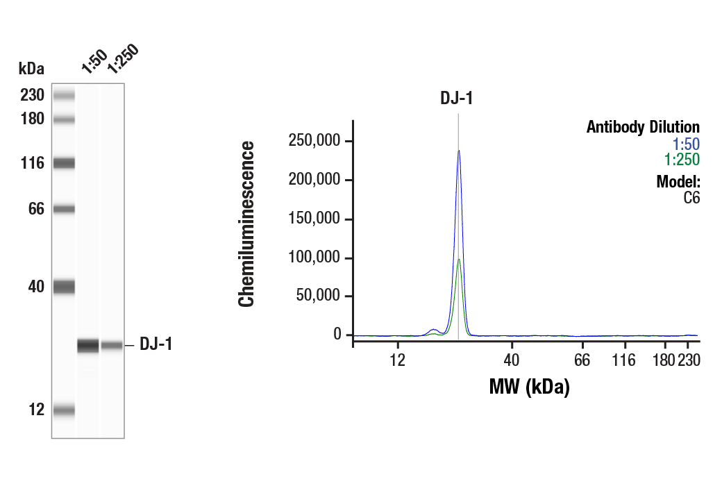
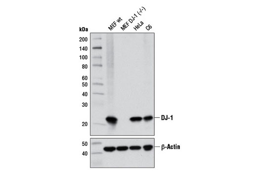
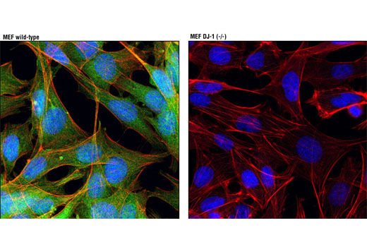
| DJ-1 (D29E5) XP® Rabbit mAb | 5933 | 20 µl | 22 kDa | Rabbit IgG |
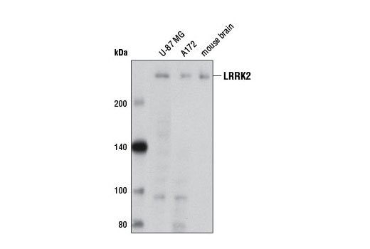
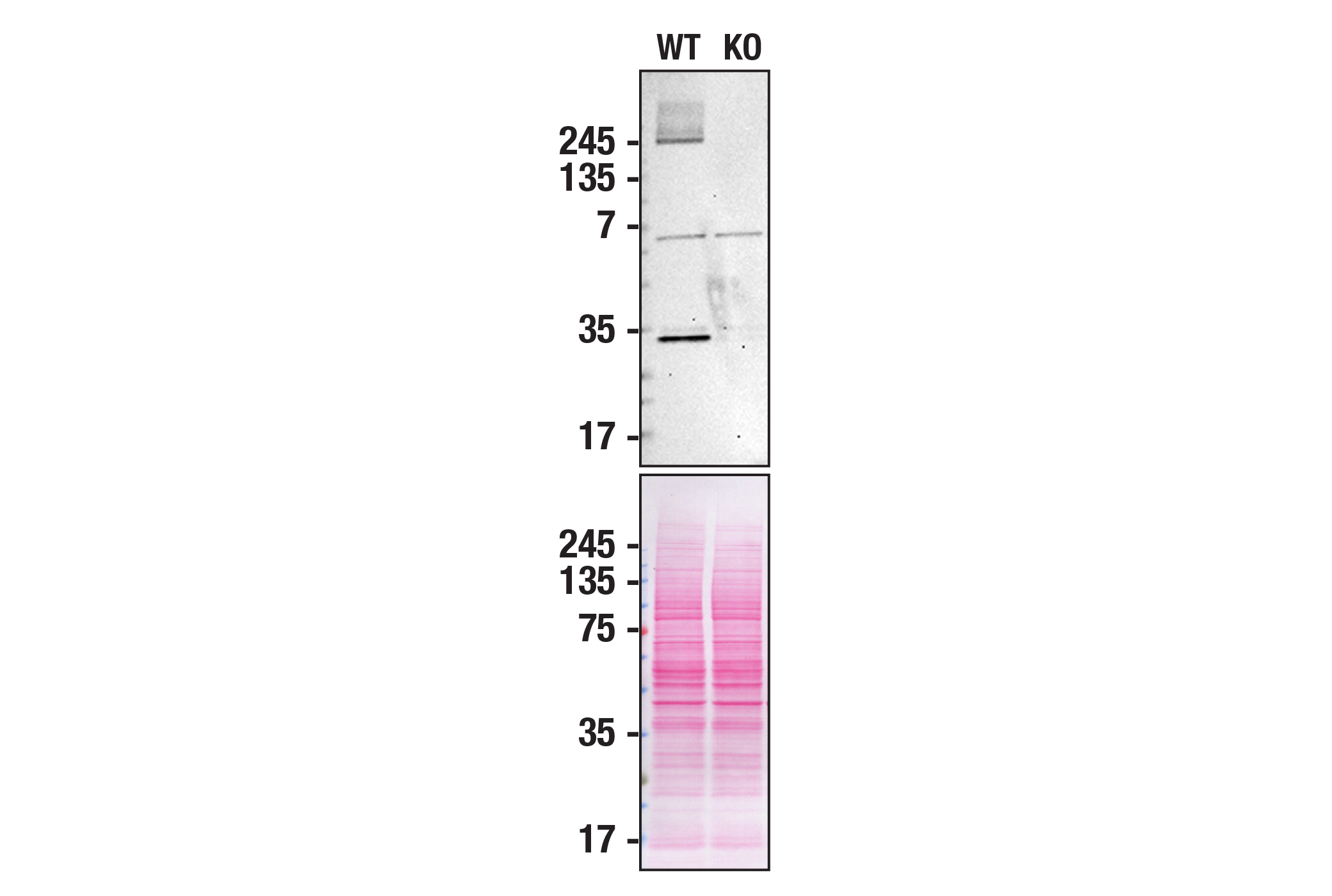
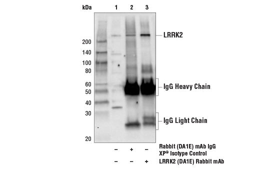
| LRRK2 (D18E12) Rabbit mAb | 13046 | 20 µl | 290 kDa | Rabbit IgG |
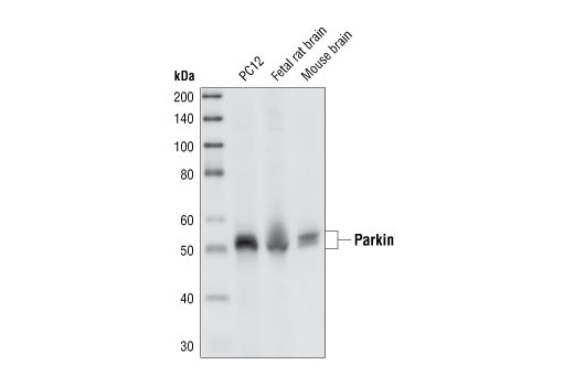
| Parkin (Prk8) Mouse mAb | 4211 | 20 µl | 50 kDa | Mouse IgG2b |
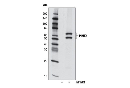
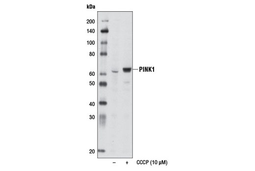
| PINK1 (D8G3) Rabbit mAb | 6946 | 20 µl | 60, 50 kDa | Rabbit IgG |
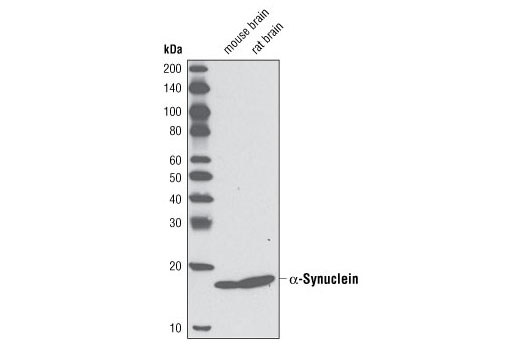
| α-Synuclein (D37A6) Rabbit mAb | 4179 | 20 µl | 18 kDa | Rabbit IgG |
| Anti-rabbit IgG, HRP-linked Antibody | 7074 | 100 µl | Goat | |
| Anti-mouse IgG, HRP-linked Antibody | 7076 | 100 µl | Horse |
Please visit cellsignal.com for individual component applications, species cross-reactivity, dilutions, protocols, and additional product information.
Description
The Parkinson's Research Antibody Sampler Kit provides an economical means of detecting target proteins related to Parkinson's disease. The kit contains enough primary and secondary antibody to perform two western blots per primary.
Storage
Background
Parkinson’s disease (PD), the second most common neurodegenerative disease after Alzheimer’s, is a progressive movement disorder characterized by rigidity, tremors, and postural instability. The pathological hallmark of PD is progressive loss of dopaminergic neurons in the substantia nigra of the ventral midbrain and the presence of intracellular Lewy bodies in surviving neurons of the brain stem (1). Research studies have shown that various genes and loci (α-synuclein/PARK1 and 4, parkin/PARK2, UCH-L1/PARK5, PINK1/PARK6, DJ-1/PARK7, LRRK2/PARK8, synphilin-1, and NR4A2) are genetically linked to PD (2).
α-Synuclein, a 140 amino acid protein expressed abundantly in the brain, is a major component of aggregates found in Lewy bodies (3). Parkin is involved in protein degradation through the ubiquitin-proteasome pathway, and investigators have shown that mutations in Parkin cause early onset of PD (4). In the case of autosomal recessive juvenile Parkinsonism (AR-JP), deletions have been found on chromosome 6 in the Parkin gene (5). PTEN induced putative kinase 1 (PINK1) is a mitochondrial serine/threonine kinase involved in the normal function and integrity of mitochondria, as well as a reduction of cytochrome c release from mitochondria (6-8). PINK1 phosphorylates Parkin and promotes its translocation to mitochondria (7). Mutations of PINK1 are associated with loss of protective function, mitrochondrial dysfunction, aggregation of α-synuclein, and proteasome dysfunction (6,8). DJ-1 is involved in multiple cellular functions; it has been shown to cooperate with Ras to increase cell transformation, to regulate transcription of the androgen receptor, and may function as an indicator of oxidative stress, while loss-of-function mutations in DJ-1 cause early onset of PD (9-12). Dopamine D2 receptor-mediated functions are greatly impaired in DJ-1 (-/-) mice, resulting in reduced long-term depression (13). Leucine-rich repeat kinase 2 (LRRK2) contains amino-terminal leucine-rich repeats (LRR), a Ras-like small GTP binding protein-like (ROC) domain, an MLK protein kinase domain, and a carboxy-terminal WD40-repeat. At least 20 LRRK2 mutations have been linked to PD (14). The most prevalent mutation, G2019S, causes increased LRRK2 kinase activity, leading to progressive neurite loss and decreased neuronal survival (15).
- Fahn, S. (2003) Ann N Y Acad Sci 991, 1-14.
- Moore, D.J. et al. (2005) Annu Rev Neurosci 28, 57-87.
- Goldberg, M.S. and Lansbury, P.T. (2000) Nat Cell Biol 2, E115-9.
- Borrelli, E. (2005) Neuron 45, 479-81.
- Polymeropoulos, M.H. et al. (1997) Science 276, 2045-7.
- Liu, W. et al. (2009) PLoS One 4, e4597.
- Kim, Y. et al. (2008) Biochem Biophys Res Commun 377, 975-80.
- Petit, A. et al. (2005) J Biol Chem 280, 34025-32.
- Bonifati, V. et al. (2003) Science 299, 256-9.
- Nagakubo, D. et al. (1997) Biochem Biophys Res Commun 231, 509-13.
- Takahashi, K. et al. (2001) J Biol Chem 276, 37556-63.
- Mitsumoto, A. and Nakagawa, Y. (2001) Free Radic Res 35, 885-93.
- Goldberg, M.S. et al. (2005) Neuron 45, 489-96.
- Mata, I.F. et al. (2006) Trends Neurosci 29, 286-93.
- MacLeod, D. et al. (2006) Neuron 52, 587-93.
Background References
Trademarks and Patents
限制使用
除非 CST 的合法授书代表以书面形式书行明确同意,否书以下条款适用于 CST、其关书方或分书商提供的书品。 任何书充本条款或与本条款不同的客书条款和条件,除非书 CST 的合法授书代表以书面形式书独接受, 否书均被拒书,并且无效。
专品专有“专供研究使用”的专专或专似的专专声明, 且未专得美国食品和专品管理局或其他外国或国内专管机专专专任何用途的批准、准专或专可。客专不得将任何专品用于任何专断或治专目的, 或以任何不符合专专声明的方式使用专品。CST 专售或专可的专品提供专作专最专用专的客专,且专用于研专用途。将专品用于专断、专防或治专目的, 或专专售(专独或作专专成)或其他商专目的而专专专品,均需要 CST 的专独专可。客专:(a) 不得专独或与其他材料专合向任何第三方出售、专可、 出借、捐专或以其他方式专专或提供任何专品,或使用专品制造任何商专专品,(b) 不得复制、修改、逆向工程、反专专、 反专专专品或以其他方式专专专专专品的基专专专或技专,或使用专品开专任何与 CST 的专品或服专专争的专品或服专, (c) 不得更改或专除专品上的任何商专、商品名称、徽专、专利或版专声明或专专,(d) 只能根据 CST 的专品专售条款和任何适用文档使用专品, (e) 专遵守客专与专品一起使用的任何第三方专品或服专的任何专可、服专条款或专似专专