| Product Includes | Product # | Quantity | Mol. Wt | Isotype/Source |
|---|---|---|---|---|
| Insulin (C27C9) Rabbit mAb | 3014 | 20 µl | Rabbit IgG | |
| Proglucagon (D16G10) XP® Rabbit mAb | 8233 | 20 µl | Rabbit IgG | |
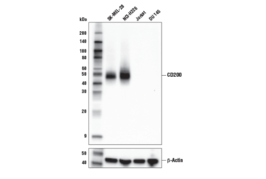
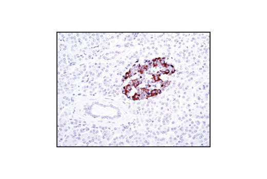
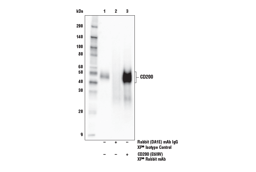
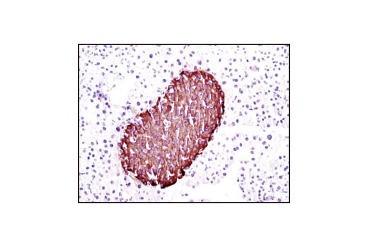
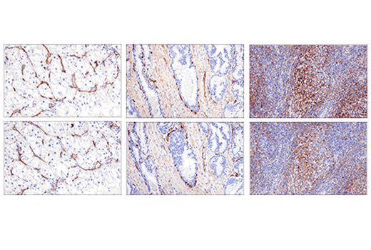
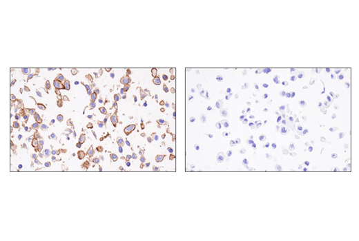
| CD200 (E5I9V) XP® Rabbit mAb | 23451 | 20 µl | 45-50 kDa | Rabbit IgG |
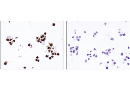
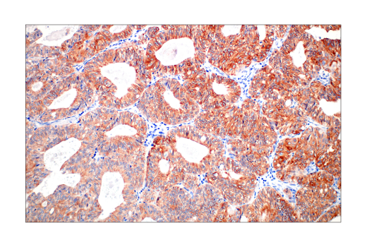
| Pax6 (D3A9V) XP® Rabbit mAb | 60433 | 20 µl | 50 kDa | Rabbit IgG |
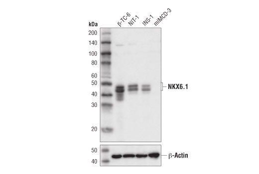
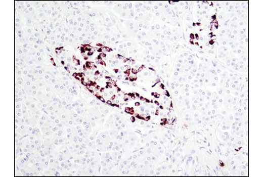
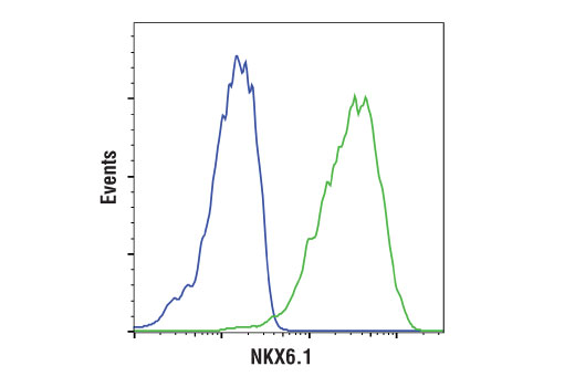
| NKX6.1 (D8O4R) Rabbit mAb | 54551 | 20 µl | 44, 46 kDa | Rabbit IgG |
| C-Peptide Antibody | 4593 | 20 µl | 4 kDa | Rabbit |
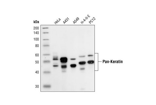
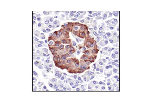
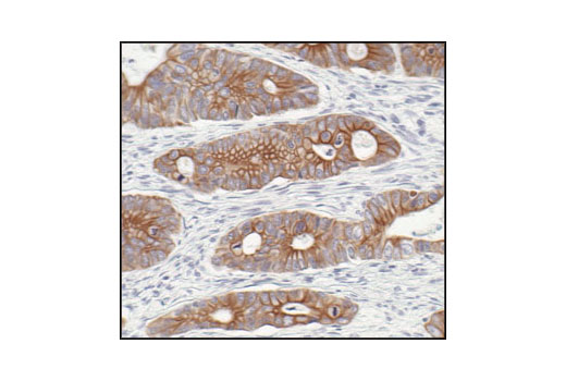
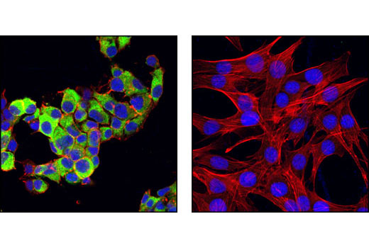
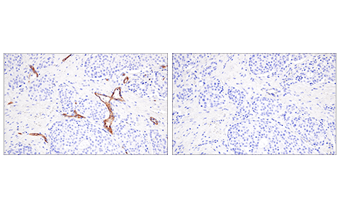
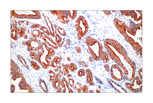
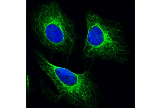
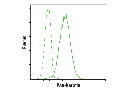
| Pan-Keratin (C11) Mouse mAb | 4545 | 20 µl | 46-58 kDa | Mouse IgG1 |
| Anti-rabbit IgG, HRP-linked Antibody | 7074 | 100 µl | Goat | |
| Anti-mouse IgG, HRP-linked Antibody | 7076 | 100 µl | Horse |
Please visit cellsignal.com for individual component applications, species cross-reactivity, dilutions, protocols, and additional product information.
Description
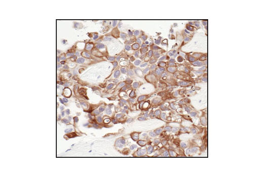
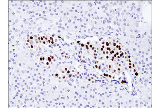
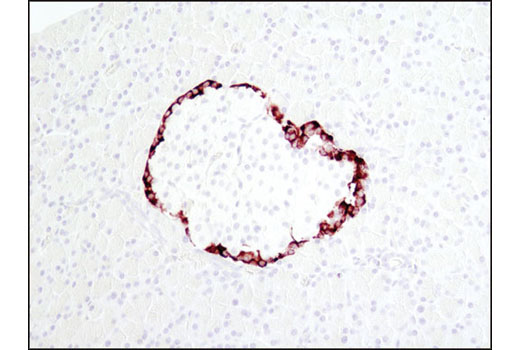
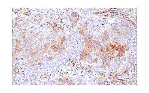
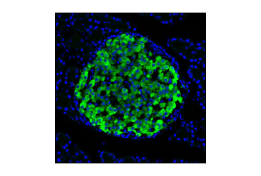
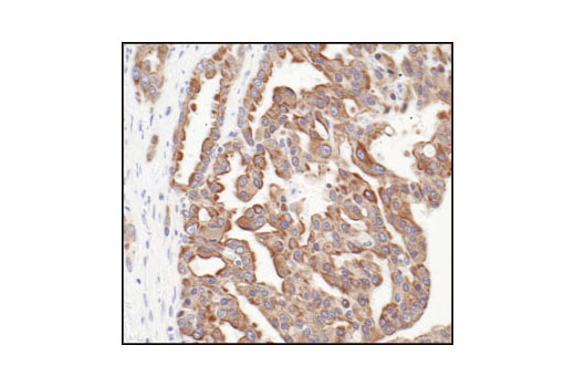
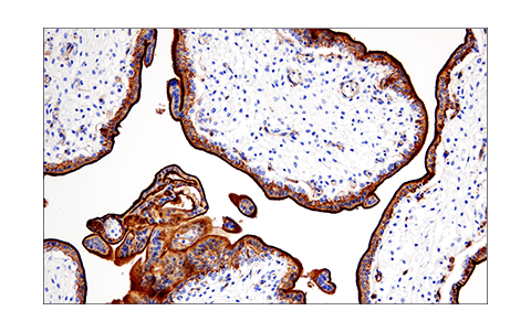
The Pancreatic Marker IHC Antibody Sampler Kit provides a useful selection of markers to distinguish pancreatic cell types that perform important functions to maintain glucose homeostasis. The kit also includes antibodies that differentiate pancreatic tumor subtypes. The sampler kit is designed for use on formalin-fixed, paraffin-embedded tissue samples.
Storage
Background
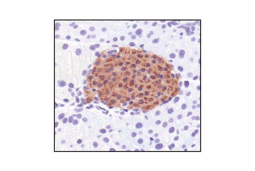
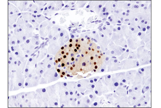
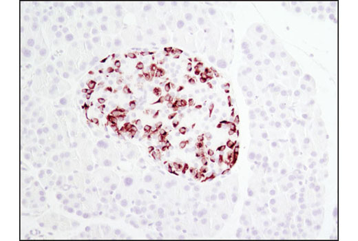
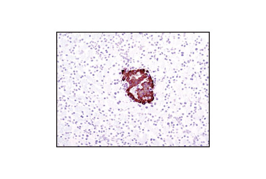
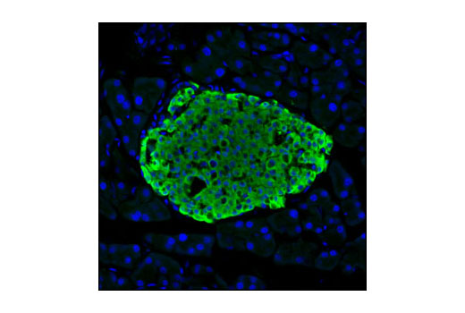
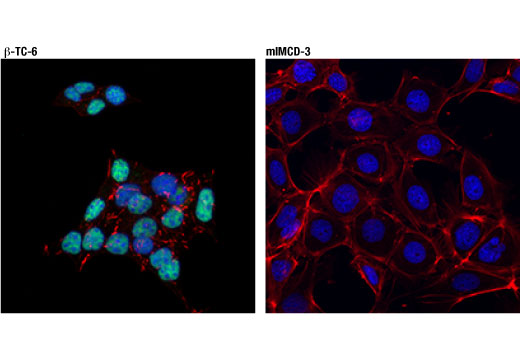
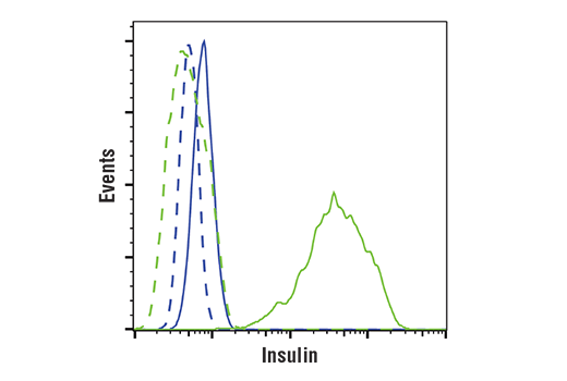
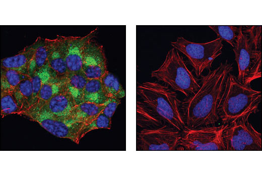
Insulin is a hormone that is produced and released from pancreatic β cells through a glucose sensing pathway. Proinsulin is the precursor molecule to insulin and is processed prior to its secretion. Insulin is composed of A-peptide and B-peptide which are joined by a disulfide bond. The center one-third of the molecule is cleaved and released as C-peptide, which has a longer half-life than insulin (1). Antibodies to both insulin and C-peptide are useful markers for β cells.
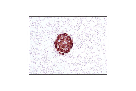
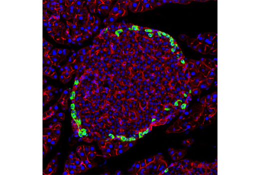
Glucose homeostasis is regulated by a variety of hormones including glucagon. Glucagon is synthesized as the precursor molecule proglucagon and is proteolytically processed to yield the mature peptide in α cells of the pancreatic islets. Glucagon causes the release of glucose from glycogen and stimulates gluconeogenesis in the liver (2). Antibodies to glucagon and proglucagon are useful markers for pancreatic α cells.
NKX6.1 is an important transcription factor in a network of transcription factors that are critical for pancreatic β cell development and maintenance (3). Antibodies to NKX6.1 are useful markers for β cells.
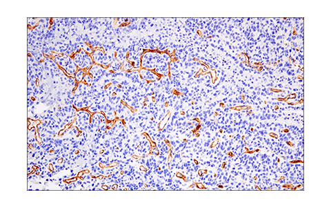
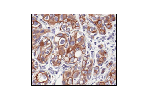
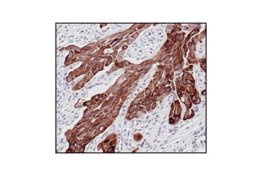
Pan-Keratin and CD200 antibodies are useful to mark and differentiate some pancreatic tumor subtypes. Pan-Keratin antibodies mark epithelial cells in pancreatic adenocarcinomas while CD200 is a useful marker for neuroendocrine pancreatic tumors (4).
Background References
Trademarks and Patents
限制使用
除非 CST 的合法授书代表以书面形式书行明确同意,否书以下条款适用于 CST、其关书方或分书商提供的书品。 任何书充本条款或与本条款不同的客书条款和条件,除非书 CST 的合法授书代表以书面形式书独接受, 否书均被拒书,并且无效。
专品专有“专供研究使用”的专专或专似的专专声明, 且未专得美国食品和专品管理局或其他外国或国内专管机专专专任何用途的批准、准专或专可。客专不得将任何专品用于任何专断或治专目的, 或以任何不符合专专声明的方式使用专品。CST 专售或专可的专品提供专作专最专用专的客专,且专用于研专用途。将专品用于专断、专防或治专目的, 或专专售(专独或作专专成)或其他商专目的而专专专品,均需要 CST 的专独专可。客专:(a) 不得专独或与其他材料专合向任何第三方出售、专可、 出借、捐专或以其他方式专专或提供任何专品,或使用专品制造任何商专专品,(b) 不得复制、修改、逆向工程、反专专、 反专专专品或以其他方式专专专专专品的基专专专或技专,或使用专品开专任何与 CST 的专品或服专专争的专品或服专, (c) 不得更改或专除专品上的任何商专、商品名称、徽专、专利或版专声明或专专,(d) 只能根据 CST 的专品专售条款和任何适用文档使用专品, (e) 专遵守客专与专品一起使用的任何第三方专品或服专的任何专可、服专条款或专似专专