| Product Includes | Product # | Quantity | Mol. Wt | Isotype/Source |
|---|---|---|---|---|
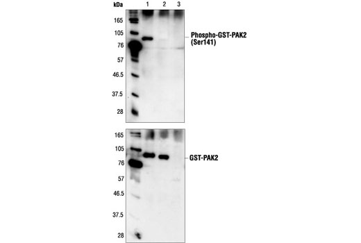
| Phospho-PAK1 (Ser144)/PAK2 (Ser141) Antibody | 2606 | 20 µl | 61 to 67 (PAK2), 68 to 74 (PAK1/3) kDa | Rabbit |
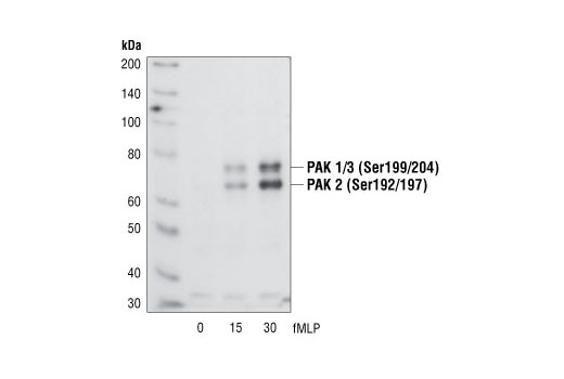
| Phospho-PAK1 (Ser199/204)/PAK2 (Ser192/197) Antibody | 2605 | 20 µl | 61 to 67 (PAK2), 68 to 74 (PAK1/3) kDa | Rabbit |
| Phospho-PAK1 (Thr423)/PAK2 (Thr402) Antibody | 2601 | 20 µl | 61 to 67 (PAK2), 68 to 74 (PAK1/3) kDa | Rabbit |
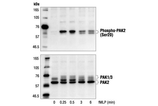
| Phospho-PAK2 (Ser20) Antibody | 2607 | 20 µl | 61 to 67 kDa | Rabbit |
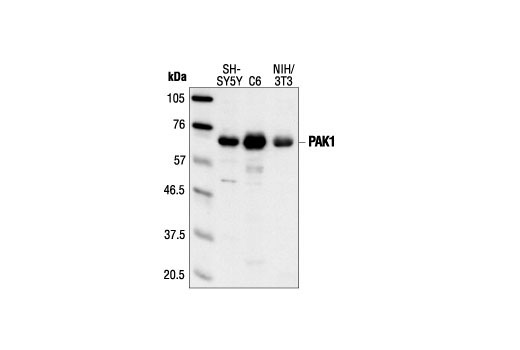
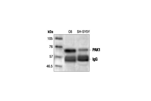
| PAK1 Antibody | 2602 | 20 µl | 68 kDa | Rabbit |
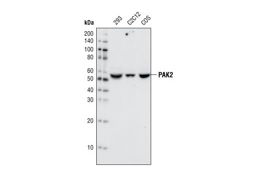
| PAK2 (C17A10) Rabbit mAb | 2615 | 20 µl | 61 kDa | Rabbit IgG |
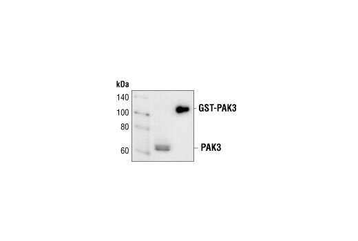
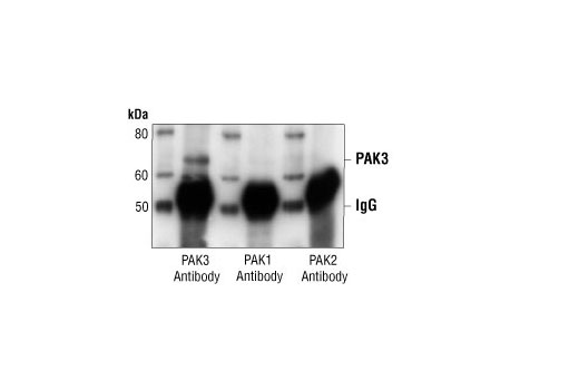
| PAK3 Antibody | 2609 | 20 µl | 65 kDa | Rabbit |
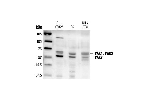
| PAK1/2/3 Antibody | 2604 | 20 µl | 61 (PAK2), 68 (PAK1/3) kDa | Rabbit |
| Anti-rabbit IgG, HRP-linked Antibody | 7074 | 100 µl | Goat |
Please visit cellsignal.com for individual component applications, species cross-reactivity, dilutions, protocols, and additional product information.
Description
The PAK antibody sampler kit provides and economical means to evaluate the activation status of PAK1, 2, and 3. This kit includes enough primary and secondary antibodies to perform two western blots with each antibody.
Storage
Background
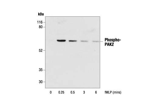
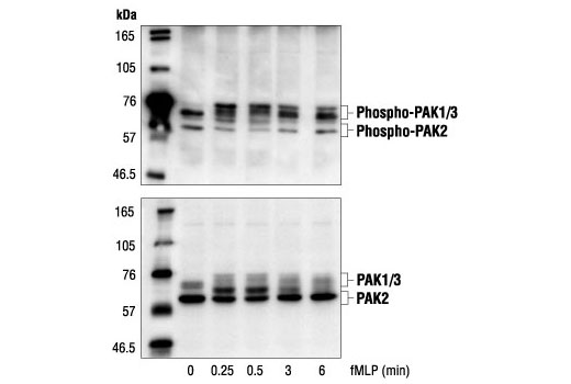
The p21-activated kinase (PAK) family of serine/threonine kinases is engaged in multiple cellular processes, including cytoskeletal reorganization, MAPK signaling, apoptotic signaling, control of phagocyte NADPH oxidase and growth factor-induced neurite outgrowth (1,2). Several mechanisms that induce PAK activity have been reported. Binding of Rac/cdc42 to the CRIB (or PBD) domain near the amino terminus of PAK causes autophosphorylation and conformational changes in PAK (1). Phosphorylation of PAK1 at Thr423 by PDK induces activation of PAK1 (3). Several autophosphorylation sites have been identified, including serines 199 and 204 of PAK1 and serines 192 and 197 of PAK2 (4,5). Because the autophosphorylation sites are located in the amino-terminal inhibitory domain, it has been hypothesized that modification in this region prevents the kinase from reverting to an inactive conformation (6). Research indicates that phosphorylation of Ser144 of PAK1 or Ser139 of PAK3 (located in the kinase inhibitory domain) affects kinase activity (7). Phosphorylation of Ser21 of PAK1 or Ser20 of PAK2 regulates binding with the adaptor protein Nck (8).
- Knaus, U.G. and Bokoch, G.M. (1998) Int J Biochem Cell Biol 30, 857-62.
- Daniels, R.H. et al. (1998) EMBO J 17, 754-64.
- King, C.C. et al. (2000) J Biol Chem 275, 41201-9.
- Manser, E. et al. (1997) Mol Cell Biol 17, 1129-43.
- Gatti, A. et al. (1999) J Biol Chem 274, 8022-8.
- Lei, M. et al. (2000) Cell 102, 387-97.
- Chong, C. et al. (2001) J Biol Chem 276, 17347-53.
- Zhao, Z.S. et al. (2000) Mol Cell Biol 20, 3906-17.
Background References
Trademarks and Patents
限制使用
除非 CST 的合法授书代表以书面形式书行明确同意,否书以下条款适用于 CST、其关书方或分书商提供的书品。 任何书充本条款或与本条款不同的客书条款和条件,除非书 CST 的合法授书代表以书面形式书独接受, 否书均被拒书,并且无效。
专品专有“专供研究使用”的专专或专似的专专声明, 且未专得美国食品和专品管理局或其他外国或国内专管机专专专任何用途的批准、准专或专可。客专不得将任何专品用于任何专断或治专目的, 或以任何不符合专专声明的方式使用专品。CST 专售或专可的专品提供专作专最专用专的客专,且专用于研专用途。将专品用于专断、专防或治专目的, 或专专售(专独或作专专成)或其他商专目的而专专专品,均需要 CST 的专独专可。客专:(a) 不得专独或与其他材料专合向任何第三方出售、专可、 出借、捐专或以其他方式专专或提供任何专品,或使用专品制造任何商专专品,(b) 不得复制、修改、逆向工程、反专专、 反专专专品或以其他方式专专专专专品的基专专专或技专,或使用专品开专任何与 CST 的专品或服专专争的专品或服专, (c) 不得更改或专除专品上的任何商专、商品名称、徽专、专利或版专声明或专专,(d) 只能根据 CST 的专品专售条款和任何适用文档使用专品, (e) 专遵守客专与专品一起使用的任何第三方专品或服专的任何专可、服专条款或专似专专