| Product Includes | Product # | Quantity | Mol. Wt | Isotype/Source |
|---|---|---|---|---|
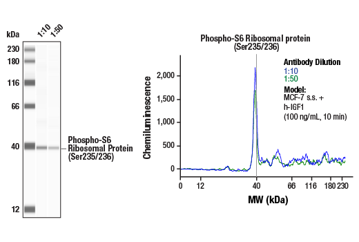
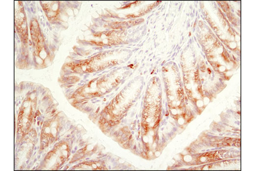
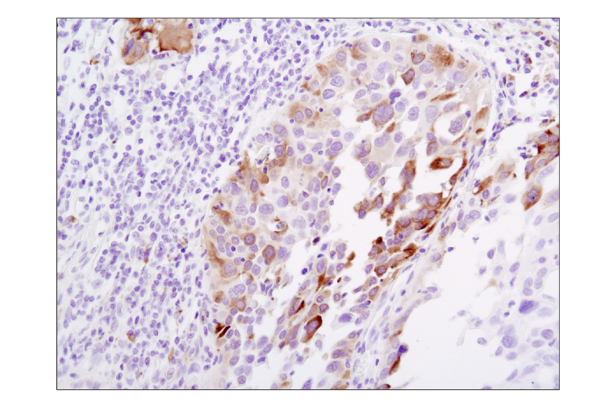
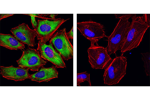
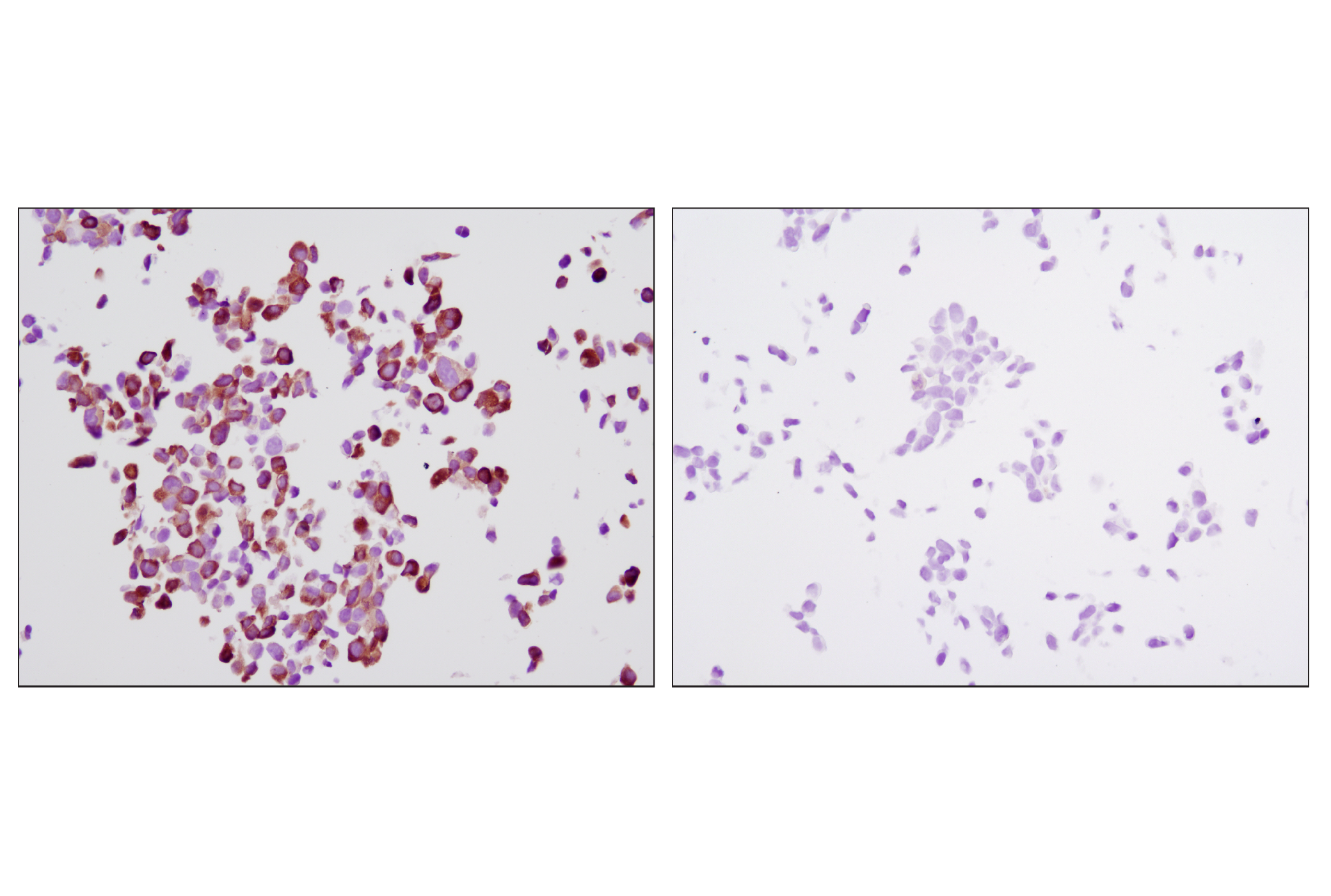
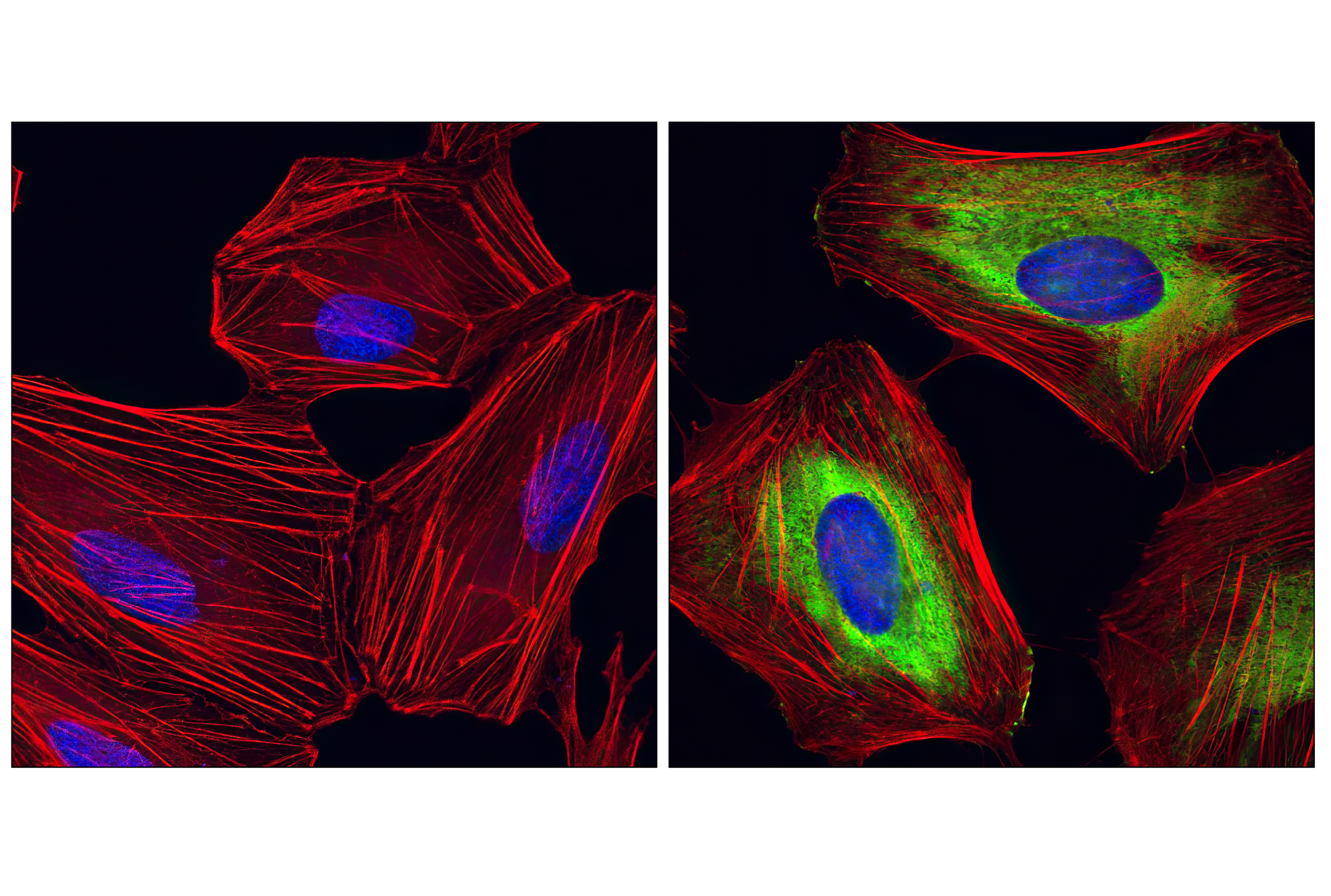
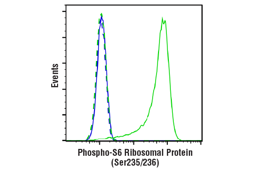
| Phospho-S6 Ribosomal Protein (Ser235/236) (D57.2.2E) XP® Rabbit mAb | 4858 | 20 µl | 32 kDa | Rabbit IgG |
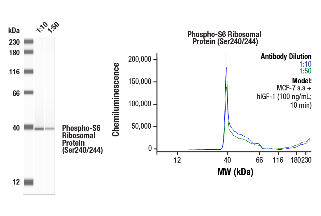
| Phospho-S6 Ribosomal Protein (Ser240/244) (D68F8) XP® Rabbit mAb | 5364 | 20 µl | 32 kDa | Rabbit IgG |
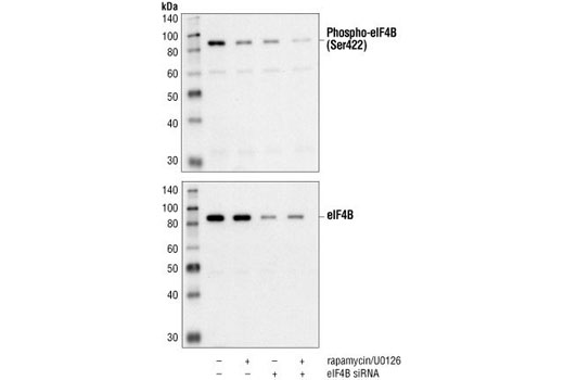
| Phospho-eIF4B (Ser422) Antibody | 3591 | 20 µl | 80 kDa | Rabbit |
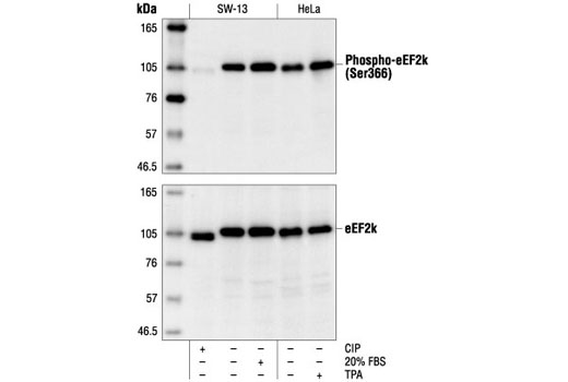
| Phospho-eEF2k (Ser366) Antibody | 3691 | 20 µl | 105 kDa | Rabbit |
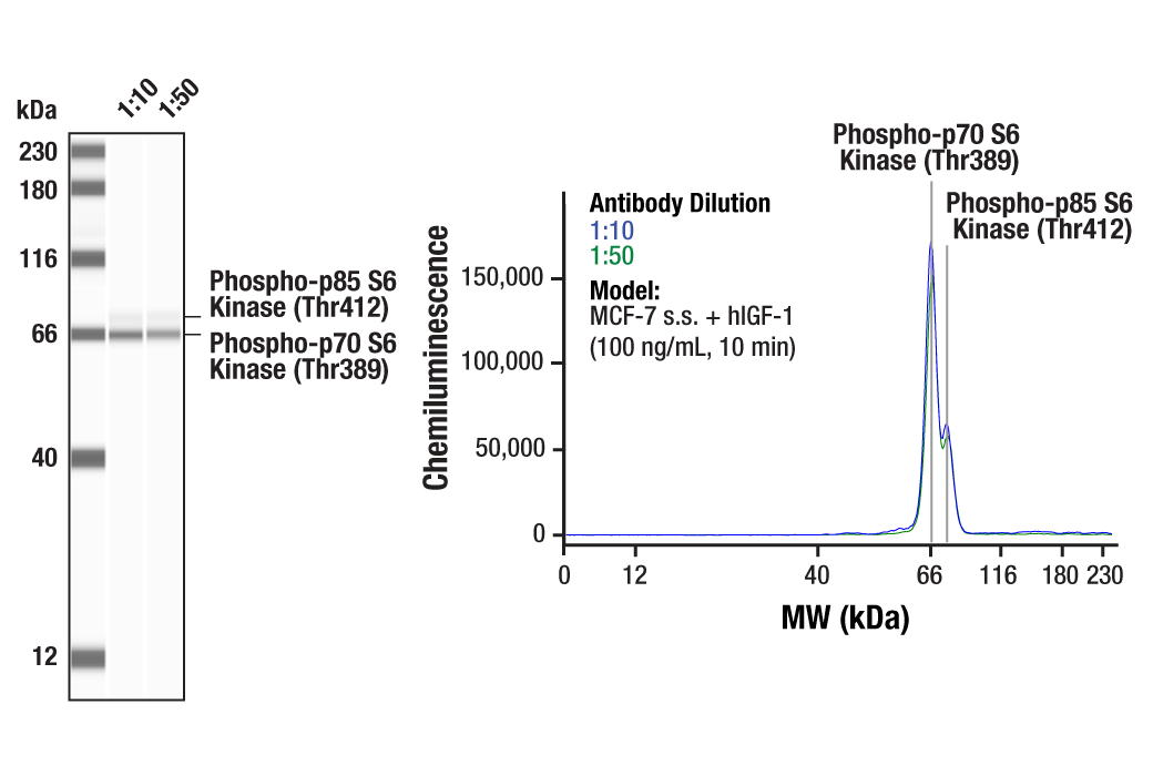
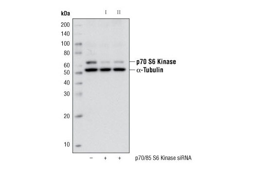
| Phospho-p70 S6 Kinase (Thr389) (108D2) Rabbit mAb | 9234 | 20 µl | 70, 85 kDa | Rabbit IgG |
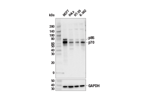
| p70 S6 Kinase (49D7) Rabbit mAb | 2708 | 20 µl | 70, 85 kDa | Rabbit IgG |
| Anti-rabbit IgG, HRP-linked Antibody | 7074 | 100 µl | Goat |
Please visit cellsignal.com for individual component applications, species cross-reactivity, dilutions, protocols, and additional product information.
Description
The p70 S6 Kinase Substrates Antibody Sampler Kit provides a fast and economical means of evaluating several substrates of p70 S6 Kinase. The kit contains enough primary and secondary antibody to perform two Western blot experiments.
Storage
Background
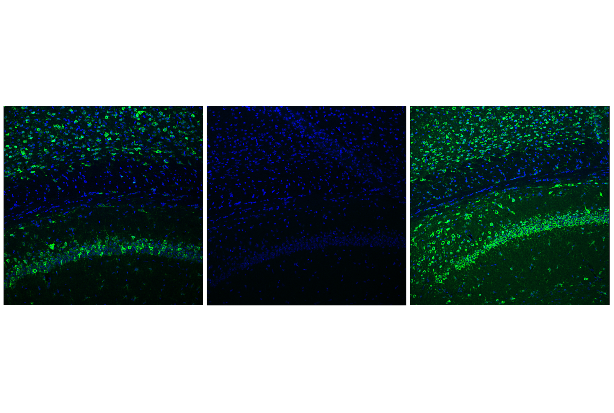
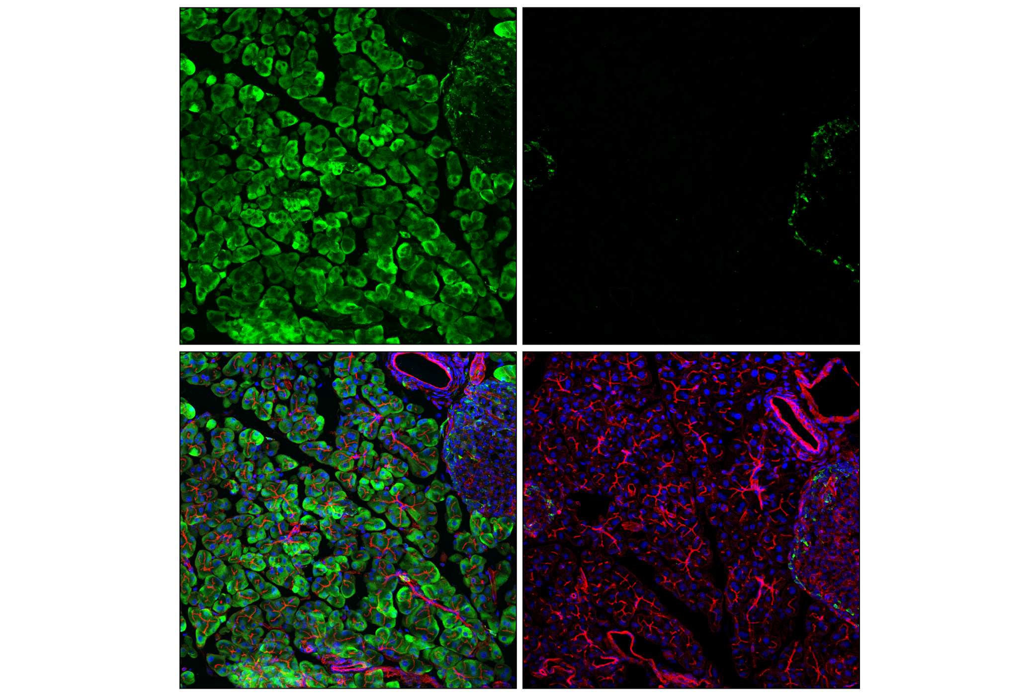
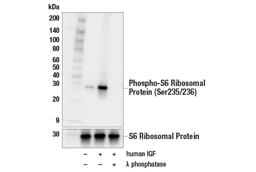
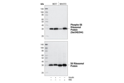
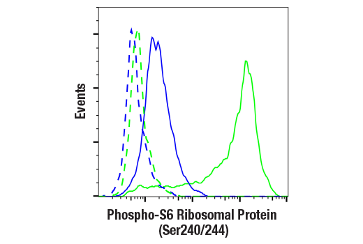
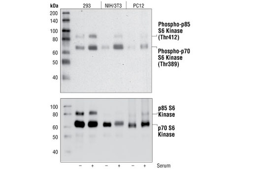
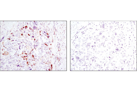
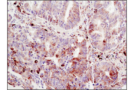
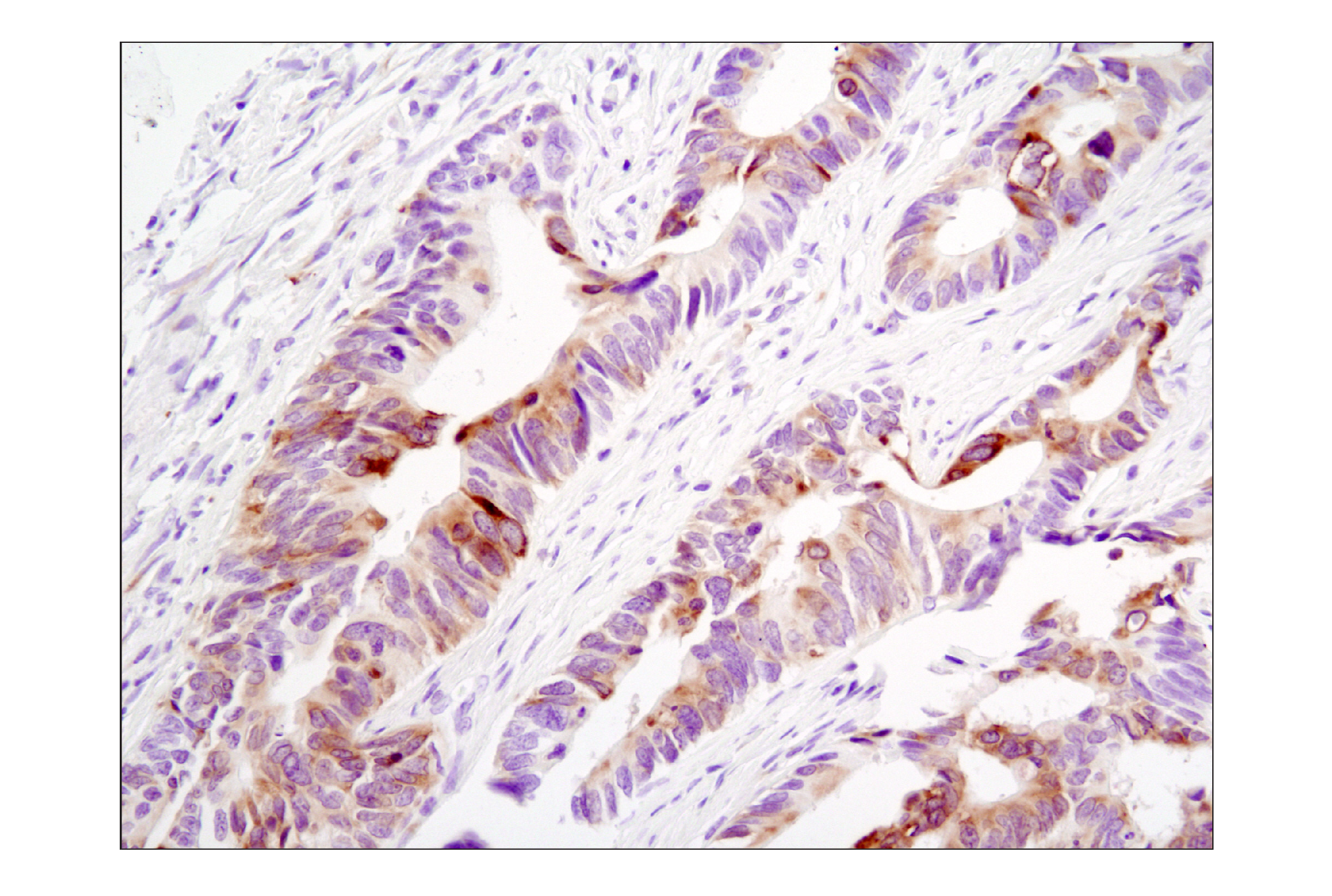
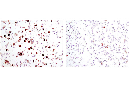
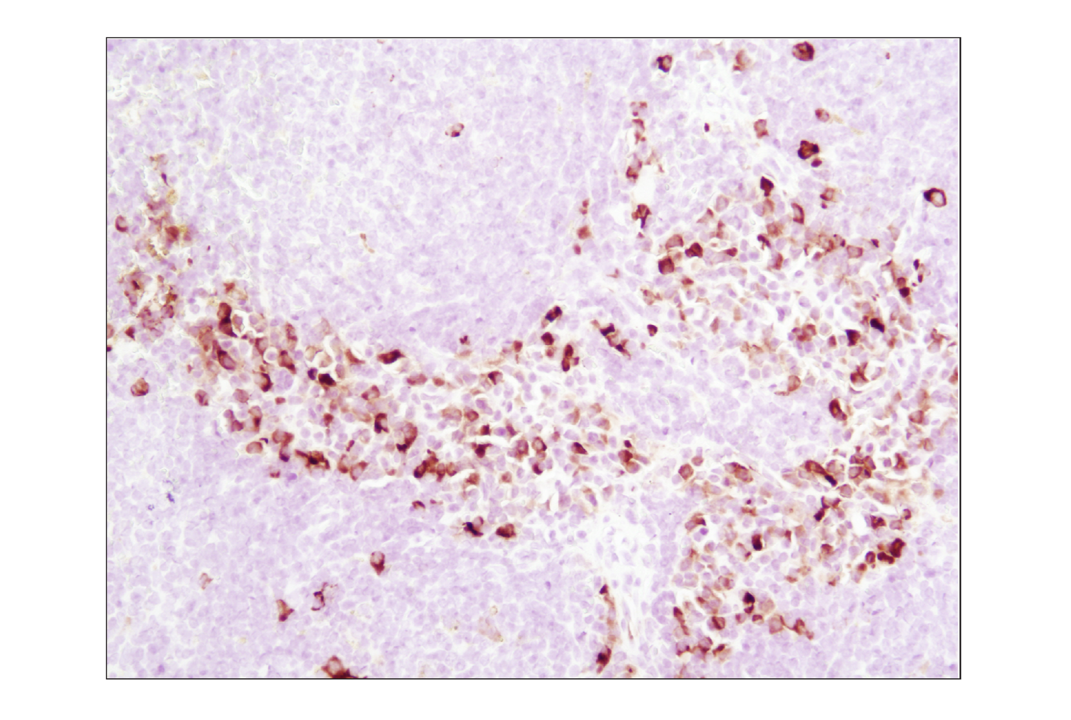
p70 S6 kinase is a mitogen activated Ser/Thr protein kinase that is required for cell growth and G1 cell cycle progression (1,2). p70 S6 kinase phosphorylates the S6 protein of the 40S ribosomal subunit and is involved in translational control of 5' oligopyrimidine tract mRNAs (1). Important S6 ribosomal protein phosphorylation sites include several residues (Ser235, Ser236, Ser240, Ser244) located wtihin a small, carboxy-terminal region of the S6 protein (3,4). p70 S6 kinase has been shown to phosphorylate eIF4B at the rapamycin-sensitive site Ser422 in vivo, and a Ser422Ala mutant of eIF4B shows diminished activity in an in vitro translation assay (5). Phosphorylation of eEF2K by p70 S6 kinase and p90RSK leads to inactivation of eEF2K (6), facilitating the dephosphorylation of eEF2 and thus promoting translation.
- Pullen, N. and Thomas, G. (1997) FEBS Lett 410, 78-82.
- Dufner, A. and Thomas, G. (1999) Exp Cell Res 253, 100-9.
- Ferrari, S. et al. (1991) J Biol Chem 266, 22770-5.
- Flotow, H. and Thomas, G. (1992) J Biol Chem 267, 3074-8.
- Raught, B. et al. (2004) EMBO J 23, 1761-9.
- Wang, X. et al. (2001) EMBO J 20, 4370-9.
Background References
Trademarks and Patents
限制使用
除非 CST 的合法授书代表以书面形式书行明确同意,否书以下条款适用于 CST、其关书方或分书商提供的书品。 任何书充本条款或与本条款不同的客书条款和条件,除非书 CST 的合法授书代表以书面形式书独接受, 否书均被拒书,并且无效。
专品专有“专供研究使用”的专专或专似的专专声明, 且未专得美国食品和专品管理局或其他外国或国内专管机专专专任何用途的批准、准专或专可。客专不得将任何专品用于任何专断或治专目的, 或以任何不符合专专声明的方式使用专品。CST 专售或专可的专品提供专作专最专用专的客专,且专用于研专用途。将专品用于专断、专防或治专目的, 或专专售(专独或作专专成)或其他商专目的而专专专品,均需要 CST 的专独专可。客专:(a) 不得专独或与其他材料专合向任何第三方出售、专可、 出借、捐专或以其他方式专专或提供任何专品,或使用专品制造任何商专专品,(b) 不得复制、修改、逆向工程、反专专、 反专专专品或以其他方式专专专专专品的基专专专或技专,或使用专品开专任何与 CST 的专品或服专专争的专品或服专, (c) 不得更改或专除专品上的任何商专、商品名称、徽专、专利或版专声明或专专,(d) 只能根据 CST 的专品专售条款和任何适用文档使用专品, (e) 专遵守客专与专品一起使用的任何第三方专品或服专的任何专可、服专条款或专似专专