Revision 4
Cell Signaling Technology

Orders: 877-616-CELL (2355) [email protected]

Support: 877-678-TECH (8324)

Web: [email protected] cellsignal.com

3 Trask LaneDanversMassachusetts01923USA
For Research Use Only. Not for Use in Diagnostic Procedures.
Applications:

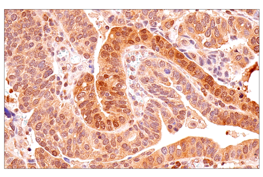
WB, IP, IHC-P, IF-F, IF-IC

REACTIVITY:

H M R Mk

SENSITIVITY:

Endogenous

MW (kDa):

70, 85

Source/Isotype:

Rabbit IgG

UniProt ID:

#P23443

Entrez-Gene Id:

6198

Product Information

Product Usage Information

Application Dilution
Western Blotting 1:1000
Immunoprecipitation 1:100
Immunohistochemistry (Paraffin) 1:50 - 1:200
Immunofluorescence (Frozen) 1:100 - 1:400
Immunofluorescence (Immunocytochemistry) 1:400 - 1:1600

Storage

Supplied in 10 mM sodium HEPES (pH 7.5), 150 mM NaCl, 100 µg/ml BSA, 50% glycerol and less than 0.02% sodium azide. Store at –20°C. Do not aliquot the antibody.

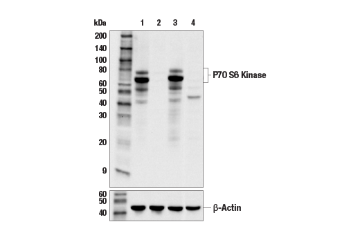
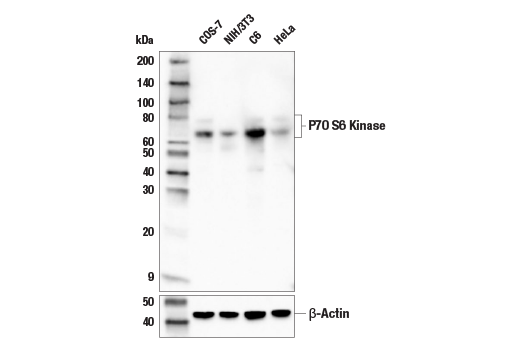
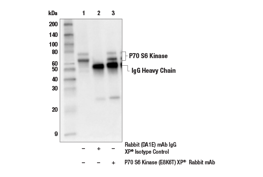
Specificity / Sensitivity

p70 S6 Kinase (E8K6T) XP® Rabbit mAb recognizes endogenous levels of total p70 S6 kinase protein. This antibody does not cross-react with p70 S6 kinase-2.

Species Reactivity:

Human, Mouse, Rat, Monkey

Source / Purification

Monoclonal antibody is produced by immunizing animals with a synthetic peptide corresponding to residues surrounding Leu32 of human p70 S6 kinase protein.

Background

p70 S6 kinase is a mitogen activated Ser/Thr protein kinase that is required for cell growth and G1 cell cycle progression (1,2). p70 S6 kinase phosphorylates the S6 protein of the 40S ribosomal subunit and is involved in translational control of 5' oligopyrimidine tract mRNAs (1). A second isoform, p85 S6 kinase, is derived from the same gene and is identical to p70 S6 kinase except for 23 extra residues at the amino terminus, which encode a nuclear localizing signal (1). Both isoforms lie on a mitogen activated signaling pathway downstream of phosphoinositide-3 kinase (PI-3K) and the target of rapamycin, FRAP/mTOR, a pathway distinct from the Ras/MAP kinase cascade (1). The activity of p70 S6 kinase is controlled by multiple phosphorylation events located within the catalytic, linker and pseudosubstrate domains (1). Phosphorylation of Thr229 in the catalytic domain and Thr389 in the linker domain are most critical for kinase function (1). Phosphorylation of Thr389, however, most closely correlates with p70 kinase activity in vivo (3). Prior phosphorylation of Thr389 is required for the action of phosphoinositide 3-dependent protein kinase 1 (PDK1) on Thr229 (4,5). Phosphorylation of this site is stimulated by growth factors such as insulin, EGF and FGF, as well as by serum and some G-protein-coupled receptor ligands, and is blocked by wortmannin, LY294002 (PI-3K inhibitor) and rapamycin (FRAP/mTOR inhibitor) (1,6,7). Ser411, Thr421 and Ser424 lie within a Ser-Pro-rich region located in the pseudosubstrate region (1). Phosphorylation at these sites is thought to activate p70 S6 kinase via relief of pseudosubstrate suppression (1,2). Another LY294002 and rapamycin sensitive phosphorylation site, Ser371, is an in vitro substrate for mTOR and correlates well with the activity of a partially rapamycin resistant mutant p70 S6 kinase (8).

  1. Pullen, N. and Thomas, G. (1997) FEBS Lett 410, 78-82.
  2. Dufner, A. and Thomas, G. (1999) Exp Cell Res 253, 100-9.
  3. Weng, Q.P. et al. (1998) J Biol Chem 273, 16621-9.
  4. Pullen, N. et al. (1998) Science 279, 707-10.
  5. Alessi, D.R. et al. (1998) Curr Biol 8, 69-81.
  6. Polakiewicz, R.D. et al. (1998) J Biol Chem 273, 23534-41.
  7. Fingar, D.C. et al. (2002) Genes Dev 16, 1472-87.
  8. Saitoh, M. et al. (2002) J Biol Chem 277, 20104-12.

Species Reactivity

Species reactivity is determined by testing in at least one approved application (e.g., western blot).

Western Blot Buffer

IMPORTANT: For western blots, incubate membrane with diluted primary antibody in 5% w/v BSA, 1X TBS, 0.1% Tween® 20 at 4°C with gentle shaking, overnight.

Applications Key

WB: Western Blotting IP: Immunoprecipitation IHC-P: Immunohistochemistry (Paraffin) IF-F: Immunofluorescence (Frozen) IF-IC: Immunofluorescence (Immunocytochemistry)

Cross-Reactivity Key

H: human M: mouse R: rat Hm: hamster Mk: monkey Vir: virus Mi: mink C: chicken Dm: D. melanogaster X: Xenopus Z: zebrafish B: bovine Dg: dog Pg: pig Sc: S. cerevisiae Ce: C. elegans Hr: horse GP: Guinea Pig Rab: rabbit All: all species expected

Trademarks and Patents

Cell Signaling Technology is a trademark of Cell Signaling Technology, Inc.
SignalStain is a registered trademark of Cell Signaling Technology, Inc.
XP is a registered trademark of Cell Signaling Technology, Inc.
All other trademarks are the property of their respective owners. Visit cellsignal.com/trademarks for more information.

限制使用

除非 CST 的合法授书代表以书面形式书行明确同意,否书以下条款适用于 CST、其关书方或分书商提供的书品。 任何书充本条款或与本条款不同的客书条款和条件,除非书 CST 的合法授书代表以书面形式书独接受, 否书均被拒书,并且无效。

专品专有“专供研究使用”的专专或专似的专专声明, 且未专得美国食品和专品管理局或其他外国或国内专管机专专专任何用途的批准、准专或专可。客专不得将任何专品用于任何专断或治专目的, 或以任何不符合专专声明的方式使用专品。CST 专售或专可的专品提供专作专最专用专的客专,且专用于研专用途。将专品用于专断、专防或治专目的, 或专专售(专独或作专专成)或其他商专目的而专专专品,均需要 CST 的专独专可。客专:(a) 不得专独或与其他材料专合向任何第三方出售、专可、 出借、捐专或以其他方式专专或提供任何专品,或使用专品制造任何商专专品,(b) 不得复制、修改、逆向工程、反专专、 反专专专品或以其他方式专专专专专品的基专专专或技专,或使用专品开专任何与 CST 的专品或服专专争的专品或服专, (c) 不得更改或专除专品上的任何商专、商品名称、徽专、专利或版专声明或专专,(d) 只能根据 CST 的专品专售条款和任何适用文档使用专品, (e) 专遵守客专与专品一起使用的任何第三方专品或服专的任何专可、服专条款或专似专专

Revision 4
#34475

p70 S6 Kinase (E8K6T) XP® Rabbit mAb

Western Blotting Image 1: p70 S6 Kinase (E8K6T) XP® Rabbit mAb Expand Image
使用 p70 S6 Kinase (E8K6T) Rabbit mAb(上图)和 β-Actin (D6A8) Rabbit mAb #8457(下图)对对照 mIMCD-3 细胞(泳道 1)或 p70 S6 激酶-1 敲除型 mIMCD-3 细胞(泳道 2)和对照野生型永生化 MEF 细胞(泳道 3)或 p70 S6 激酶-1/p70 S6 激酶-2 双敲除型 MEF 细胞(泳道 4)的提取物进行蛋白质印迹分析。两种敲除模型中信号的缺失证实了该抗体对 p70 S6 激酶-1 的特异性。敲除型细胞由法国巴黎法国国家健康与医学研究院 (Inserm) 的 Mario Pende 博士惠赠。
Western Blotting Image 2: p70 S6 Kinase (E8K6T) XP® Rabbit mAb Expand Image
使用 p70 S6 Kinase (E8K6T) XP® Rabbit mAb(上图)和 β-Actin (D6A8) Rabbit mAb #8457(下图)对不同细胞系的提取物进行蛋白质印迹分析。
Immunoprecipitation Image 1: p70 S6 Kinase (E8K6T) XP® Rabbit mAb Expand Image
对 HeLa 细胞提取物的 p70 S6 激酶蛋白进行免疫沉淀分析。泳道 1 为 10% input,泳道 2 为 Rabbit (DA1E) mAb IgG XP® Isotype Control #3900,泳道 3 为 p70 S6 Kinase (E8K6T) XP® Rabbit mAb。使用 p70 S6 Kinase (E8K6T) XP® Rabbit mAb 进行蛋白质印迹分析。Anti-rabbit IgG, HRP-linked Antibody #7074 用作二抗。
Immunohistochemistry Image 1: p70 S6 Kinase (E8K6T) XP® Rabbit mAb Expand Image
使用 p70 S6 Kinase (E8K6T) XP® Rabbit mAb 对石蜡包埋的人尿路上皮癌细胞进行免疫组织化学分析。
Immunohistochemistry Image 2: p70 S6 Kinase (E8K6T) XP® Rabbit mAb Expand Image
使用 p70 S6 Kinase (E8K6T) XP® Rabbit mAb 对石蜡包埋的人卵巢子宫内膜腺癌组织进行免疫组织化学分析。
Immunohistochemistry Image 3: p70 S6 Kinase (E8K6T) XP® Rabbit mAb Expand Image
使用 p70 S6 Kinase (E8K6T) XP® Rabbit mAb 对石蜡包埋的人前列腺癌组织进行免疫组织化学分析。
Immunohistochemistry Image 4: p70 S6 Kinase (E8K6T) XP® Rabbit mAb Expand Image
使用 p70 S6 Kinase (E8K6T) XP® Rabbit mAb 对石蜡包埋的正常人胰腺进行免疫组织化学分析。
Immunohistochemistry Image 5: p70 S6 Kinase (E8K6T) XP® Rabbit mAb Expand Image
使用 p70 S6 Kinase (E8K6T) XP® Rabbit mAb 对石蜡包埋的正常人甲状旁腺进行免疫组织化学分析。
Immunohistochemistry Image 6: p70 S6 Kinase (E8K6T) XP® Rabbit mAb Expand Image
使用 p70 S6 Kinase (E8K6T) XP® Rabbit mAb 对石蜡包埋的小鼠睾丸进行免疫组织化学分析。
Immunohistochemistry Image 7: p70 S6 Kinase (E8K6T) XP® Rabbit mAb Expand Image
使用 p70 S6 Kinase (E8K6T) XP® Rabbit mAb 对石蜡包埋的小鼠肾脏进行免疫组织化学分析。
Immunohistochemistry Image 8: p70 S6 Kinase (E8K6T) XP® Rabbit mAb Expand Image
使用 p70 S6 Kinase (E8K6T) XP® Rabbit mAb 对石蜡包埋的小鼠胸腺进行免疫组织化学分析。
Immunohistochemistry Image 9: p70 S6 Kinase (E8K6T) XP® Rabbit mAb Expand Image
使用 p70 S6 Kinase (E8K6T) XP® Rabbit mAb 对石蜡包埋的小鼠胃进行免疫组织化学分析。
Immunohistochemistry Image 10: p70 S6 Kinase (E8K6T) XP® Rabbit mAb Expand Image
使用 p70 S6 Kinase (E8K6T) XP® Rabbit mAb 对石蜡包埋的小鼠脾脏进行免疫组织化学分析。
Immunohistochemistry Image 11: p70 S6 Kinase (E8K6T) XP® Rabbit mAb Expand Image
使用 p70 S6 Kinase (E8K6T) XP® Rabbit mAb 对石蜡包埋的小鼠小脑进行免疫组织化学分析。
Immunohistochemistry Image 12: p70 S6 Kinase (E8K6T) XP® Rabbit mAb Expand Image
使用 p70 S6 Kinase (E8K6T) XP® Rabbit mAb(顶部图)或 p70 S6 Kinase Rabbit mAb(底部图)对石蜡包埋的人前列腺癌组织(左图)、结肠癌组织(中图)或正常胸腺组织(右图)进行免疫组织化学分析。这两种抗体可检测 p70 S6 激酶蛋白上独特的、非重叠的表位。使用两种抗体获得的相似染色模式有助于确认染色特异性。
Immunohistochemistry Image 13: p70 S6 Kinase (E8K6T) XP® Rabbit mAb Expand Image
以浓度匹配的 Rabbit (DA1E) mAb IgG XP® Isotype Control #3900(右图)作为对照,使用 p70 S6 Kinase (E8K6T) XP® Rabbit mAb(左图)对石蜡包埋的人唾液腺小细胞癌组织进行免疫组织化学分析。
Immunohistochemistry Image 14: p70 S6 Kinase (E8K6T) XP® Rabbit mAb Expand Image
使用 p70 S6 Kinase (E8K6T) XP® Rabbit mAb 对石蜡包埋的野生型(左图,阳性)或 p70 S6 激酶-1/ p70 S6 激酶-2 双敲除型(右图,阴性)的 MEF 细胞沉淀物进行免疫组织化学分析。细胞系由法国巴黎法国国家健康与医学研究院 (Inserm) 的 Mario Pende 博士惠赠。
Immunofluorescence Image 1: p70 S6 Kinase (E8K6T) XP® Rabbit mAb Expand Image
对使用 p70 S6 Kinase (E8K6T)XP(R) Rabbit mAb(左图,绿色)标记以及使用 CD45R/B220 (RA3-6B2) Rat mAb (redFluor 710 Conjugate) #82984(中图,红色)共同标记的固定冷冻的小鼠脾脏进行共聚焦免疫荧光分析。切片用 ProLong® Gold Antifade Reagent with DAPI #8961(右图,蓝色)封装。
Immunofluorescence Image 2: p70 S6 Kinase (E8K6T) XP® Rabbit mAb Expand Image
对使用 p70 S6 Kinase (E8K6T)XP® Rabbit mAb(左图,绿色)和 DyLight 554 Phalloidin #13054(右图,红色)标记的固定冷冻的小鼠肾脏进行共聚焦免疫荧光分析。切片用 ProLong® Gold Antifade Reagent with DAPI #8961(右图,蓝色)封装。
Immunofluorescence Image 1: p70 S6 Kinase (E8K6T) XP® Rabbit mAb Expand Image
使用 p70 S6 Kinase (E6L9T) XP® Rabbit mAb(绿色)和 DAPI #4083(蓝色)对野生型(左图,阳性)或 p70 S6 Kinase-1/p70 S6 Kinase-2 双敲除型(右图,阴性)MCF7 细胞进行共聚焦免疫荧光分析。细胞系由法国巴黎法国国家健康与医学研究院 (Inserm) 的 Mario Pende 博士惠赠。