Revision 6

#13109Store at -20C

Cell Signaling Technology

Orders: 877-616-CELL (2355) [email protected]

Support: 877-678-TECH (8324)

Web: [email protected] cellsignal.com

3 Trask LaneDanversMassachusetts01923USA
For Research Use Only. Not for Use in Diagnostic Procedures.
Applications:

WB, IP, IF-IC, FC-FP, ChIP, ChIP-seq

REACTIVITY:

H

SENSITIVITY:

Endogenous

MW (kDa):

75

Source/Isotype:

Rabbit IgG

UniProt ID:

#Q9H3D4

Entrez-Gene Id:

8626

Product Information

Product Usage Information

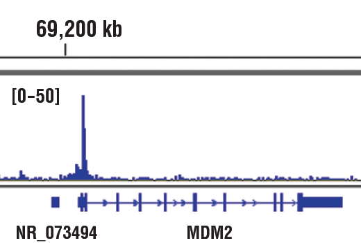
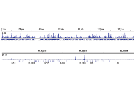
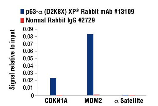
For optimal ChIP and ChIP-seq results, use 5 μl of antibody and 10 μg of chromatin (approximately 4 x 10⁶ cells) per IP. This antibody has been validated using SimpleChIP® Enzymatic Chromatin IP Kits.

Application Dilution
Western Blotting 1:1000
Immunoprecipitation 1:100
Immunofluorescence (Immunocytochemistry) 1:400 - 1:1600
Flow Cytometry (Fixed/Permeabilized) 1:800 - 1:3200
Chromatin IP 1:100
Chromatin IP-seq 1:100

Storage

Supplied in 10 mM sodium HEPES (pH 7.5), 150 mM NaCl, 100 µg/ml BSA, 50% glycerol and less than 0.02% sodium azide. Store at –20°C. Do not aliquot the antibody.

For a carrier free (BSA and azide free) version of this product see product #49248.

Specificity / Sensitivity

p63-α (D2K8X) XP® Rabbit mAb recognizes endogenous levels of total p63-α protein. This antibody will detect both the full-length isoform (TAp63-α) and the truncated δNp63-α isoform, but will not recognize the p63-β or p63-γ isoform.

Species Reactivity:

Human

Source / Purification

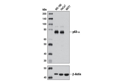
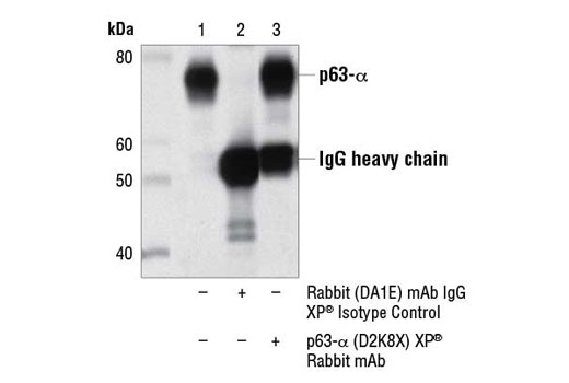
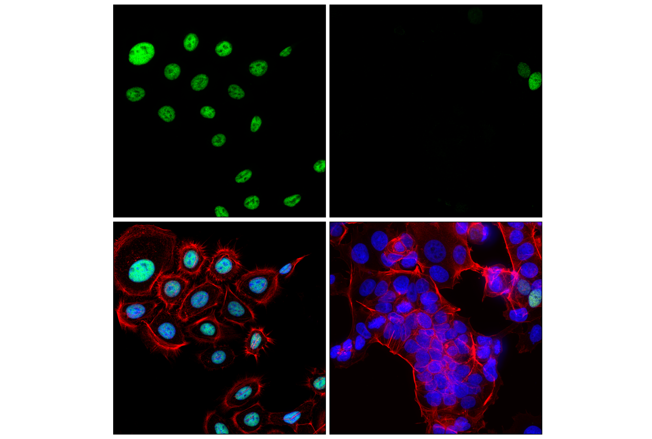
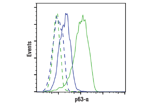
Monoclonal antibody is produced by immunizing animals with a synthetic peptide corresponding to residues near the carboxy terminus of human p63-α protein.

Background

The p53 tumor suppressor protein plays a major role in cellular response to DNA damage and other genomic aberrations. Activation of p53 can lead to either cell cycle arrest and DNA repair or apoptosis (1). In addition to p53, mammalian cells contain two p53 family members, p63 and p73, which are similar to p53 in both structure and function (2). While p63 can induce p53-responsive genes and apoptosis, mutation of p63 rarely results in tumors (2). Research investigators frequently observe amplification of the p63 gene in squamous cell carcinomas of the lung, head and neck (2,3). The p63 gene contains an alternative transcription initiation site that yields a truncated ΔNp63 lacking the transactivation domain, and alternative splicing at the carboxy-terminus yields the α, β, and γ isoforms (3,4).

  1. Levine, A.J. (1997) Cell 88, 323-31.
  2. Waltermann, A. et al. (2003) Oncogene 22, 5686-93.
  3. Hibi, K. et al. (2000) Proc Natl Acad Sci U S A 97, 5462-7.
  4. Yang, A. et al. (1999) Nature 398, 714-8.

Species Reactivity

Species reactivity is determined by testing in at least one approved application (e.g., western blot).

Western Blot Buffer

IMPORTANT: For western blots, incubate membrane with diluted primary antibody in 5% w/v BSA, 1X TBS, 0.1% Tween® 20 at 4°C with gentle shaking, overnight.

Applications Key

WB: Western Blotting IP: Immunoprecipitation IF-IC: Immunofluorescence (Immunocytochemistry) FC-FP: Flow Cytometry (Fixed/Permeabilized) ChIP: Chromatin IP ChIP-seq: Chromatin IP-seq

Cross-Reactivity Key

H: human M: mouse R: rat Hm: hamster Mk: monkey Vir: virus Mi: mink C: chicken Dm: D. melanogaster X: Xenopus Z: zebrafish B: bovine Dg: dog Pg: pig Sc: S. cerevisiae Ce: C. elegans Hr: horse GP: Guinea Pig Rab: rabbit All: all species expected

Trademarks and Patents

Cell Signaling Technology is a trademark of Cell Signaling Technology, Inc.
XP is a registered trademark of Cell Signaling Technology, Inc.
All other trademarks are the property of their respective owners. Visit cellsignal.com/trademarks for more information.

限制使用

除非 CST 的合法授书代表以书面形式书行明确同意,否书以下条款适用于 CST、其关书方或分书商提供的书品。 任何书充本条款或与本条款不同的客书条款和条件,除非书 CST 的合法授书代表以书面形式书独接受, 否书均被拒书,并且无效。

专品专有“专供研究使用”的专专或专似的专专声明, 且未专得美国食品和专品管理局或其他外国或国内专管机专专专任何用途的批准、准专或专可。客专不得将任何专品用于任何专断或治专目的, 或以任何不符合专专声明的方式使用专品。CST 专售或专可的专品提供专作专最专用专的客专,且专用于研专用途。将专品用于专断、专防或治专目的, 或专专售(专独或作专专成)或其他商专目的而专专专品,均需要 CST 的专独专可。客专:(a) 不得专独或与其他材料专合向任何第三方出售、专可、 出借、捐专或以其他方式专专或提供任何专品,或使用专品制造任何商专专品,(b) 不得复制、修改、逆向工程、反专专、 反专专专品或以其他方式专专专专专品的基专专专或技专,或使用专品开专任何与 CST 的专品或服专专争的专品或服专, (c) 不得更改或专除专品上的任何商专、商品名称、徽专、专利或版专声明或专专,(d) 只能根据 CST 的专品专售条款和任何适用文档使用专品, (e) 专遵守客专与专品一起使用的任何第三方专品或服专的任何专可、服专条款或专似专专

Revision 6
#13109

p63-α (D2K8X) XP® Rabbit mAb

Western Blotting Image 1: p63-α (D2K8X) XP® Rabbit mAb Expand Image
使用 p63-α (D2K8X) XP® Rabbit mAb(上图)或 β-Actin (D6A8) Rabbit mAb #8457(下图),对 ME-180 (+)、HaCaT (+) 和 MCF7 (-) 细胞的提取物进行蛋白质印迹分析。
Immunoprecipitation Image 1: p63-α (D2K8X) XP® Rabbit mAb Expand Image
使用 Rabbit (DA1E) mAb IgG XP®2 Isotype Control #(泳道 )或 p63-α (D2K8X) XP®3 Rabbit mAb(泳道 3900)对 Me180 细胞提取物 p63-α 进行免疫沉淀分析。泳道 1 是 10% 输入对照。使用 p63-α (D2K8X) XP® Rabbit mAb 进行蛋白质印迹分析。
Immunofluorescence Image 1: p63-α (D2K8X) XP® Rabbit mAb Expand Image
Confocal immunofluorescent analysis of ME-180 cells (left, positive) and MCF7 cells (right, negative excluding a subpopulation of presumed cancer stem cells) using p63-α (D2K8X) XP® Rabbit mAb (green), DyLight 554 Phalloidin #13054 (red), and DAPI #4083 (blue).
Flow Cytometry Image 1: p63-α (D2K8X) XP® Rabbit mAb Expand Image
使用 p63-α (D2K8X) XP® Rabbit mAb(实线)或浓度匹配的 Rabbit (DA1E) mAb IgG XP® Isotype Control #3900(虚线)对 MCF7 细胞(蓝色)和 ME-180 细胞(绿色)进行流式细胞分析。Anti-rabbit IgG (H+L), F(ab')2 Fragment (PE Conjugate) #79408 用作二抗。
Chromatin Immunoprecipitation Image 1: p63-α (D2K8X) XP® Rabbit mAb Expand Image
使用 SimpleChIP® 酶解染色质免疫共沉淀试剂盒(磁珠法)#9005 对 HaCaT 细胞的交联染色质和 p63-α (D2K8X) XP® 兔单克隆抗体 进行染色质免疫沉淀。使用 DNA Library Prep Kit for Illumina® (ChIP-seq, CUT&RUN) #56795 制备 DNA 库。结果图显示在 p63-α 的已知靶基因 MDM2 内结合(参见包含 ChIP-qPCR 数据的其他结果图)。
Chromatin Immunoprecipitation Image 2: p63-α (D2K8X) XP® Rabbit mAb Expand Image
使用 SimpleChIP® 酶解染色质免疫共沉淀试剂盒(磁珠法)#9005 对 HaCaT 细胞的交联染色质和 p63-α (D2K8X) XP® 兔单克隆抗体 进行染色质免疫沉淀。使用 DNA Library Prep Kit for Illumina® (ChIP-seq, CUT&RUN) #56795 制备 DNA 库。结果图显示在染色体 12(上图)内的结合,包括 p63-α 的已知靶标基因 MDM2(下图)(参见包含 ChIP-qPCR 数据的其他结果图)。
Chromatin Immunoprecipitation Image 3: p63-α (D2K8X) XP® Rabbit mAb Expand Image
使用 SimpleChIP® Enzymatic Chromatin IP Kit (Magnetic Beads) #9003,对 HaCaT 细胞中提取的交联染色质,在加入 p63-α (D2K8X) XP® Rabbit mAb 或Normal Rabbit IgG #2729 后进行染色质免疫沉淀分析。采用 SimpleChIP® Human CDKN1A Promoter Primers #6449、human MDM2 intron 2 primers 和 SimpleChIP® Human α Satellite Repeat Primers #4486,通过实时定量 PCR 对富集的 DNA 进行定量分析。将每份样品中免疫沉淀的 DNA 的量表现为相对于所输入染色质总量(等于 1)的信号。