| Product Includes | Product # | Quantity | Mol. Wt | Isotype/Source |
|---|---|---|---|---|
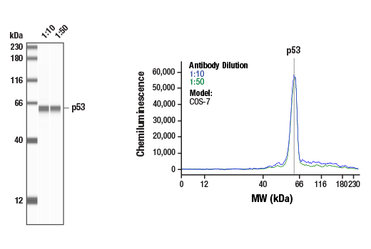
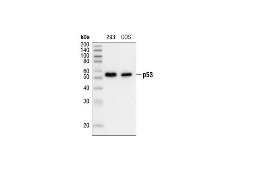
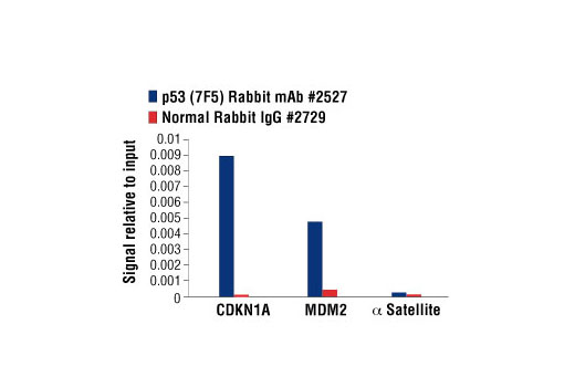
| p53 (7F5) Rabbit mAb | 2527 | 20 µl | 53 kDa | Rabbit IgG |
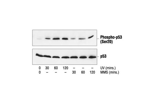
| Phospho-p53 (Ser20) Antibody | 9287 | 20 µl | 53 kDa | Rabbit |
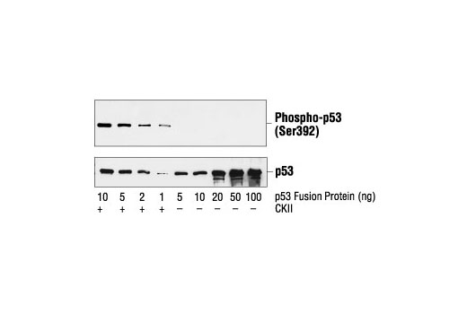
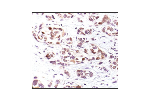
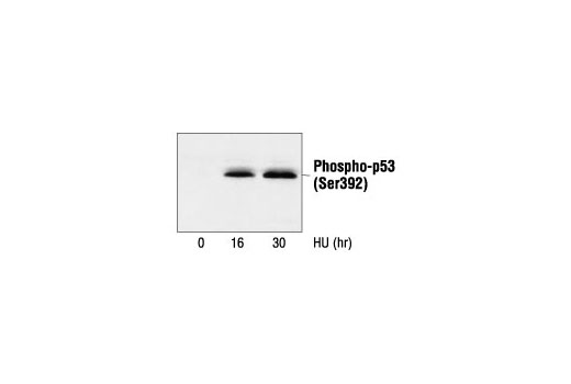
| Phospho-p53 (Ser392) Antibody | 9281 | 20 µl | 53 kDa | Rabbit |
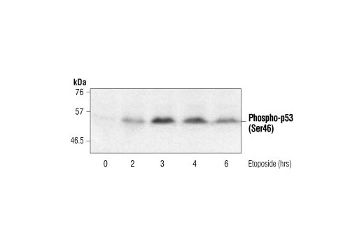
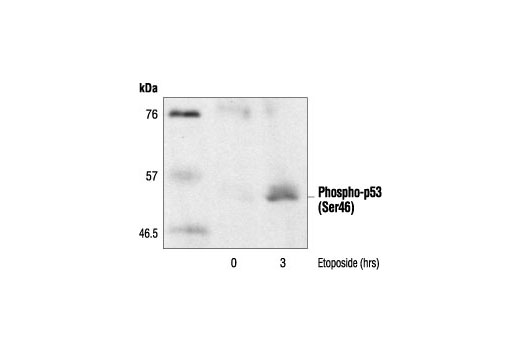
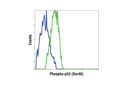
| Phospho-p53 (Ser46) Antibody | 2521 | 20 µl | 53 kDa | Rabbit |
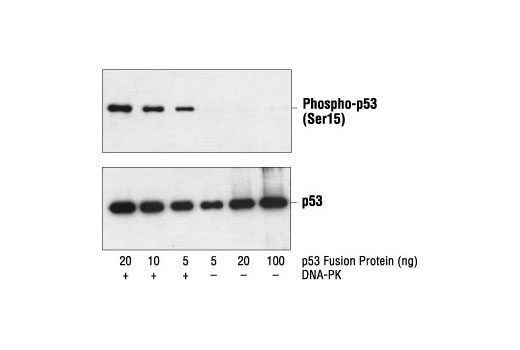
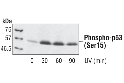
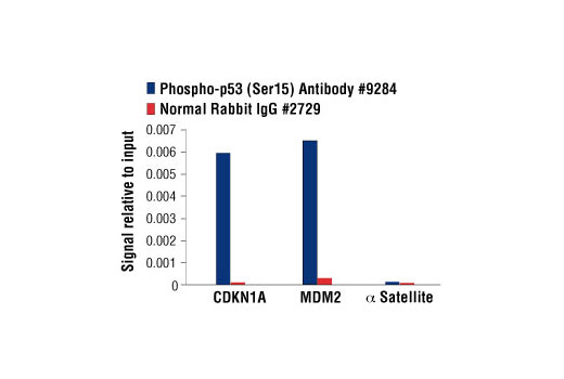
| Phospho-p53 (Ser15) Antibody | 9284 | 20 µl | 53 kDa | Rabbit |
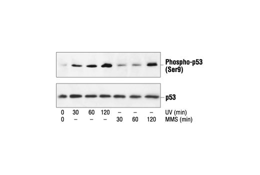
| Phospho-p53 (Ser9) Antibody | 9288 | 20 µl | 53 kDa | Rabbit |
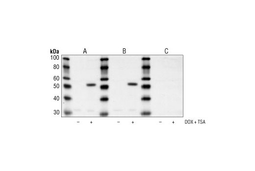
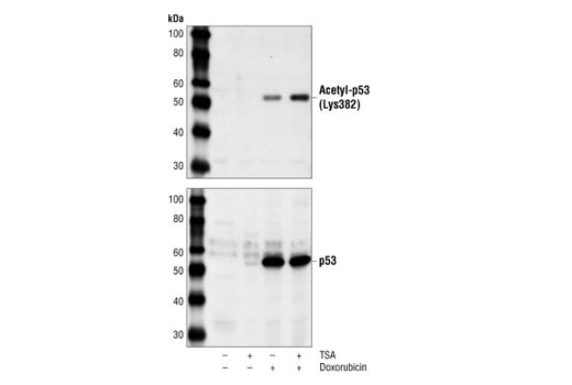
| Acetyl-p53 (Lys382) Antibody | 2525 | 20 µl | 53 kDa | Rabbit |
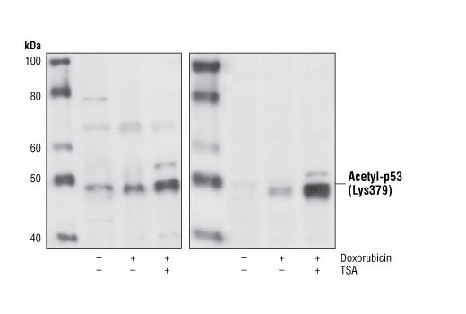
| Acetyl-p53 (Lys379) Antibody | 2570 | 20 µl | 53 kDa | Rabbit |
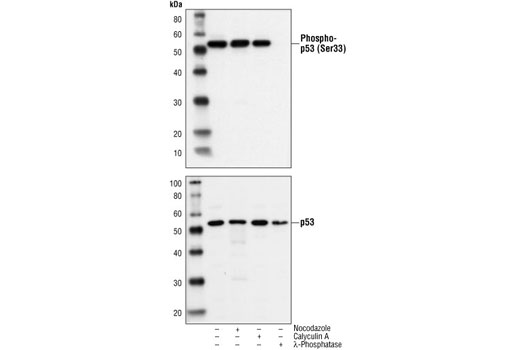
| Phospho-p53 (Ser33) Antibody | 2526 | 20 µl | 53 kDa | Rabbit |
| Anti-rabbit IgG, HRP-linked Antibody | 7074 | 100 µl | Goat |
Please visit cellsignal.com for individual component applications, species cross-reactivity, dilutions, protocols, and additional product information.
Description
The p53 Antibody Sampler Kit provides an economical means of detecting p53 activity using modification-specific and control antibodies. The kit includes enough antibody to perform two western blot experiments with each primary antibody.
Storage
Background
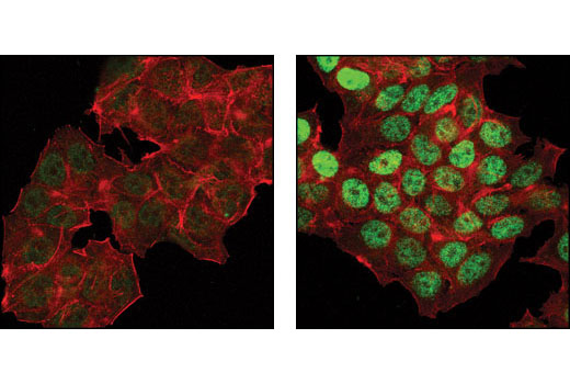
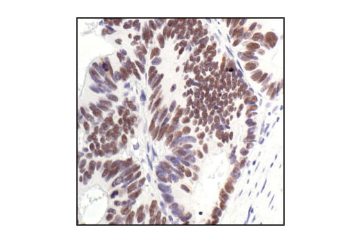
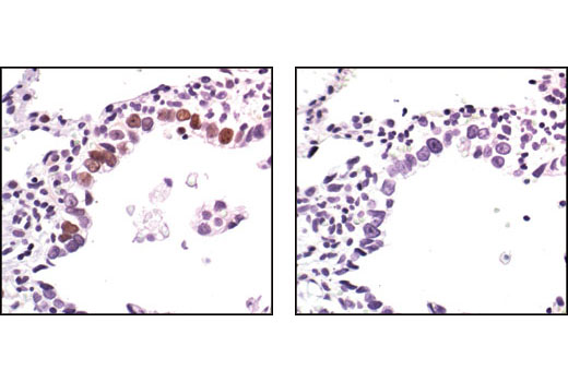
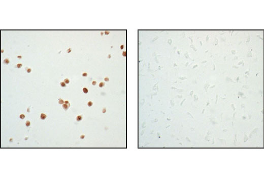
The p53 tumor suppressor protein plays a major role in cellular response to DNA damage and other genomic aberrations. Activation of p53 can lead to either cell cycle arrest and DNA repair or apoptosis (1). p53 is phosphorylated at multiple sites in vivo and by several different protein kinases in vitro (2,3). DNA damage induces phosphorylation of p53 at Ser15 and Ser20 and leads to a reduced interaction between p53 and its negative regulator, the oncoprotein MDM2 (4). MDM2 inhibits p53 accumulation by targeting it for ubiquitination and proteasomal degradation (5,6). p53 can be phosphorylated by ATM, ATR, and DNA-PK at Ser15 and Ser37. Phosphorylation impairs the ability of MDM2 to bind p53, promoting both the accumulation and activation of p53 in response to DNA damage (4,7). Chk2 and Chk1 can phosphorylate p53 at Ser20, enhancing its tetramerization, stability, and activity (8,9). p53 is phosphorylated at Ser392 in vivo (10,11) and by CAK in vitro (11). Phosphorylation of p53 at Ser392 is increased in human tumors (12) and has been reported to influence the growth suppressor function, DNA binding, and transcriptional activation of p53 (10,13,14). p53 is phosphorylated at Ser6 and Ser9 by CK1δ and CK1ε both in vitro and in vivo (13,15). Phosphorylation of p53 at Ser46 regulates the ability of p53 to induce apoptosis (16). Acetylation of p53 is mediated by p300 and CBP acetyltransferases. Inhibition of deacetylation suppressing MDM2 from recruiting HDAC1 complex by p19 (ARF) stabilizes p53. Acetylation appears to play a positive role in the accumulation of p53 protein in stress response (17). Following DNA damage, human p53 becomes acetylated at Lys382 (Lys379 in mouse) in vivo to enhance p53-DNA binding (18). Deacetylation of p53 occurs through interaction with the SIRT1 protein, a deacetylase that may be involved in cellular aging and the DNA damage response (19).
- Levine, A.J. (1997) Cell 88, 323-31.
- Meek, D.W. (1994) Semin Cancer Biol 5, 203-10.
- Milczarek, G.J. et al. (1997) Life Sci 60, 1-11.
- Shieh, S.Y. et al. (1997) Cell 91, 325-34.
- Chehab, N.H. et al. (1999) Proc Natl Acad Sci U S A 96, 13777-82.
- Honda, R. et al. (1997) FEBS Lett 420, 25-7.
- Tibbetts, R.S. et al. (1999) Genes Dev 13, 152-7.
- Shieh, S.Y. et al. (1999) EMBO J 18, 1815-23.
- Hirao, A. et al. (2000) Science 287, 1824-7.
- Hao, M. et al. (1996) J Biol Chem 271, 29380-5.
- Lu, H. et al. (1997) Mol Cell Biol 17, 5923-34.
- Ullrich, S.J. et al. (1993) Proc Natl Acad Sci U S A 90, 5954-8.
- Kohn, K.W. (1999) Mol Biol Cell 10, 2703-34.
- Lohrum, M. and Scheidtmann, K.H. (1996) Oncogene 13, 2527-39.
- Knippschild, U. et al. (1997) Oncogene 15, 1727-36.
- Oda, K. et al. (2000) Cell 102, 849-62.
- Ito, A. et al. (2001) EMBO J 20, 1331-40.
- Sakaguchi, K. et al. (1998) Genes Dev 12, 2831-41.
- Solomon, J.M. et al. (2006) Mol Cell Biol 26, 28-38.
Background References
Trademarks and Patents
限制使用
除非 CST 的合法授书代表以书面形式书行明确同意,否书以下条款适用于 CST、其关书方或分书商提供的书品。 任何书充本条款或与本条款不同的客书条款和条件,除非书 CST 的合法授书代表以书面形式书独接受, 否书均被拒书,并且无效。
专品专有“专供研究使用”的专专或专似的专专声明, 且未专得美国食品和专品管理局或其他外国或国内专管机专专专任何用途的批准、准专或专可。客专不得将任何专品用于任何专断或治专目的, 或以任何不符合专专声明的方式使用专品。CST 专售或专可的专品提供专作专最专用专的客专,且专用于研专用途。将专品用于专断、专防或治专目的, 或专专售(专独或作专专成)或其他商专目的而专专专品,均需要 CST 的专独专可。客专:(a) 不得专独或与其他材料专合向任何第三方出售、专可、 出借、捐专或以其他方式专专或提供任何专品,或使用专品制造任何商专专品,(b) 不得复制、修改、逆向工程、反专专、 反专专专品或以其他方式专专专专专品的基专专专或技专,或使用专品开专任何与 CST 的专品或服专专争的专品或服专, (c) 不得更改或专除专品上的任何商专、商品名称、徽专、专利或版专声明或专专,(d) 只能根据 CST 的专品专售条款和任何适用文档使用专品, (e) 专遵守客专与专品一起使用的任何第三方专品或服专的任何专可、服专条款或专似专专