| Product Includes | Product # | Quantity | Mol. Wt | Isotype/Source |
|---|---|---|---|---|
| ADAM9 (D64B5) Rabbit mAb | 4151 | 20 µl | 100-115, 75-80 kDa | Rabbit IgG |
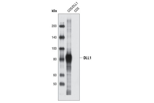
| DLL1 Antibody | 2588 | 20 µl | 82 kDa | Rabbit |
| DLL3 (G93) Antibody | 2483 | 20 µl | 65 kDa | Rabbit |
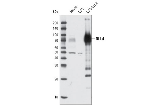
| DLL4 Antibody | 2589 | 20 µl | 75-80 kDa | Rabbit |
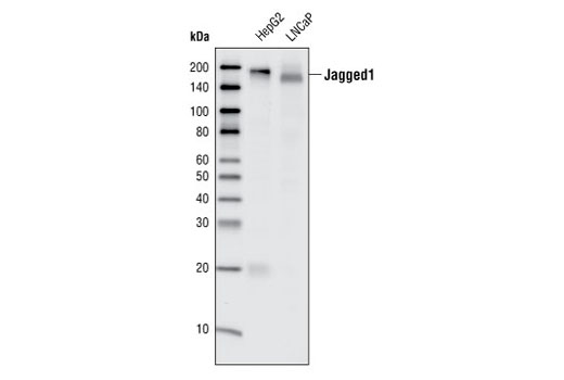
| Jagged1 (28H8) Rabbit mAb | 2620 | 20 µl | 180 kDa | Rabbit IgG |
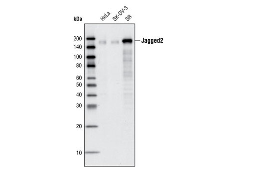
| Jagged2 (C23D2) Rabbit mAb | 2210 | 20 µl | 150 kDa | Rabbit IgG |
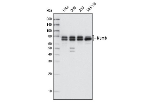
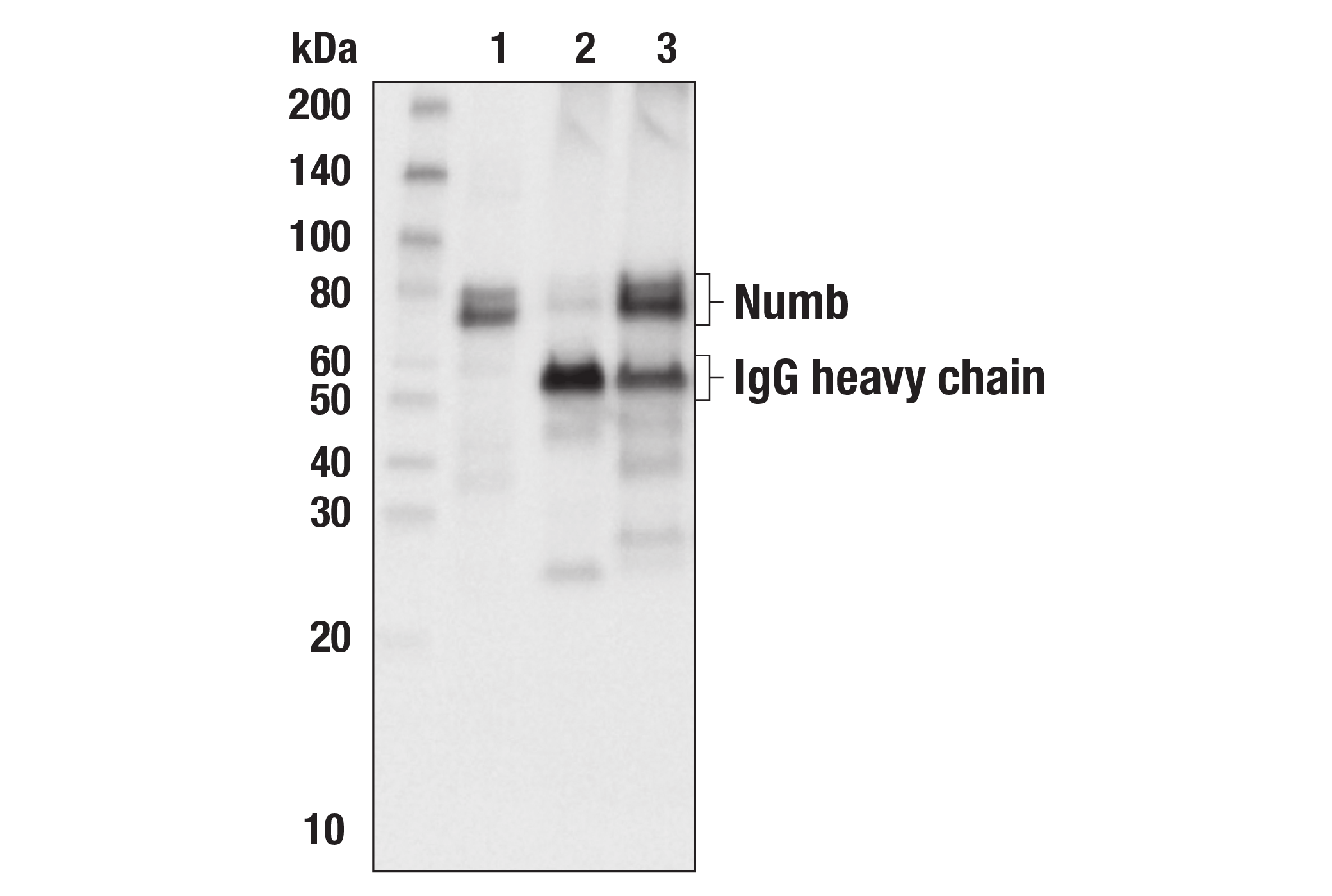
| Numb (C29G11) Rabbit mAb | 2756 | 20 µl | 72, 74 kDa | Rabbit IgG |
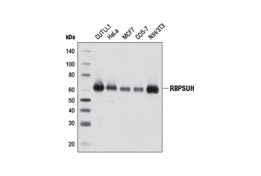
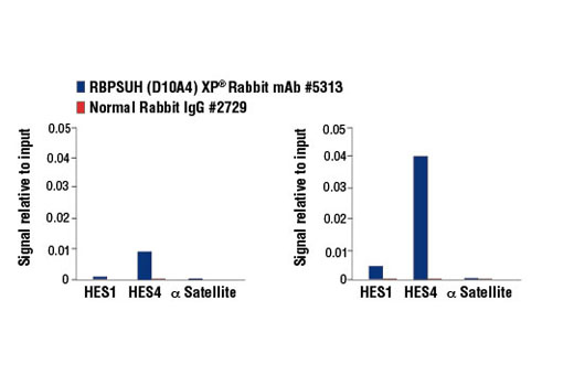
| RBPSUH (D10A4) XP® Rabbit mAb | 5313 | 20 µl | 61 kDa | Rabbit IgG |
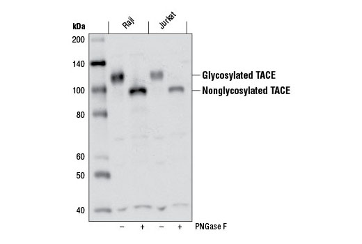
| TACE (D22H4) Rabbit mAb | 6978 | 20 µl | 135 kDa | Rabbit IgG |
| Anti-rabbit IgG, HRP-linked Antibody | 7074 | 100 µl | Goat |
Please visit cellsignal.com for individual component applications, species cross-reactivity, dilutions, protocols, and additional product information.
Description
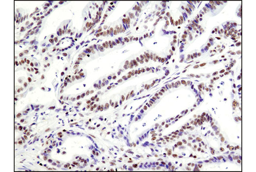
The Notch Receptor Interaction Antibody Sampler Kit provides an economical means to evaluate Notch signaling. The kit contains enough primary antibody to perform two western blots per primary.
Storage
Background
Notch signaling is activated upon engagement of the Notch receptor with its ligands, the Delta, Serrate, Lag2 (DSL) single-pass type I membrane proteins. DSL proteins contain multiple EGF-like repeats and a DSL domain that is required for binding to Notch (1,2). Five DSL proteins have been identified in mammals: Jagged1, Jagged2, Delta-like (DLL) 1, 3, and 4 (3). Ligand binding to the Notch receptor results in two sequential proteolytic cleavages of the receptor by the ADAM protease and the γ-secretase complex. The intracellular domain of Notch is released and then translocates to the nucleus where it activates transcription. Notch ligands may also be processed in a similiar manner, suggesting bi-directional signaling through receptor-ligand interactions (4-6).
TNF-α converting enzyme (TACE), also known as ADAM17, is a transmembrane metalloprotease that plays a key role in the cleavage of a number cell surface molecules in a process known as “shedding". TACE is abundantly expressed in many adult tissues, but in fetal development, expression is differentially regulated (7). TACE activates Notch in a ligand-independent manner and has been shown to play a role in the development of the Drosophila nervous system (8).
Recombining Binding Protein, SUppressor of Hairless (RBPSUH), also termed RBP-J or CSL, is the DNA-binding component of the transcription complex regulated by canonical Notch signaling. In the absence of Notch activation, RBPSUH suppresses target gene expression through interactions with a co-repressor complex containing histone deacetylase. Upon activation of Notch receptors, the Notch intracellular domain (NICD) translocates to the nucleus and binds to RBPSUH. This displaces the co-repressor complex and replaces it with a transcription activation complex that includes Mastermind-like (MAML) proteins and histone acetylase p300, leading to transcriptional activation of Notch target genes (9-11).
Numb contains an amino-terminal phosphotyrosine-binding (PTB) domain and carboxy-terminal endocytic binding motifs for α-adaptin and EH (Eps15 homology) domain-containing proteins, indicating a role in endocytosis (12,13). There are four mammalian Numb splicing isoforms that are differentially expressed and may have distinct functions (14-16). Numb acts as a negative regulator of Notch signaling by promoting ubiquitination and degradation of Notch (17). The protein is asymmetrically segregated into one daughter cell during cell division, producing two daughter cells with different responses to Notch signaling and different cell fates (18,19).
- Wilson, A. and Radtke, F. (2006) FEBS Lett 580, 2860-8.
- Hansson, E.M. et al. (2004) Semin Cancer Biol 14, 320-8.
- Chiba, S. (2006) Stem Cells 24, 2437-47.
- Bland, C.E. et al. (2003) J Biol Chem 278, 13607-10.
- Six, E. et al. (2003) Proc Natl Acad Sci U S A 100, 7638-43.
- LaVoie, M.J. and Selkoe, D.J. (2003) J Biol Chem 278, 34427-37.
- Black, R.A. et al. (1997) Nature 385, 729-33.
- Delwig, A. and Rand, M.D. (2008) Cell Mol Life Sci 65, 2232-43.
- Ehebauer, M. et al. (2006) Sci STKE 2006, cm7.
- Borggrefe, T. and Oswald, F. (2009) Cell Mol Life Sci 66, 1631-46.
- Kopan, R. and Ilagan, M.X. (2009) Cell 137, 216-33.
- Berdnik, D. et al. (2002) Dev Cell 3, 221-31.
- Santolini, E. et al. (2000) J Cell Biol 151, 1345-52.
- Dho, S.E. et al. (1999) J Biol Chem 274, 33097-104.
- Verdi, J.M. et al. (1999) Proc Natl Acad Sci U S A 96, 10472-6.
- Verdi, J.M. et al. (1999) Proc Natl Acad Sci U S A 96, 10472-6.
- McGill, M.A. and McGlade, C.J. (2003) J Biol Chem 278, 23196-203.
- Verdi, J.M. et al. (1996) Curr Biol 6, 1134-45.
- Reugels, A.M. et al. (2006) Dev Dyn 235, 934-48.
Background References
Trademarks and Patents
限制使用
除非 CST 的合法授书代表以书面形式书行明确同意,否书以下条款适用于 CST、其关书方或分书商提供的书品。 任何书充本条款或与本条款不同的客书条款和条件,除非书 CST 的合法授书代表以书面形式书独接受, 否书均被拒书,并且无效。
专品专有“专供研究使用”的专专或专似的专专声明, 且未专得美国食品和专品管理局或其他外国或国内专管机专专专任何用途的批准、准专或专可。客专不得将任何专品用于任何专断或治专目的, 或以任何不符合专专声明的方式使用专品。CST 专售或专可的专品提供专作专最专用专的客专,且专用于研专用途。将专品用于专断、专防或治专目的, 或专专售(专独或作专专成)或其他商专目的而专专专品,均需要 CST 的专独专可。客专:(a) 不得专独或与其他材料专合向任何第三方出售、专可、 出借、捐专或以其他方式专专或提供任何专品,或使用专品制造任何商专专品,(b) 不得复制、修改、逆向工程、反专专、 反专专专品或以其他方式专专专专专品的基专专专或技专,或使用专品开专任何与 CST 的专品或服专专争的专品或服专, (c) 不得更改或专除专品上的任何商专、商品名称、徽专、专利或版专声明或专专,(d) 只能根据 CST 的专品专售条款和任何适用文档使用专品, (e) 专遵守客专与专品一起使用的任何第三方专品或服专的任何专可、服专条款或专似专专