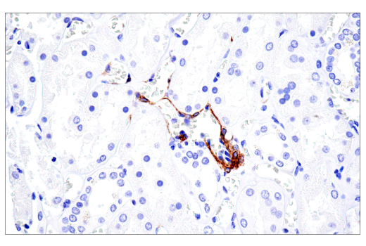
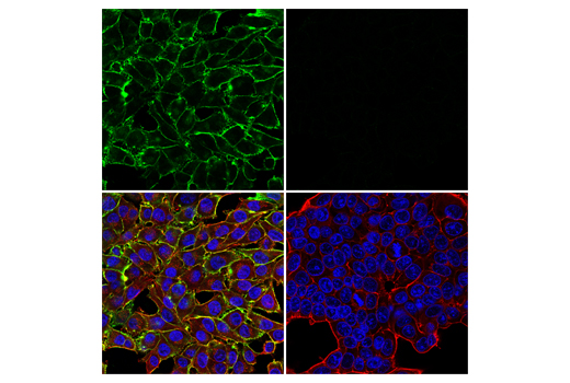
Revision 1
Cell Signaling Technology

Orders: 877-616-CELL (2355) [email protected]

Support: 877-678-TECH (8324)

Web: [email protected] cellsignal.com

3 Trask LaneDanversMassachusetts01923USA
For Research Use Only. Not for Use in Diagnostic Procedures.
Applications:

WB, IHC-P, IF-IC

REACTIVITY:

H

SENSITIVITY:

Endogenous

MW (kDa):

250, 450

Source/Isotype:

Rabbit IgG

UniProt ID:

#Q6UVK1

Entrez-Gene Id:

1464

Product Information

Product Usage Information

This product is the carrier-free version of product #43916. All data were generated using the same antibody clone in the standard formulation which contains BSA and glycerol. This formulation is ideal for use with technologies requiring specialized or custom antibody labeling, including fluorophores, metals, lanthanides, and oligonucleotides. It is not recommended for ChIP, ChIP-seq, CUT&RUN, or CUT&Tag assays. If you require a carrier-free formulation for chromatin profiling, please contact us. Optimal dilutions/concentrations should be determined by the end user.

Formulation

Supplied in 1X PBS, BSA and Azide Free.

For standard formulation of this product see product #43916.

Storage

Store at -20°C. This product will freeze at -20°C so it is recommended to aliquot into single-use vials to avoid multiple freeze/thaw cycles. A slight precipitate may be present and can be dissolved by gently vortexing. This will not interfere with antibody performance.

Specificity / Sensitivity

NG2/CSPG4 (E3B3G) XP® Rabbit mAb (BSA and Azide Free) recognizes endogenous levels of total NG2/CSPG4 protein.

Species Reactivity:

Human

Source / Purification

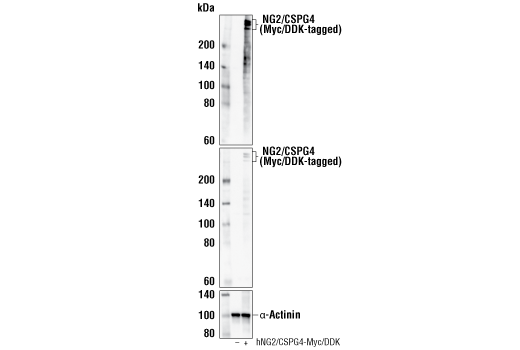
Monoclonal antibody is produced by immunizing animals with a synthetic peptide corresponding to residues surrounding Asp1410 of human NG2/CSPG4 protein.

Background

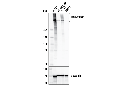
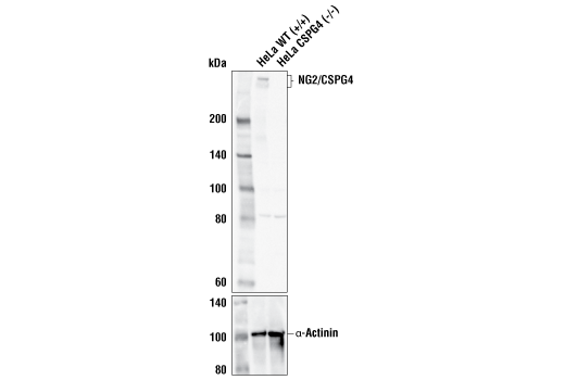
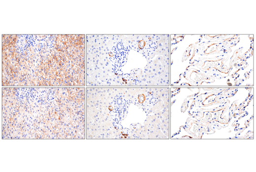
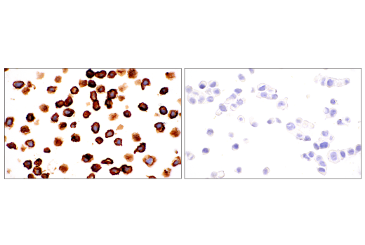
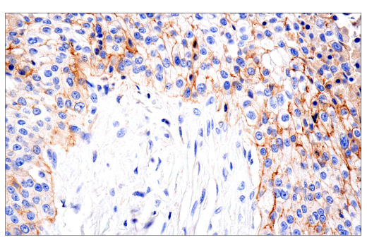
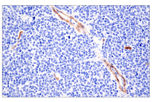
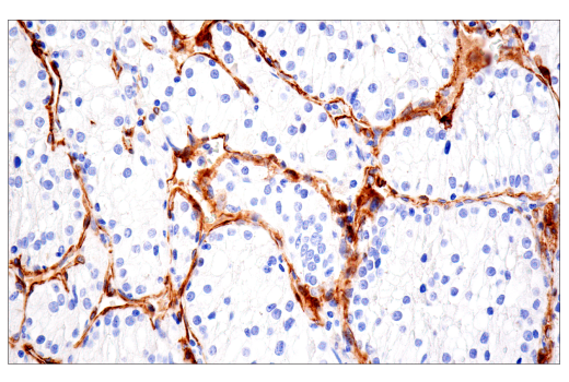
The chondroitin sulfate proteoglycan NG2 is a type I membrane protein expressed by subpopulations of glia including oligodendroglial precursor cells and a variety of tumor cells. Normal precursor cells and malignant tumor cells migrate and proliferate, but there is evidence that cells may not be able to engage in both activities at the same time. However, NG2 is involved in promoting both proliferation and motility (1). The extracellular domain of NG2 sequesters growth factors and binds to both growth factor receptors and extracellular matrix ligands such as fibronectin, collagens and laminin. The cytoplasmic domain is involved in activating Rac, Cdc42, and p130 Cas (2). PKCα phosphorylates NG2 at Thr2256, triggering the redistribution of NG2 from apical microprocesses to lamellipodia accompanied by enhanced cell motility (3). ERK phosphorylates NG2 at Thr2314, stimulating cell proliferation (4).

  1. Karram, K. et al. (2005) J Anat 207, 735-44.
  2. Eisenmann, K.M. et al. (1999) Nat Cell Biol 1, 507-13.
  3. Makagiansar, I.T. et al. (2004) J Biol Chem 279, 55262-70.
  4. Makagiansar, I.T. et al. (2007) J Cell Biol 178, 155-65.

Species Reactivity

Species reactivity is determined by testing in at least one approved application (e.g., western blot).

Applications Key

WB: Western Blotting IHC-P: Immunohistochemistry (Paraffin) IF-IC: Immunofluorescence (Immunocytochemistry)

Cross-Reactivity Key

H: human M: mouse R: rat Hm: hamster Mk: monkey Vir: virus Mi: mink C: chicken Dm: D. melanogaster X: Xenopus Z: zebrafish B: bovine Dg: dog Pg: pig Sc: S. cerevisiae Ce: C. elegans Hr: horse GP: Guinea Pig Rab: rabbit All: all species expected

Trademarks and Patents

Cell Signaling Technology is a trademark of Cell Signaling Technology, Inc.
XP is a registered trademark of Cell Signaling Technology, Inc.
All other trademarks are the property of their respective owners. Visit cellsignal.com/trademarks for more information.

限制使用

除非 CST 的合法授书代表以书面形式书行明确同意,否书以下条款适用于 CST、其关书方或分书商提供的书品。 任何书充本条款或与本条款不同的客书条款和条件,除非书 CST 的合法授书代表以书面形式书独接受, 否书均被拒书,并且无效。

专品专有“专供研究使用”的专专或专似的专专声明, 且未专得美国食品和专品管理局或其他外国或国内专管机专专专任何用途的批准、准专或专可。客专不得将任何专品用于任何专断或治专目的, 或以任何不符合专专声明的方式使用专品。CST 专售或专可的专品提供专作专最专用专的客专,且专用于研专用途。将专品用于专断、专防或治专目的, 或专专售(专独或作专专成)或其他商专目的而专专专品,均需要 CST 的专独专可。客专:(a) 不得专独或与其他材料专合向任何第三方出售、专可、 出借、捐专或以其他方式专专或提供任何专品,或使用专品制造任何商专专品,(b) 不得复制、修改、逆向工程、反专专、 反专专专品或以其他方式专专专专专品的基专专专或技专,或使用专品开专任何与 CST 的专品或服专专争的专品或服专, (c) 不得更改或专除专品上的任何商专、商品名称、徽专、专利或版专声明或专专,(d) 只能根据 CST 的专品专售条款和任何适用文档使用专品, (e) 专遵守客专与专品一起使用的任何第三方专品或服专的任何专可、服专条款或专似专专

Revision 1
#25162

NG2/CSPG4 (E3B3G) XP® Rabbit mAb (BSA and Azide Free)

Western Blotting Image 1: NG2/CSPG4 (E3B3G) XP® Rabbit mAb (BSA and Azide Free) Expand Image
使用 NG2/CSPG4 (E3B3G) XP® Rabbit mAb(上图)、DYKDDDDK Tag Antibody #2368(中图)或 α-Actinin (D6F6) XP® Rabbit mAb #6487(下图),对转染空载 (-) 或转染带有 Myc/DDK 标签的全长人 NG2/CSPG4 蛋白 (hNG2/CSPG4-Myc/DDK; +) 表达载体的 293T 细胞提取物进行蛋白质印迹法分析。数据的生成使用的是该产品的常规组分产品。
Western Blotting Image 2: NG2/CSPG4 (E3B3G) XP® Rabbit mAb (BSA and Azide Free) Expand Image
使用 NG2/CSPG4 (E3B3G) XP® Rabbit mAb(上图)或 α-Actinin (D6F6) XP® Rabbit mAb #6487(下图)对不同细胞系的提取物进行蛋白质印迹法分析。如同预期,NG2/CSPG4 蛋白在 MCF7 细胞中不表达。数据的生成使用的是该产品的常规组分产品。
Western Blotting Image 3: NG2/CSPG4 (E3B3G) XP® Rabbit mAb (BSA and Azide Free) Expand Image
使用 NG2/CSPG4 (E3B3G) XP® Rabbit mAb(上图)或 α-Actinin (D6F6) XP® Rabbit mAb #6487(下图)对野生型 (+/+) 或 CSPG4 敲除型 (-/-) HeLa 细胞的提取物进行蛋白质印迹法分析。数据的生成使用的是该产品的常规组分产品。
Immunohistochemistry Image 1: NG2/CSPG4 (E3B3G) XP® Rabbit mAb (BSA and Azide Free) Expand Image
使用 NG2/CSPG4 (E3B3G) XP® Rabbit mAb 对石蜡包埋的正常人肾进行免疫组织化学分析。数据的生成使用的是该产品的常规组分产品。
Immunohistochemistry Image 2: NG2/CSPG4 (E3B3G) XP® Rabbit mAb (BSA and Azide Free) Expand Image
使用 NG2/CSPG4 (E3B3G) XP® Rabbit mAb(顶图)或 NG2/CSPG4 Rabbit mAb(底图)石蜡包埋的人胃肠道基质肿瘤(左)、正常肝脏(中间)、或正常肺(右)进行免疫组织化学分析。这两种抗体可检测人 NG2/CSPG4 上的独立、独特表位。使用两种抗体获得的相似染色模式有助于确认染色特异性。数据的生成使用的是该产品的常规组分产品。
Immunohistochemistry Image 3: NG2/CSPG4 (E3B3G) XP® Rabbit mAb (BSA and Azide Free) Expand Image
使用 NG2/CSPG4 (E3B3G) XP® Rabbit mAb 对石蜡包埋的 A-375 细胞沉淀物(左图,阳性)或 MCF7 细胞沉淀物(右图,阴性)进行免疫组织化学分析。数据的生成使用的是该产品的常规组分产品。
Immunohistochemistry Image 4: NG2/CSPG4 (E3B3G) XP® Rabbit mAb (BSA and Azide Free) Expand Image
使用 NG2/CSPG4 (E3B3G) XP® Rabbit mAb 对石蜡包埋的人鳞状细胞口腔癌进行免疫组织化学分析。数据的生成使用的是该产品的常规组分产品。
Immunohistochemistry Image 5: NG2/CSPG4 (E3B3G) XP® Rabbit mAb (BSA and Azide Free) Expand Image
使用 NG2/CSPG4 (E3B3G) XP® Rabbit mAb 对石蜡包埋的人 B 细胞非霍奇金氏淋巴瘤进行免疫组织化学分析。数据的生成使用的是该产品的常规组分产品。
Immunohistochemistry Image 6: NG2/CSPG4 (E3B3G) XP® Rabbit mAb (BSA and Azide Free) Expand Image
使用 NG2/CSPG4 (E3B3G) XP® Rabbit mAb 对石蜡包埋的人结肠癌进行免疫组织化学分析。数据的生成使用的是该产品的常规组分产品。
Immunohistochemistry Image 7: NG2/CSPG4 (E3B3G) XP® Rabbit mAb (BSA and Azide Free) Expand Image
使用 NG2/CSPG4 (E3B3G) XP® Rabbit mAb 对石蜡包埋的人肾细胞癌进行免疫组织化学分析。数据的生成使用的是该产品的常规组分产品。
Immunofluorescence Image 1: NG2/CSPG4 (E3B3G) XP® Rabbit mAb (BSA and Azide Free) Expand Image
使用 NG2/CSPG4 (E3B3G) XP® Rabbit mAb(绿色)和 β-Actin (8H10D10) Mouse mAb #3700(红色)对 A-375 细胞(左,阳性)或 MCF7 细胞(右,阴性)进行共聚焦免疫荧光分析。切片用 ProLong® Gold Antifade Reagent with DAPI #8961(蓝色)封装。数据的生成使用的是该产品的常规组分产品。