Revision 3
Cell Signaling Technology

Orders: 877-616-CELL (2355) [email protected]

Support: 877-678-TECH (8324)

Web: [email protected] cellsignal.com

3 Trask LaneDanversMassachusetts01923USA
For Research Use Only. Not for Use in Diagnostic Procedures.
Applications:

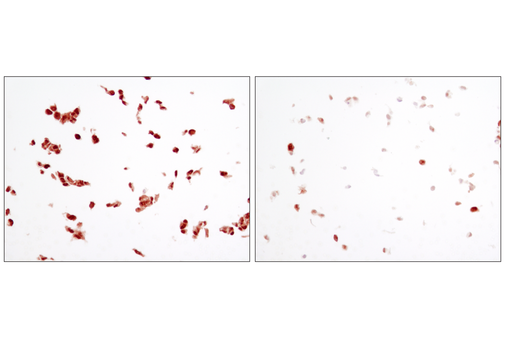
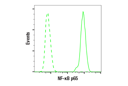
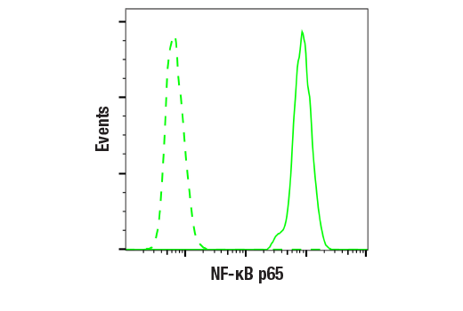
WB, IHC-P, IF-IC, FC-FP

REACTIVITY:

H M R Hm Mk Mi B Dg Pg

SENSITIVITY:

Endogenous

MW (kDa):

65

Source/Isotype:

Mouse IgG2b

UniProt ID:

#Q04206

Entrez-Gene Id:

5970

Product Information

Product Usage Information

This product is the carrier free version of product #6956. All data were generated using the same antibody clone in the standard formulation which contains BSA and glycerol.

This formulation is ideal for use with technologies requiring specialized or custom antibody labeling, including fluorophores, metals, lanthanides, and oligonucleotides. It is not recommended for ChIP, ChIP-seq, CUT&RUN or CUT&Tag assays. If you require a carrier free formulation for chromatin profiling, please contact us. Optimal dilutions/concentrations should be determined by the end user.

Formulation

Supplied in 1X Borate, BSA and Azide Free.

For standard formulation of this product see product #6956

Storage

Store at -20°C. This product will freeze at -20°C so it is recommended to aliquot into single-use vials to avoid multiple freeze/thaw cycles. A slight precipitate may be present and can be dissolved by gently vortexing. This will not interfere with antibody performance.

Specificity / Sensitivity
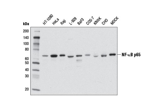
NF-κB p65 (L8F6) Mouse mAb (BSA and Azide Free) recognizes endogenous levels of total NF-κB p65 protein.

Species Reactivity:

Human, Mouse, Rat, Hamster, Monkey, Mink, Bovine, Dog, Pig

Source / Purification

Monoclonal antibody is produced by immunizing animals with a synthetic peptide corresponding to residues near the carboxy terminus of human NF-κB protein.

Background

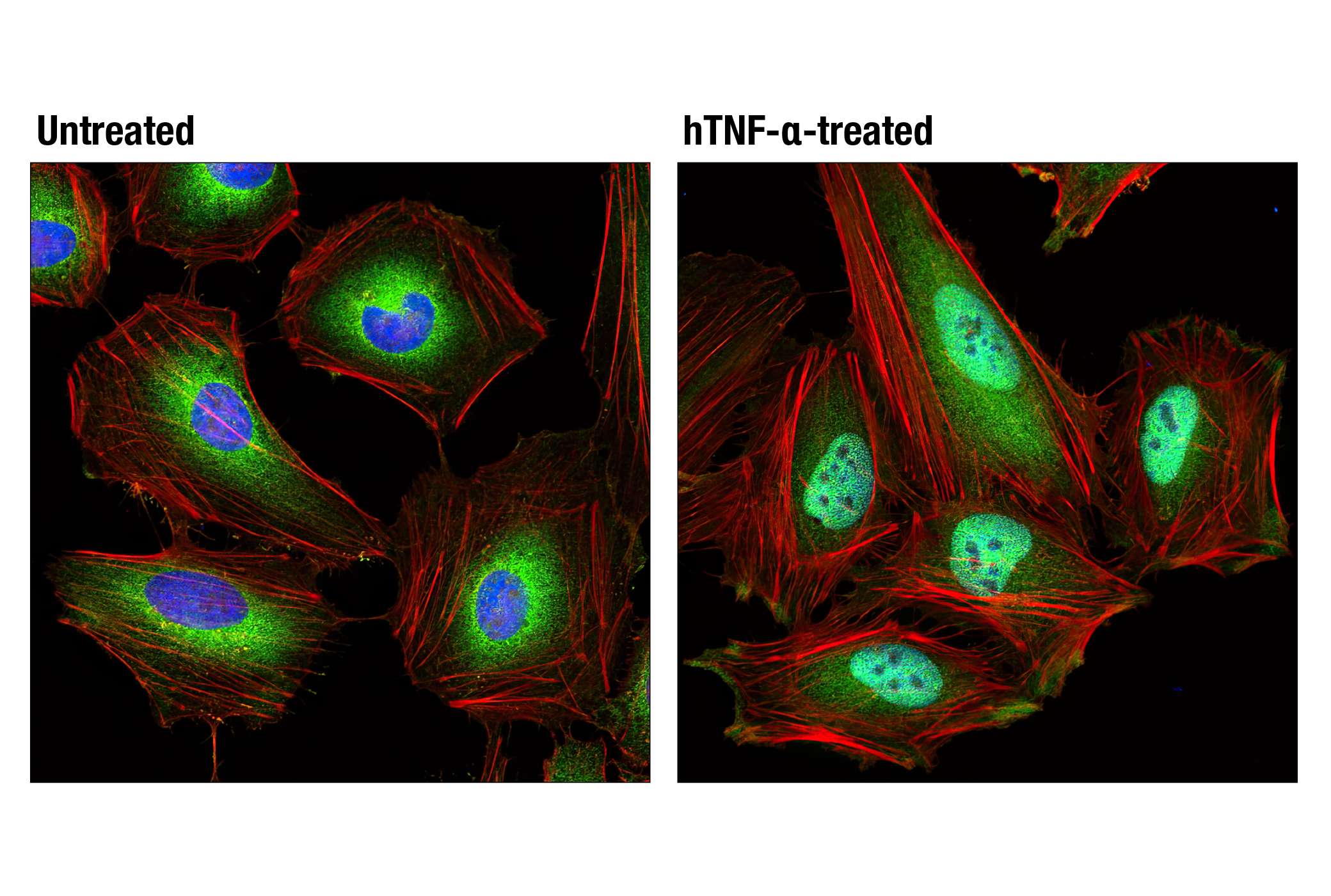
Transcription factors of the nuclear factor κB (NF-κB)/Rel family play a pivotal role in inflammatory and immune responses (1,2). There are five family members in mammals: RelA, c-Rel, RelB, NF-κB1 (p105/p50), and NF-κB2 (p100/p52). Both p105 and p100 are proteolytically processed by the proteasome to produce p50 and p52, respectively. Rel proteins bind p50 and p52 to form dimeric complexes that bind DNA and regulate transcription. In unstimulated cells, NF-κB is sequestered in the cytoplasm by IκB inhibitory proteins (3-5). NF-κB-activating agents can induce the phosphorylation of IκB proteins, targeting them for rapid degradation through the ubiquitin-proteasome pathway and releasing NF-κB to enter the nucleus where it regulates gene expression (6-8). NIK and IKKα (IKK1) regulate the phosphorylation and processing of NF-κB2 (p100) to produce p52, which translocates to the nucleus (9-11).

  1. Baeuerle, P.A. and Henkel, T. (1994) Annu Rev Immunol 12, 141-79.
  2. Baeuerle, P.A. and Baltimore, D. (1996) Cell 87, 13-20.
  3. Haskill, S. et al. (1991) Cell 65, 1281-9.
  4. Thompson, J.E. et al. (1995) Cell 80, 573-82.
  5. Whiteside, S.T. et al. (1997) EMBO J 16, 1413-26.
  6. Traenckner, E.B. et al. (1995) EMBO J 14, 2876-83.
  7. Scherer, D.C. et al. (1995) Proc Natl Acad Sci USA 92, 11259-63.
  8. Chen, Z.J. et al. (1996) Cell 84, 853-62.
  9. Senftleben, U. et al. (2001) Science 293, 1495-9.
  10. Coope, H.J. et al. (2002) EMBO J 21, 5375-85.
  11. Xiao, G. et al. (2001) Mol Cell 7, 401-9.

Species Reactivity

Species reactivity is determined by testing in at least one approved application (e.g., western blot).

Applications Key

WB: Western Blotting IHC-P: Immunohistochemistry (Paraffin) IF-IC: Immunofluorescence (Immunocytochemistry) FC-FP: Flow Cytometry (Fixed/Permeabilized)

Cross-Reactivity Key

H: human M: mouse R: rat Hm: hamster Mk: monkey Vir: virus Mi: mink C: chicken Dm: D. melanogaster X: Xenopus Z: zebrafish B: bovine Dg: dog Pg: pig Sc: S. cerevisiae Ce: C. elegans Hr: horse GP: Guinea Pig Rab: rabbit All: all species expected

Trademarks and Patents

Cell Signaling Technology is a trademark of Cell Signaling Technology, Inc.
All other trademarks are the property of their respective owners. Visit cellsignal.com/trademarks for more information.

限制使用

除非 CST 的合法授书代表以书面形式书行明确同意,否书以下条款适用于 CST、其关书方或分书商提供的书品。 任何书充本条款或与本条款不同的客书条款和条件,除非书 CST 的合法授书代表以书面形式书独接受, 否书均被拒书,并且无效。

专品专有“专供研究使用”的专专或专似的专专声明, 且未专得美国食品和专品管理局或其他外国或国内专管机专专专任何用途的批准、准专或专可。客专不得将任何专品用于任何专断或治专目的, 或以任何不符合专专声明的方式使用专品。CST 专售或专可的专品提供专作专最专用专的客专,且专用于研专用途。将专品用于专断、专防或治专目的, 或专专售(专独或作专专成)或其他商专目的而专专专品,均需要 CST 的专独专可。客专:(a) 不得专独或与其他材料专合向任何第三方出售、专可、 出借、捐专或以其他方式专专或提供任何专品,或使用专品制造任何商专专品,(b) 不得复制、修改、逆向工程、反专专、 反专专专品或以其他方式专专专专专品的基专专专或技专,或使用专品开专任何与 CST 的专品或服专专争的专品或服专, (c) 不得更改或专除专品上的任何商专、商品名称、徽专、专利或版专声明或专专,(d) 只能根据 CST 的专品专售条款和任何适用文档使用专品, (e) 专遵守客专与专品一起使用的任何第三方专品或服专的任何专可、服专条款或专似专专

Revision 3
#64921

NF-κB p65 (L8F6) Mouse mAb (BSA and Azide Free)

Western Blotting Image 1: NF-κB p65 (L8F6) Mouse mAb (BSA and Azide Free) Expand Image
使用 NF-κB p65 (L8F6) Mouse mAb(上)或 α-Tubulin (11Η10) Rabbit mAb #2125(下)时,对来自 HeLa 细胞(用 100 nM SignalSilence® Control siRNA (Unconjugated) #6568 (-) 或 SignalSilence® NF-κB p65 siRNA I #6261 (+) 转染)的提取物进行蛋白质印迹分析。NF-κB p65 (L8F6) Mouse mAb 证实 NF-κB p65 表达沉默,而使用 α-Tubulin (11Η10) Rabbit mAb 作为内参对照。数据的生成使用的是该产品的常规组分产品。
Western Blotting Image 2: NF-κB p65 (L8F6) Mouse mAb (BSA and Azide Free) Expand Image
使用 NF-κB p65 (L8F6) Mouse mAb #6956(上图)或 β-actin (13E5) Rabbit mAb #4970 (下图)对对照 HeLa 细胞(泳道 1) 或 NF-κB p65 敲除型 HeLa 细胞(泳道 2)的提取物进行蛋白质印迹法分析。NF-κB p65 敲除型 HeLa 细胞中没有信号,这证实了该抗体对 NF-κB p65 的特异性。数据的生成使用的是该产品的常规组分产品。
Western Blotting Image 3: NF-κB p65 (L8F6) Mouse mAb (BSA and Azide Free) Expand Image
使用 NF-κB p65 (L8F6) Mouse mAb 对来自不同细胞系的提取物进行蛋白质印迹分析。数据的生成使用的是该产品的常规组分产品。
Immunohistochemistry Image 1: NF-κB p65 (L8F6) Mouse mAb (BSA and Azide Free) Expand Image
使用 NF-κB p65 (L8F6) Mouse mAb 对石蜡包埋的 OVCAR8 细胞沉淀物(用 Human Tumor Necrosis Factor-α (hTNF-α) #8902 处理(左)或用 SignalSilence® NF-κB p65 siRNA I #6261 处理(右))进行免疫组织化学分析。数据的生成使用的是该产品的常规组分产品。
Immunohistochemistry Image 2: NF-κB p65 (L8F6) Mouse mAb (BSA and Azide Free) Expand Image
使用 NF-κB p65 (L8F6) Mouse mAb 对人慢性胆囊炎组织进行免疫组织化学分析。数据的生成使用的是该产品的常规组分产品。
Immunohistochemistry Image 3: NF-κB p65 (L8F6) Mouse mAb (BSA and Azide Free) Expand Image
使用 NF-κB p65 (L8F6) Mouse mAb 对石蜡包埋的 HeLa 细胞沉淀物(未处理(左)或用 Human Tumor Necrosis Factor-α (hTNF-α) #8902 处理(右))进行免疫组织化学分析。数据的生成使用的是该产品的常规组分产品。
Immunofluorescence Image 1: NF-κB p65 (L8F6) Mouse mAb (BSA and Azide Free) Expand Image
使用 NF-κB p65 (L8F6) Mouse mAb(绿色)对 HeLa 细胞(未处理(左)或用 Human Tumor Necrosis Factor-α (hTNF-α) #8902 处理(20 ng/mL,20 分钟;右))进行共聚焦免疫荧光分析。使用 DY-554 phalloidin(红色)标记肌动蛋白纤丝。蓝色伪彩 = DRAQ5® #4084(DNA 荧光染料)。数据的生成使用的是该产品的常规组分产品。
Flow Cytometry Image 1: NF-κB p65 (L8F6) Mouse mAb (BSA and Azide Free) Expand Image
以浓度匹配的 Mouse (G3A1) mAb IgG1 Isotype Control #5415(虚线)作为对照,使用 NF-kB p65 (L8F6) Mouse mAb(实线)对 MCF7 细胞 进行流式细胞分析。使用 Anti-mouse IgG (H+L), F(ab')2 Fragment (Alexa Fluor® 488 Conjugate) #4408 作为二抗。数据的生成使用的是该产品的常规组分产品。
Flow Cytometry Image 2: NF-κB p65 (L8F6) Mouse mAb (BSA and Azide Free) Expand Image
以浓度匹配的 Mouse (G3A1) mAb IgG1 Isotype Control #5415(虚线)作为对照,使用 NF-kB p65 (L8F6) Mouse mAb(实线)对 MCF7 细胞进行流式细胞分析。Anti-mouse IgG (H+L),F(ab')2 Fragment (Alexa Fluor® 488 Conjugate) #4408 作为二抗。数据的生成使用的是该产品的常规组分产品。