Revision 9
Cell Signaling Technology

Orders: 877-616-CELL (2355) [email protected]

Support: 877-678-TECH (8324)

Web: [email protected] cellsignal.com

3 Trask LaneDanversMassachusetts01923USA
For Research Use Only. Not for Use in Diagnostic Procedures.
Applications:

WB, W-S, IP, IHC-P, IF-IC, FC-FP, ChIP, ChIP-seq, C&R

REACTIVITY:

H M R Hm Mk Dg

SENSITIVITY:

Endogenous

MW (kDa):

65

Source/Isotype:

Rabbit IgG

UniProt ID:

#Q04206

Entrez-Gene Id:

5970

Product Information

Product Usage Information

For optimal ChIP and ChIP-seq results, use 5 μl of antibody and 10 μg of chromatin (approximately 4 x 10⁶ cells) per IP. This antibody has been validated using SimpleChIP® Enzymatic Chromatin IP Kits.

The CUT&RUN dilution was determined using CUT&RUN Assay Kit #86652.
Application Dilution
Western Blotting 1:1000
Simple Western™ 1:10 - 1:50
Immunoprecipitation 1:100
Immunohistochemistry (Paraffin) 1:400 - 1:1600
Immunofluorescence (Immunocytochemistry) 1:400 - 1:1600
Flow Cytometry (Fixed/Permeabilized) 1:400 - 1:1600
Chromatin IP 1:100
Chromatin IP-seq 1:100
CUT&RUN 1:100

Storage

Supplied in 10 mM sodium HEPES (pH 7.5), 150 mM NaCl, 100 µg/ml BSA, 50% glycerol and less than 0.02% sodium azide. Store at –20°C. Do not aliquot the antibody.

For a carrier free (BSA and azide free) version of this product see product #69994.

Specificity / Sensitivity

NF-κB p65 (D14E12) XP® Rabbit mAb recognizes endogenous levels of total NF-κB p65/RelA protein. It does not cross react with other NF-κB/Rel family members.

Species Reactivity:

Human, Mouse, Rat, Hamster, Monkey, Dog

Source / Purification

Monoclonal antibody is produced by immunizing animals with a synthetic peptide corresponding to residues surrounding Glu498 of human NF-κB p65/RelA protein.

Background

Transcription factors of the nuclear factor κB (NF-κB)/Rel family play a pivotal role in inflammatory and immune responses (1,2). There are five family members in mammals: RelA, c-Rel, RelB, NF-κB1 (p105/p50), and NF-κB2 (p100/p52). Both p105 and p100 are proteolytically processed by the proteasome to produce p50 and p52, respectively. Rel proteins bind p50 and p52 to form dimeric complexes that bind DNA and regulate transcription. In unstimulated cells, NF-κB is sequestered in the cytoplasm by IκB inhibitory proteins (3-5). NF-κB-activating agents can induce the phosphorylation of IκB proteins, targeting them for rapid degradation through the ubiquitin-proteasome pathway and releasing NF-κB to enter the nucleus where it regulates gene expression (6-8). NIK and IKKα (IKK1) regulate the phosphorylation and processing of NF-κB2 (p100) to produce p52, which translocates to the nucleus (9-11).

  1. Baeuerle, P.A. and Henkel, T. (1994) Annu Rev Immunol 12, 141-79.
  2. Baeuerle, P.A. and Baltimore, D. (1996) Cell 87, 13-20.
  3. Haskill, S. et al. (1991) Cell 65, 1281-9.
  4. Thompson, J.E. et al. (1995) Cell 80, 573-82.
  5. Whiteside, S.T. et al. (1997) EMBO J 16, 1413-26.
  6. Traenckner, E.B. et al. (1995) EMBO J 14, 2876-83.
  7. Scherer, D.C. et al. (1995) Proc Natl Acad Sci USA 92, 11259-63.
  8. Chen, Z.J. et al. (1996) Cell 84, 853-62.
  9. Senftleben, U. et al. (2001) Science 293, 1495-9.
  10. Coope, H.J. et al. (2002) EMBO J 21, 5375-85.
  11. Xiao, G. et al. (2001) Mol Cell 7, 401-9.

Species Reactivity

Species reactivity is determined by testing in at least one approved application (e.g., western blot).

Western Blot Buffer

IMPORTANT: For western blots, incubate membrane with diluted primary antibody in 5% w/v BSA, 1X TBS, 0.1% Tween® 20 at 4°C with gentle shaking, overnight.

Applications Key

WB: Western Blotting W-S: Simple Western™ IP: Immunoprecipitation IHC-P: Immunohistochemistry (Paraffin) IF-IC: Immunofluorescence (Immunocytochemistry) FC-FP: Flow Cytometry (Fixed/Permeabilized) ChIP: Chromatin IP ChIP-seq: Chromatin IP-seq C&R: CUT&RUN

Cross-Reactivity Key

H: human M: mouse R: rat Hm: hamster Mk: monkey Vir: virus Mi: mink C: chicken Dm: D. melanogaster X: Xenopus Z: zebrafish B: bovine Dg: dog Pg: pig Sc: S. cerevisiae Ce: C. elegans Hr: horse GP: Guinea Pig Rab: rabbit All: all species expected

Trademarks and Patents

Cell Signaling Technology is a trademark of Cell Signaling Technology, Inc.
XP is a registered trademark of Cell Signaling Technology, Inc.
All other trademarks are the property of their respective owners. Visit cellsignal.com/trademarks for more information.

限制使用

除非 CST 的合法授书代表以书面形式书行明确同意,否书以下条款适用于 CST、其关书方或分书商提供的书品。 任何书充本条款或与本条款不同的客书条款和条件,除非书 CST 的合法授书代表以书面形式书独接受, 否书均被拒书,并且无效。

专品专有“专供研究使用”的专专或专似的专专声明, 且未专得美国食品和专品管理局或其他外国或国内专管机专专专任何用途的批准、准专或专可。客专不得将任何专品用于任何专断或治专目的, 或以任何不符合专专声明的方式使用专品。CST 专售或专可的专品提供专作专最专用专的客专,且专用于研专用途。将专品用于专断、专防或治专目的, 或专专售(专独或作专专成)或其他商专目的而专专专品,均需要 CST 的专独专可。客专:(a) 不得专独或与其他材料专合向任何第三方出售、专可、 出借、捐专或以其他方式专专或提供任何专品,或使用专品制造任何商专专品,(b) 不得复制、修改、逆向工程、反专专、 反专专专品或以其他方式专专专专专品的基专专专或技专,或使用专品开专任何与 CST 的专品或服专专争的专品或服专, (c) 不得更改或专除专品上的任何商专、商品名称、徽专、专利或版专声明或专专,(d) 只能根据 CST 的专品专售条款和任何适用文档使用专品, (e) 专遵守客专与专品一起使用的任何第三方专品或服专的任何专可、服专条款或专似专专
Revision 9
#8242

NF-κB p65 (D14E12) XP® Rabbit mAb

Western Blotting Image 1: NF-κB p65 (D14E12) XP® Rabbit mAb Expand Image
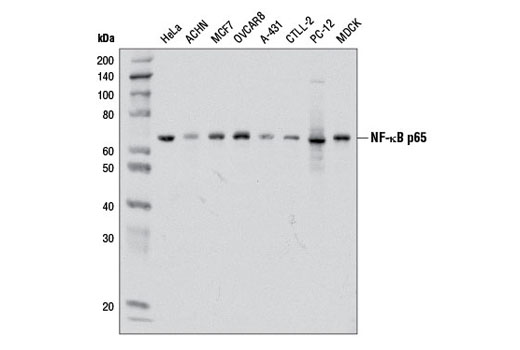
使用 NF-κB p65 (D14E12) XP® Rabbit mAb 对来自不同细胞系的提取物进行蛋白质印迹分析。
Western Blotting Image 1: NF-κB p65 (D14E12) XP® Rabbit mAb Expand Image
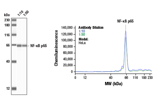
使用 NF-κB p65 (D14E12) XP® Rabbit mAb #8242 对 HeLa 细胞的裂解物 (1 mg/mL) 进行 Simple Western™ 分析。虚拟泳道式图像(左图)显示一抗稀释比例为 1:10 和 1:50 时的单一靶标条带(如图所示)。对应的电泳图(右图)为一抗稀释比例在 1:10(蓝线)和 1:50(绿线)时沿毛细血管内分子量的化学发光结果。在还原条件下,使用 12-230 kDa 分离模块在 ProteinSimple(BioTechne 品牌)的 Jess™ Simple Western 仪器上进行该实验。
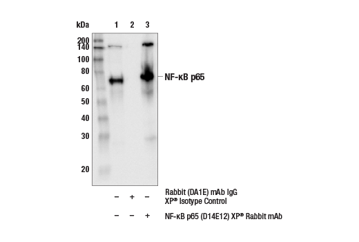
Immunoprecipitation Image 1: NF-κB p65 (D14E12) XP® Rabbit mAb Expand Image
对 CHO 细胞提取物的 NF-kB p65 进行免疫沉淀分析。泳道 1 为 10% input,泳道 2 用 Rabbit (DA1E) mAb IgG XP® Isotype Control #3900 进行沉淀,泳道 3 为 NF-κB p65 (D14E12) XP® Rabbit mAb #8242。使用 NF-κB p65 (L8F6) Mouse mAb #6956 进行蛋白质印迹法分析。
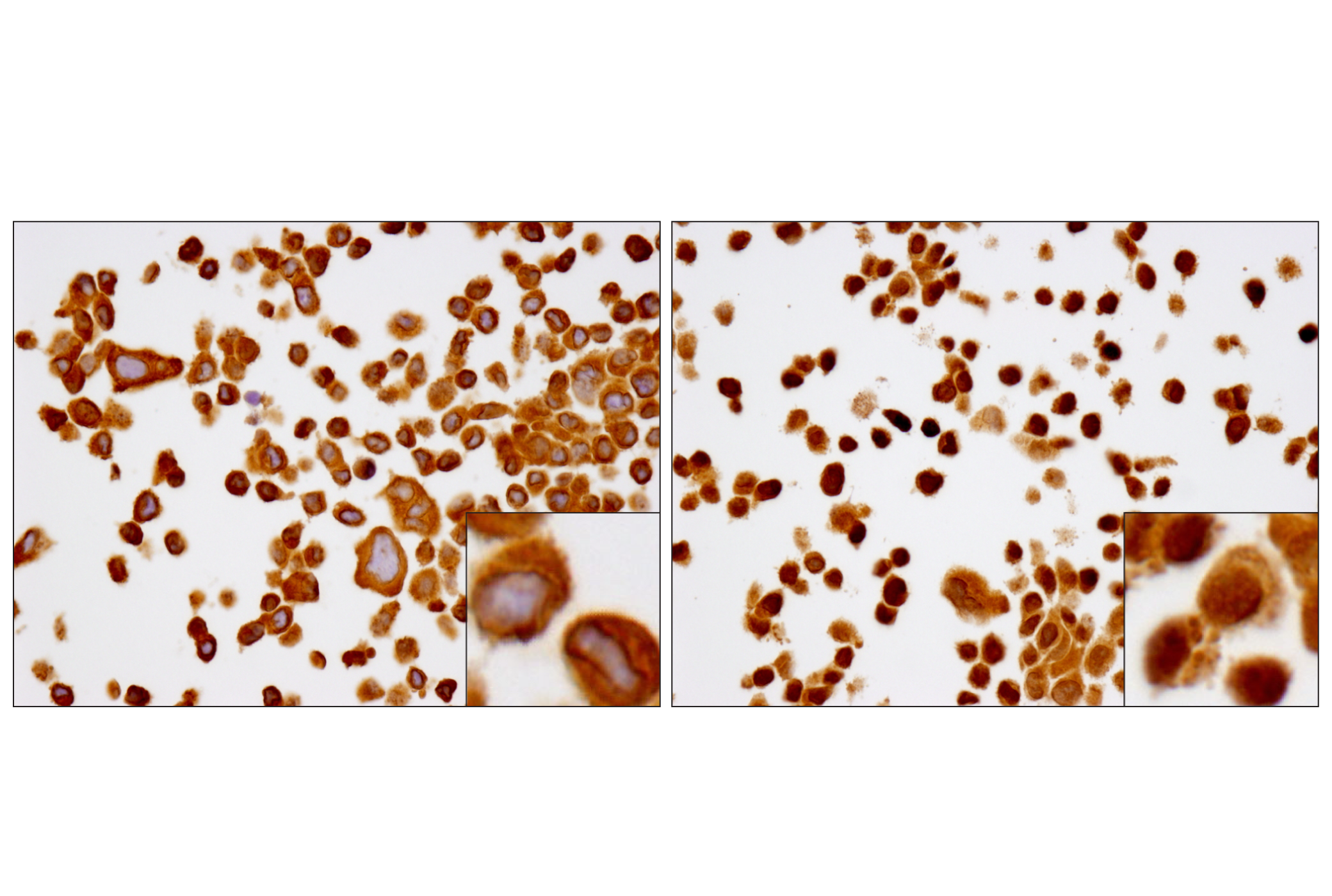
Immunohistochemistry Image 1: NF-κB p65 (D14E12) XP® Rabbit mAb Expand Image
在 SignalSlide® NF-κB p65 IHC Controls #12873(石蜡包埋的 HCT116 细胞(未处理(左)或用 hTNF-α #8902 处理(右))上使用 NF-κB p65 (D14E12) XP® Rabbit mAb 进行免疫组织化学分析。
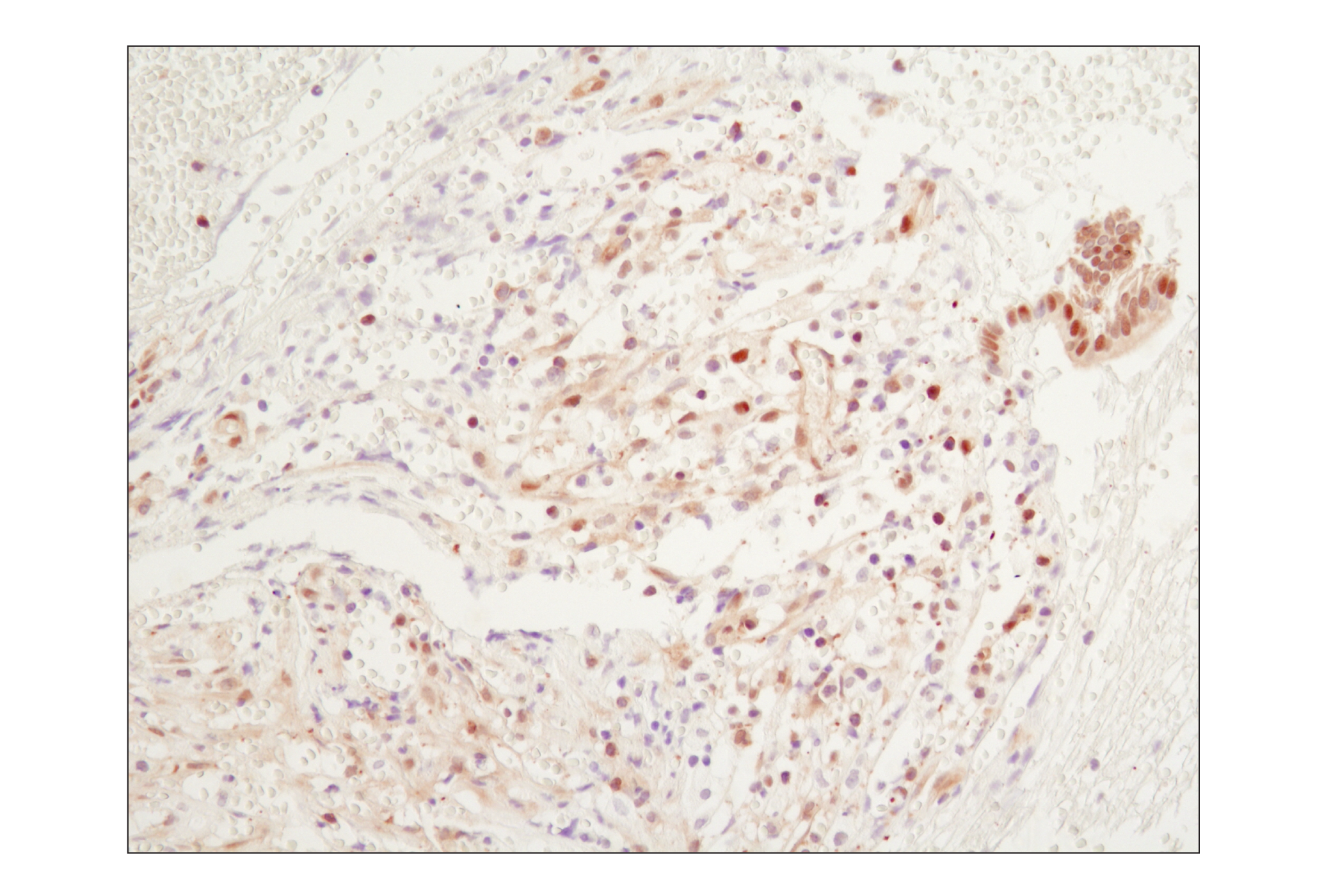
Immunohistochemistry Image 2: NF-κB p65 (D14E12) XP® Rabbit mAb Expand Image
使用 NF-κB p65 (D14E12) XP® Rabbit mAb 对石蜡包埋的人慢性胆囊炎进行免疫组织化学分析。
Immunofluorescence Image 1: NF-κB p65 (D14E12) XP® Rabbit mAb Expand Image
使用 NF-κB p65(D14E12) XP® Rabbit mAb(绿色)对 HT-1080 细胞(未处理(左)或用 hTNF-α #8902 处理(20 ng/ml,20 分钟)(右))进行共聚焦免疫荧光分析。使用 DY-554 phalloidin(红色)标记肌动蛋白纤丝。蓝色伪彩 = DRAQ5® #4084(DNA 荧光染料)。
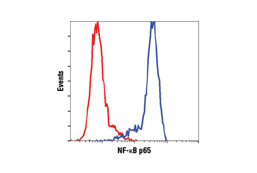
Flow Cytometry Image 1: NF-κB p65 (D14E12) XP® Rabbit mAb Expand Image
与浓度匹配的 Rabbit (DA1E) mAb IgG XP® Isotype Control #3900(红色)相比,使用 NF-κB p65 (D14E12) XP® Rabbit mAb(蓝色)对 HeLa 细胞进行流式细胞分析。
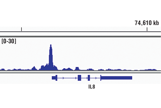
Chromatin Immunoprecipitation Image 1: NF-κB p65 (D14E12) XP® Rabbit mAb Expand Image
使用 SimpleChIP® Enzymatic Chromatin IP Kit (Magnetic Beads) #9005,对经 hTNF-α #8902(30 ng/ml,1 小时)处理的 HeLa 细胞中提取的交联染色质,在加入 NF-κB p65 (D14E12) XP® Rabbit mAb 后进行染色质免疫沉淀。使用 DNA Library Prep Kit for Illumina® (ChIP-seq, CUT&RUN) #56795 制备 DNA 库。本图显示整个 IL-8(NFκB 的一个已知靶标基因)范围内的结合作用(参见包含 ChIP-qPCR 数据的其他图)。欲知其他 ChIP-seq 情况,请下载产品说明书。
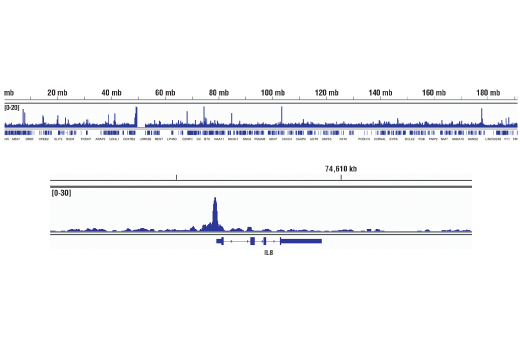
Chromatin Immunoprecipitation Image 2: NF-κB p65 (D14E12) XP® Rabbit mAb Expand Image
使用 SimpleChIP® Enzymatic Chromatin IP Kit (Magnetic Beads) #9005,对经 hTNF-α #8902(30 ng/ml,1 小时)处理的 HeLa 细胞中提取的交联染色质,在加入 NF-κB p65 (D14E12) XP® Rabbit mAb 后进行染色质免疫沉淀。使用 DNA Library Prep Kit for Illumina® (ChIP-seq, CUT&RUN) #56795 制备 DNA 库。结果图显示在染色体4(上图)内的结合,包括 NFκB 的已知靶标基因 IL-8(下图)(参见包含 ChIP-qPCR 数据的其他结果图)。
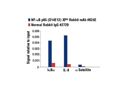
Chromatin Immunoprecipitation Image 3: NF-κB p65 (D14E12) XP® Rabbit mAb Expand Image
使用 SimpleChIP® Enzymatic Chromatin IP Kit (Magnetic Beads) #9003,对经 hTNF-α #8902(30 ng/ml,1 小时)处理的 Hela 细胞中提取的交联染色质,在加入 NF-κB p65 (D14E12) XP® Rabbit mAb 或 Normal Rabbit IgG #2729 后进行染色质免疫沉淀。使用 SimpleChIP® Human IκBα Promoter Primers #5552、人 IL-8 启动子引物和 SimpleChIP® Human α Satellite Repeat Primers #4486,通过实时 PCR 对富集的 DNA 进行定量分析。将每份样品中免疫沉淀的 DNA 的量表现为相对于所输入染色质总量(等于 1)的信号。
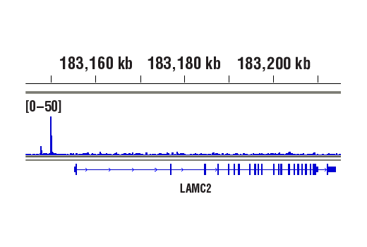
CUT and RUN Image 1: NF-κB p65 (D14E12) XP® Rabbit mAb Expand Image
使用 CUT&RUN Assay Kit #86652 对经 hTNF-α #8902(30 ng/ml,1 小时)处理的 HeLa 细胞和 NF-κB p65 (D14E12) XP® Rabbit mAb 进行 CUT&RUN。€‚使用 DNA Library Prep Kit for Illumina® (ChIP-seq, CUT&RUN) #56795 制备 DNA 库。结果图显示在 NF-κB p65 的已知靶基因 LAMC2 内结合(参见包含 CUT&RUN-qPCR 数据的其他结果图)。
CUT and RUN Image 2: NF-κB p65 (D14E12) XP® Rabbit mAb Expand Image
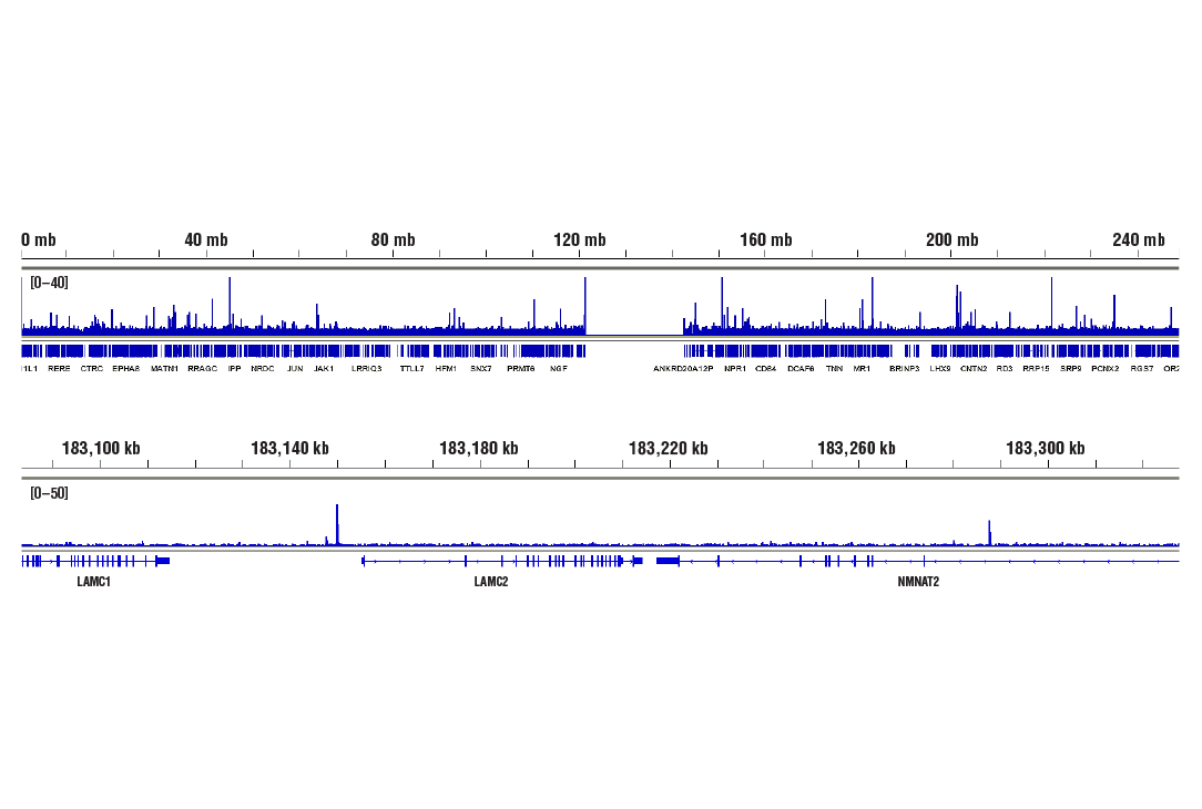
使用 CUT&RUN Assay Kit #86652 对经 hTNF-α #8902(30 ng/ml,1 小时)处理的 HeLa 细胞和 NF-κB p65 (D14E12) XP® Rabbit mAb 进行 CUT&RUN。€‚使用 DNA Library Prep Kit for Illumina® (ChIP-seq, CUT&RUN) #56795 制备 DNA 库。结果图显示在染色体 1(上图)内结合,包括 NF-κB p65 的已知靶基因 LAMC2(下图)(参见包含 CUT&RUN-qPCR 数据的其他结果图)。
CUT and RUN Image 3: NF-κB p65 (D14E12) XP® Rabbit mAb Expand Image
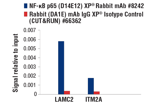
使用 CUT&RUN Assay Kit #86652 对经 hTNF-α #8902(30 ng/ml,1 小时)处理的 HeLa 细胞和 NF-κB p65 (D14E12) XP® Rabbit mAb 或 Rabbit (DA1E) mAb IgG XP® Isotype Control (CUT&RUN) #66362 进行 CUT&RUN。使用人 LAMC2 上游引物和人 ITM2A 下游引物进行实时 PCR 来对富集的 DNA 进行定量。将每份样品中免疫沉淀的 DNA 的量表现为相对于所输入染色质总量(等于 1)的信号。