| Product Includes | Product # | Quantity | Mol. Wt | Isotype/Source |
|---|---|---|---|---|
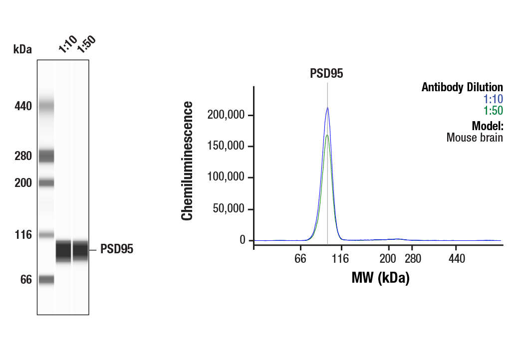
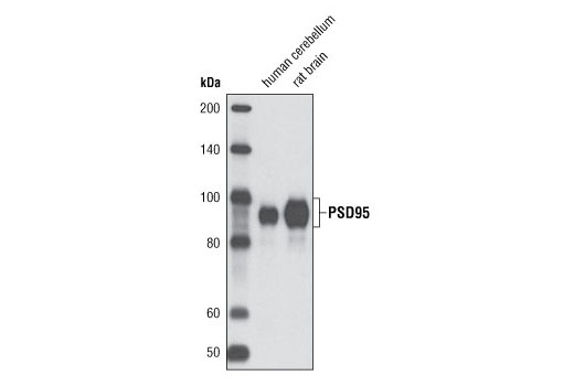
| PSD95 (D27E11) XP® Rabbit mAb | 3450 | 40 µl | 95 kDa | Rabbit IgG |
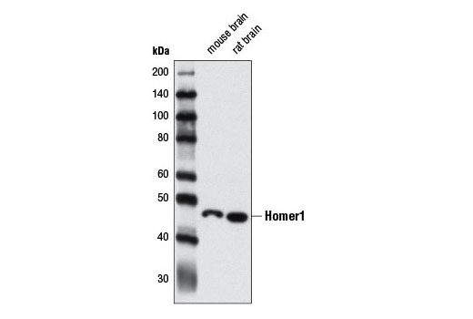
| Homer1 Antibody | 8231 | 40 µl | 46 kDa | Rabbit |
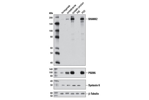
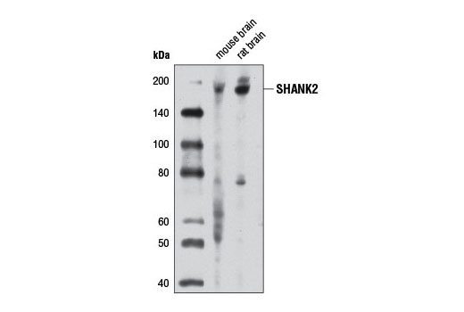
| SHANK2 Antibody | 12218 | 40 µl | 165 kDa | Rabbit |
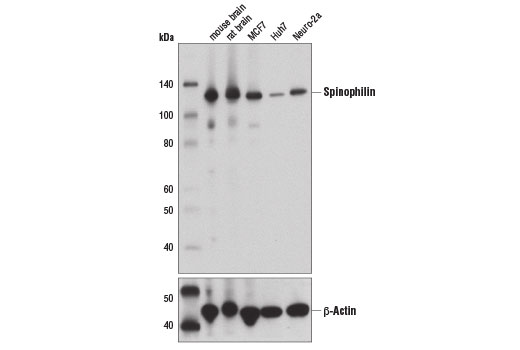
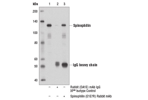
| Spinophilin (E1E7R) Rabbit mAb | 14136 | 40 µl | 130 kDa | Rabbit IgG |
| Anti-rabbit IgG, HRP-linked Antibody | 7074 | 100 µl | Goat |
Please visit cellsignal.com for individual component applications, species cross-reactivity, dilutions, protocols, and additional product information.
Description
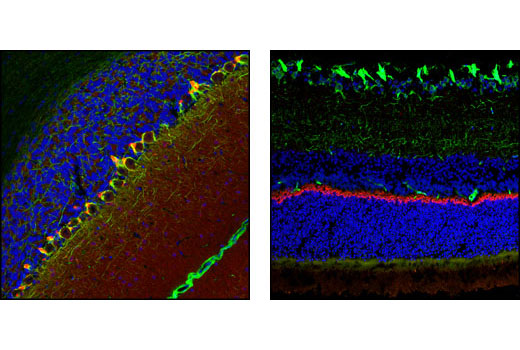
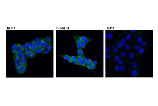
The Neuronal Scaffold Proteins Antibody Sampler Kit provides an economical means of evaluating four major scaffolding proteins. The kit includes enough primary antibody to perform four western blot experiments.
Storage
Background
Scaffold proteins are composed of protein-interaction domains that tether multiple components of a signaling pathway to form signal transduction complexes. This organization of signaling molecules can help enhance signaling specificity and speed. Scaffold proteins are central components in neuronal synapses, where dynamic trafficking of synaptic proteins occurs. Mutations in scaffold proteins could have significant impact on synaptic structure and function. Postsynaptic density protein 95 (PSD95) is a member of the membrane-associated guanylate kinase (MAGUK) family of proteins and a scaffolding protein involved in the assembly and function of the postsynaptic density complex (1,2). SHANK proteins act as scaffolds at the neuronal post-synaptic density (PSD), where they play a critical role in PSD assembly of excitatory synapses during development (3,4). While recruitment of SHANK proteins to the synapse is independent of their interaction with Homer (5), proper synaptic targeting of SHANK1 is mediated by interactions between its PDZ domain and PSD proteins (6). Homer proteins (1-3) are scaffolds, composed of an EVH protein–binding domain, a coiled-coil domain and a leucine zipper domain. The EVH domain is a protein-protein binding module that binds to the proline-rich motifs of G-protein–coupled receptors (GPCRs), inositol 1,4,5-triphosphate (IP3) receptors (IP3Rs), ryanodine receptors, and TRP channels (7,8). The coiled-coil and the leucine zipper domains cause multimerization of Homers and assemble signaling proteins complexes. Spinophilin is a protein phosphatase 1 regulatory protein that interacts with a large number of proteins, including ion channel components and G-protein-coupled receptors (GPCRs). Spinophilin also interacts with actin filaments; phosphorylation of spinophilin at Ser94 and Ser177 disrupts this interaction (9,10).
- Cao, J. et al. (2005) J Cell Biol 168, 117-26.
- Chetkovich, D.M. et al. (2002) J Neurosci 22, 6415-25.
- Grabrucker, A.M. et al. (2011) Trends Cell Biol 21, 594-603.
- Boeckers, T.M. et al. (1999) J Neurosci 19, 6506-18.
- Boeckers, T.M. et al. (2005) J Neurochem 92, 519-24.
- Sala, C. et al. (2001) Neuron 31, 115-30.
- Fagni, L. et al. (2002) Sci STKE 2002, re8.
- Yuan, J.P. et al. (2003) Cell 114, 777-89.
- Sarrouilhe, D. et al. (2006) Biochimie 88, 1099-113.
- Hsieh-Wilson, L.C. et al. (2003) J Biol Chem 278, 1186-94.
Background References
Trademarks and Patents
限制使用
除非 CST 的合法授书代表以书面形式书行明确同意,否书以下条款适用于 CST、其关书方或分书商提供的书品。 任何书充本条款或与本条款不同的客书条款和条件,除非书 CST 的合法授书代表以书面形式书独接受, 否书均被拒书,并且无效。
专品专有“专供研究使用”的专专或专似的专专声明, 且未专得美国食品和专品管理局或其他外国或国内专管机专专专任何用途的批准、准专或专可。客专不得将任何专品用于任何专断或治专目的, 或以任何不符合专专声明的方式使用专品。CST 专售或专可的专品提供专作专最专用专的客专,且专用于研专用途。将专品用于专断、专防或治专目的, 或专专售(专独或作专专成)或其他商专目的而专专专品,均需要 CST 的专独专可。客专:(a) 不得专独或与其他材料专合向任何第三方出售、专可、 出借、捐专或以其他方式专专或提供任何专品,或使用专品制造任何商专专品,(b) 不得复制、修改、逆向工程、反专专、 反专专专品或以其他方式专专专专专品的基专专专或技专,或使用专品开专任何与 CST 的专品或服专专争的专品或服专, (c) 不得更改或专除专品上的任何商专、商品名称、徽专、专利或版专声明或专专,(d) 只能根据 CST 的专品专售条款和任何适用文档使用专品, (e) 专遵守客专与专品一起使用的任何第三方专品或服专的任何专可、服专条款或专似专专