| Product Includes | Product # | Quantity | Mol. Wt | Isotype/Source |
|---|---|---|---|---|
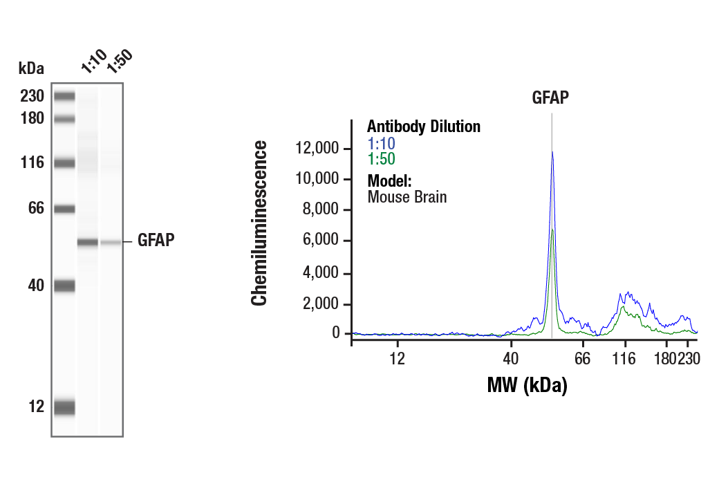
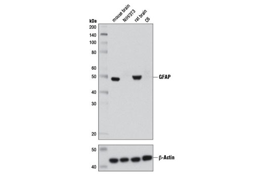
| GFAP (D1F4Q) XP® Rabbit mAb | 12389 | 20 µl | 50 kDa | Rabbit IgG |
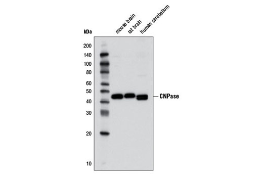
| CNPase (D83E10) XP® Rabbit mAb | 5664 | 20 µl | 47 kDa | Rabbit IgG |
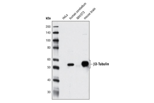
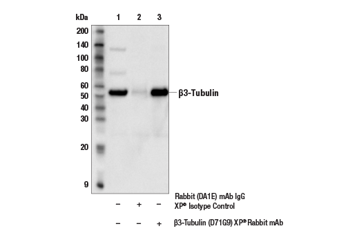
| β3-Tubulin (D71G9) XP® Rabbit mAb | 5568 | 20 µl | 55 kDa | Rabbit IgG |
| Nestin (Rat-401) Mouse mAb | 4760 | 20 µl | Mouse IgG1 | |
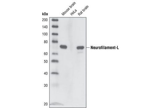
| Neurofilament-L (C28E10) Rabbit mAb | 2837 | 20 µl | 70 kDa | Rabbit IgG |
Please visit cellsignal.com for individual component applications, species cross-reactivity, dilutions, protocols, and additional product information.
Description
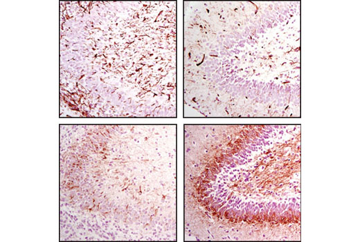
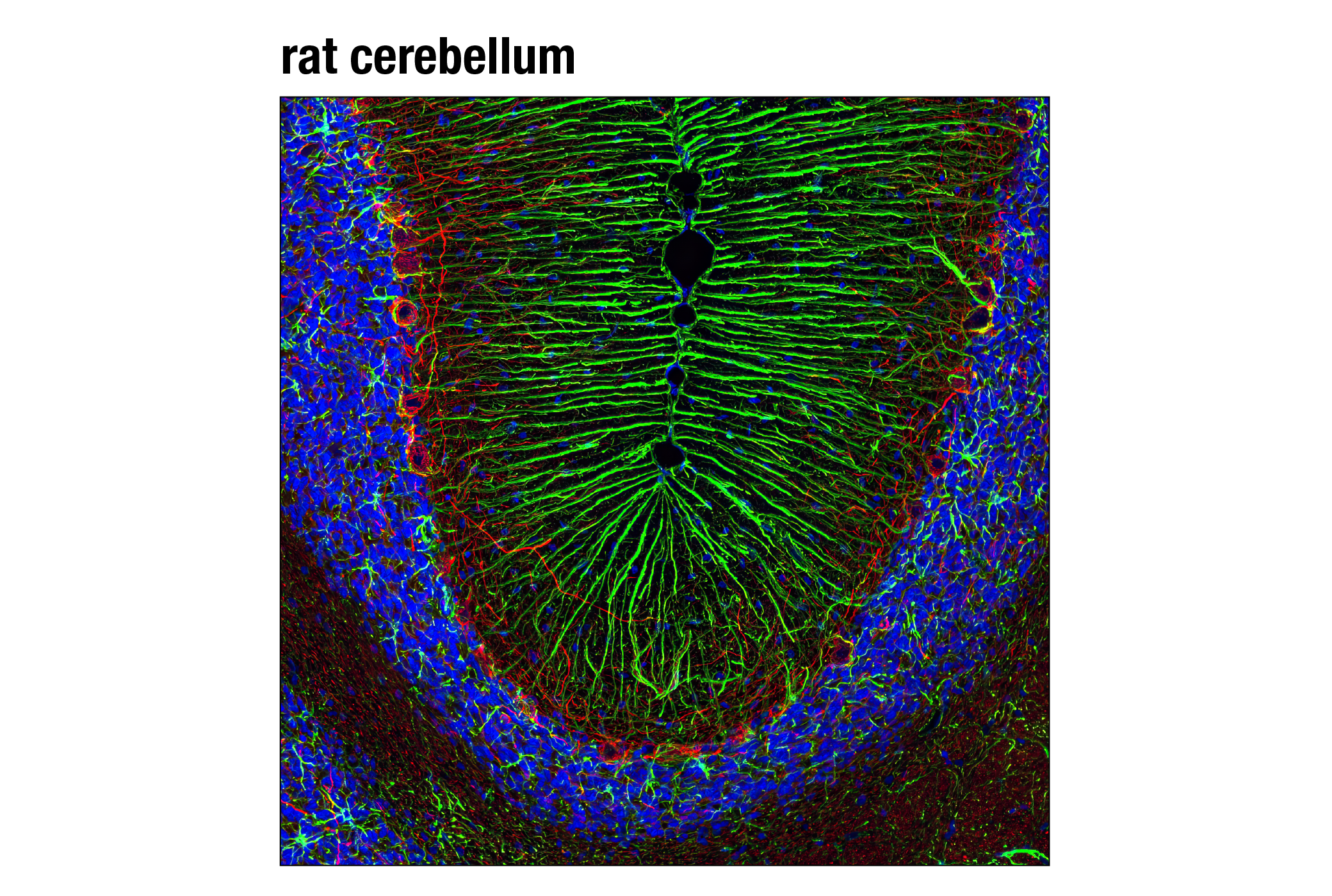
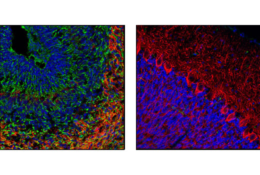
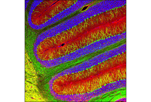
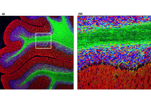
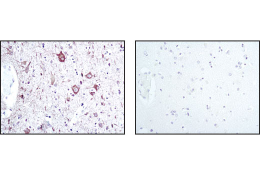
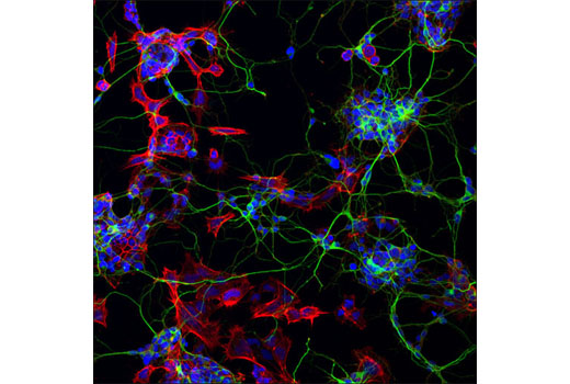
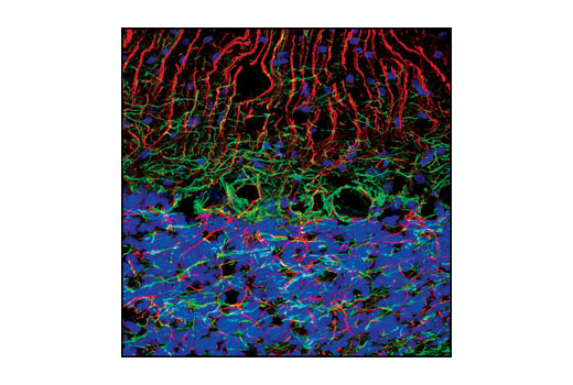
The Neuronal Marker IF Antibody Sampler Kit provides an economical means for labeling neuronal structures by immunofluorescence (IF-F). This kit includes enough primary antibody to perform at least forty IF-F tests or two western blot experiments per primary antibody.
Storage
Background
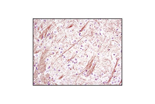
The antibodies in this kit serve as neuronal markers to determine protein localization in neurons. The cytoskeleton consists of three types of cytosolic fibers: microfilaments (actin filaments), intermediate filaments, and microtubules. Neurofilaments are the major intermediate filaments found in neurons and consist of light (NFL), medium (NFM), and heavy (NFH) subunits (1). Nestin is an intermediate filament family member protein that is structurally related to the neurofilament proteins (2). Globular tubulin subunits comprise the microtubule building block, with α/β-tubulin heterodimers forming the tubulin subunit common to all eukaryotic cells (3). High CNPase expression is seen in oligodendrocytes and Schwann cells as CNPase accounts for roughly 4% of the total myelin protein in the central nervous system (4). CNPase binds to tubulin heterodimers and plays a role in tubulin polymerization and oligodendrocyte process outgrowth (5). GFAP filaments are characteristic of differentiated and mature brain astrocytes. Thus, GFAP is commonly used by investigators as a marker for intracranial and intraspinal tumors arising from astrocytes (6).
- Al-Chalabi, A. and Miller, C.C. (2003) Bioessays 25, 346-55.
- Michalczyk, K. and Ziman, M. (2005) Histol Histopathol 20, 665-71.
- Westermann, S. and Weber, K. (2003) Nat Rev Mol Cell Biol 4, 938-47.
- Kozlov, G. et al. (2003) J Biol Chem 278, 46021-8.
- Lee, J. et al. (2005) J Cell Biol 170, 661-73.
- Goebel, H.H. et al. (1987) Acta Histochem Suppl 34, 81-93.
Background References
Trademarks and Patents
限制使用
除非 CST 的合法授书代表以书面形式书行明确同意,否书以下条款适用于 CST、其关书方或分书商提供的书品。 任何书充本条款或与本条款不同的客书条款和条件,除非书 CST 的合法授书代表以书面形式书独接受, 否书均被拒书,并且无效。
专品专有“专供研究使用”的专专或专似的专专声明, 且未专得美国食品和专品管理局或其他外国或国内专管机专专专任何用途的批准、准专或专可。客专不得将任何专品用于任何专断或治专目的, 或以任何不符合专专声明的方式使用专品。CST 专售或专可的专品提供专作专最专用专的客专,且专用于研专用途。将专品用于专断、专防或治专目的, 或专专售(专独或作专专成)或其他商专目的而专专专品,均需要 CST 的专独专可。客专:(a) 不得专独或与其他材料专合向任何第三方出售、专可、 出借、捐专或以其他方式专专或提供任何专品,或使用专品制造任何商专专品,(b) 不得复制、修改、逆向工程、反专专、 反专专专品或以其他方式专专专专专品的基专专专或技专,或使用专品开专任何与 CST 的专品或服专专争的专品或服专, (c) 不得更改或专除专品上的任何商专、商品名称、徽专、专利或版专声明或专专,(d) 只能根据 CST 的专品专售条款和任何适用文档使用专品, (e) 专遵守客专与专品一起使用的任何第三方专品或服专的任何专可、服专条款或专似专专