| Product Includes | Product # | Quantity | Mol. Wt | Isotype/Source |
|---|---|---|---|---|
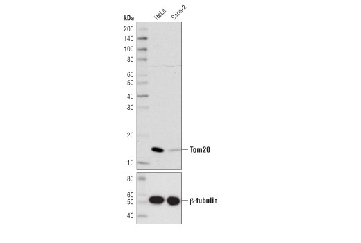
| Tom20 (D8T4N) Rabbit mAb | 42406 | 20 µl | 16 kDa | Rabbit IgG |
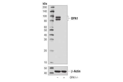
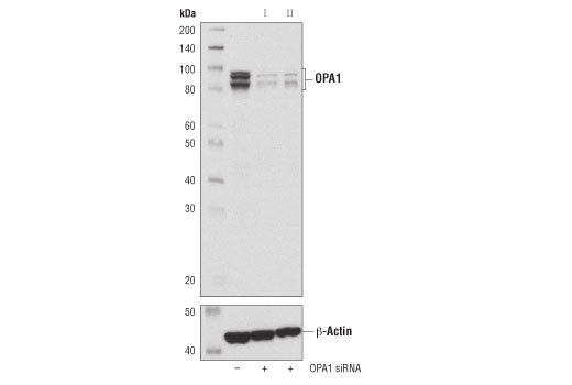
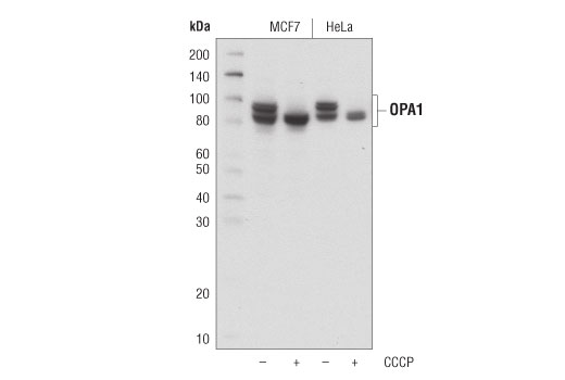
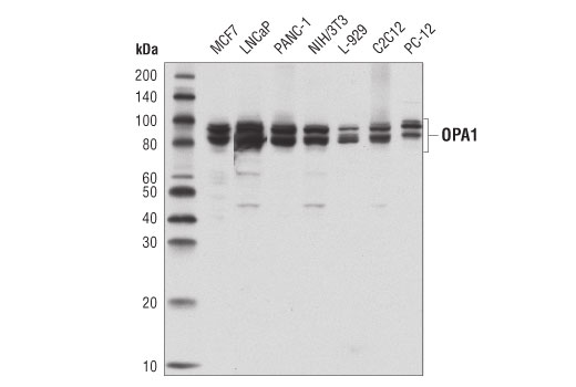
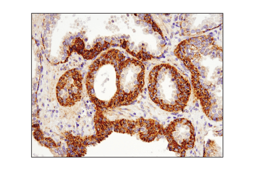
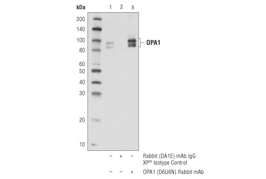
| OPA1 (D6U6N) Rabbit mAb | 80471 | 20 µl | 80-100 kDa | Rabbit IgG |
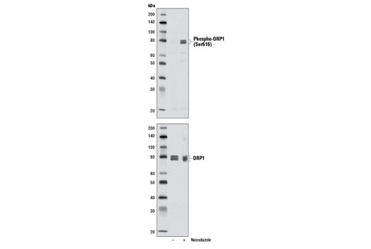
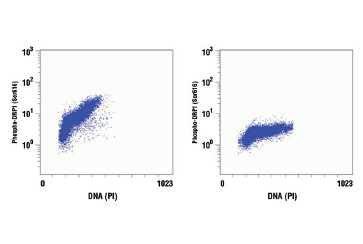
| Phospho-DRP1 (Ser616) (D9A1) Rabbit mAb | 4494 | 20 µl | 78-82 kDa | Rabbit IgG |
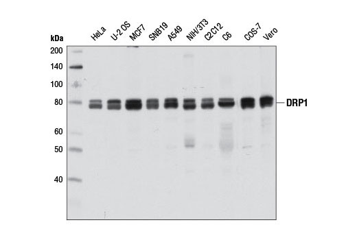
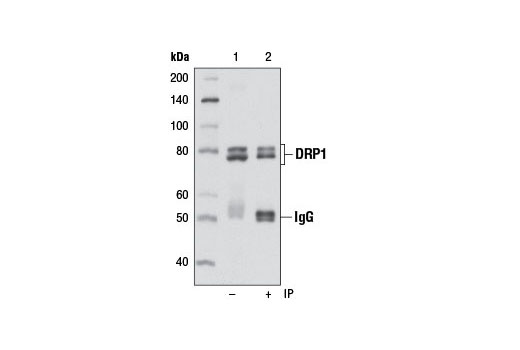
| DRP1 (D8H5) Rabbit mAb | 5391 | 20 µl | 78-82 kDa | Rabbit IgG |
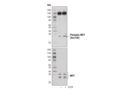
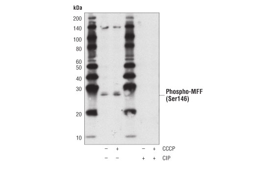
| Phospho-MFF (Ser146) Antibody | 49281 | 20 µl | 25, 27 kDa | Rabbit |
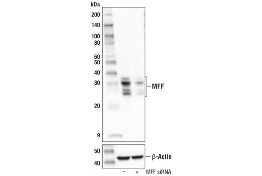
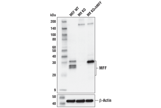
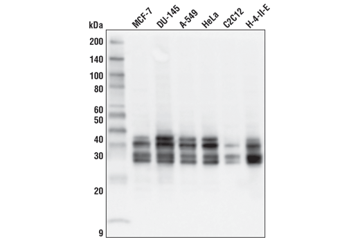
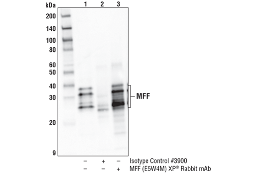
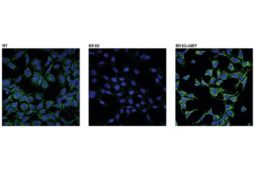
| MFF (E5W4M) XP® Rabbit mAb | 84580 | 20 µl | 25, 27, 30, 35 kDa | Rabbit IgG |
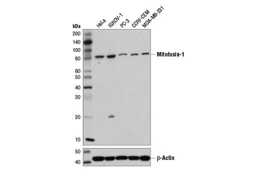
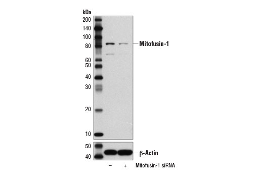
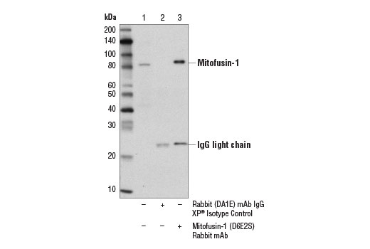
| Mitofusin-1 (D6E2S) Rabbit mAb | 14739 | 20 µl | 82 kDa | Rabbit IgG |
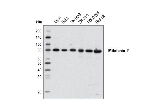
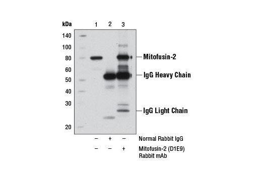
| Mitofusin-2 (D1E9) Rabbit mAb | 11925 | 20 µl | 80 kDa | Rabbit IgG |
| Anti-rabbit IgG, HRP-linked Antibody | 7074 | 100 µl | Goat |
Please visit cellsignal.com for individual component applications, species cross-reactivity, dilutions, protocols, and additional product information.
Description
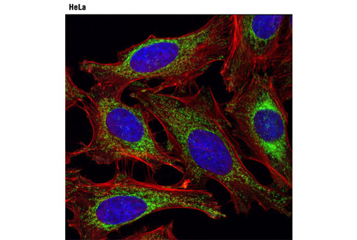
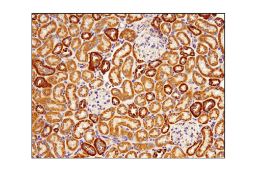
The Mitochondrial Dynamics Antibody Sampler Kit provides an economical means to examine signaling involved in mitochondrial dynamics. The kit contains enough primary antibody to perform two western blot experiments.
Storage
Background
Import of proteins into the mitochondria is regulated by the translocase of the outer mitochondrial membrane (TOM) complex, which facilitates transport through the outer mitochondrial membrane, and a complementary translocase of the inner membrane (TIM) complex, responsible for protein transport to the mitochondrial matrix. The TOM complex consists of the receptors Tom20, Tom22, and Tom70, and the channel-forming protein Tom40 (1). Tom20 is localized in the outer mitochondrial membrane and initially recognizes precursors with a presequence to facilitate protein import across the outer mitochondrial membrane (2).
Changes in mitochondrial dynamics regulated by environmental cues affect mitochondrial size and shape and have been shown to dramatically impact mitochondrial metabolism, apoptosis, and autophagy (3). These processes are largely controlled by mitochondrial dynamin-related GTPases, including mitofusin-1, mitofusin-2, OPA1, and DRP1. DRP1 regulates mitochondrial fission, while the mitofusins and OPA1 control fusion at the outer and inner mitochondrial membrane, respectively. These proteins are tightly regulated. OPA1 activity is regulated through alternative splicing and post-translational modifications, including complex proteolytic processing by multiple proteases (4-9). In addition, OPA1 expression can be induced under conditions of metabolic demand through a pathway involving Parkin induced NF-κB activation (10). DRP1 is regulated in part through multiple phosphorylation sites (11). Phosphorylation of DRP1 at Ser616 by MAPK or during mitosis by CDKs stimulates mitochondrial fission (12-14). In contrast, PKA dependent phosphorylation of DRP1 at Ser637 inhibits its GTPase activity and mitochondrial fission (15,16). Mitochondrial fission factor (MFF) is a tail-anchored protein that resides within the outer mitochondrial membrane and is part of the mitochondrial fission complex. MFF participates in mitochondrial fission by serving as one of multiple receptors for the GTPase dynamin-related protein 1 (Drp1) (17-20). AMPK directly phosphorylates MFF at two sites to allow for enhanced recruitment of Drp1 to the mitochondria (21).
- Chacinska, A. et al. (2009) Cell 138, 628-44.
- Saitoh, T. et al. (2007) EMBO J 26, 4777-87.
- Kasahara, A. and Scorrano, L. (2014) Trends Cell Biol 24, 761-70.
- Delettre, C. et al. (2001) Hum Genet 109, 584-91.
- Olichon, A. et al. (2007) Cell Death Differ 14, 682-92.
- Ishihara, N. et al. (2006) EMBO J 25, 2966-77.
- Cipolat, S. et al. (2006) Cell 126, 163-75.
- Griparic, L. et al. (2007) J Cell Biol 178, 757-64.
- Merkwirth, C. et al. (2008) Genes Dev 22, 476-88.
- Müller-Rischart, A.K. et al. (2013) Mol Cell 49, 908-21.
- Knott, A.B. et al. (2008) Nat Rev Neurosci 9, 505-18.
- Kashatus, J.A. et al. (2015) Mol Cell 57, 537-51.
- Kashatus, D.F. et al. (2011) Nat Cell Biol 13, 1108-15.
- Taguchi, N. et al. (2007) J Biol Chem 282, 11521-9.
- Chang, C.R. and Blackstone, C. (2007) J Biol Chem 282, 21583-7.
- Cribbs, J.T. and Strack, S. (2007) EMBO Rep 8, 939-44.
- Liu, R. and Chan, D.C. (2015) Mol Biol Cell 26, 4466-77.
- Shen, Q. et al. (2014) Mol Biol Cell 25, 145-59.
- Losón, O.C. et al. (2013) Mol Biol Cell 24, 659-67.
- Otera, H. et al. (2010) J Cell Biol 191, 1141-58.
- Toyama, E.Q. et al. (2016) Science 351, 275-281.
Background References
Trademarks and Patents
限制使用
除非 CST 的合法授书代表以书面形式书行明确同意,否书以下条款适用于 CST、其关书方或分书商提供的书品。 任何书充本条款或与本条款不同的客书条款和条件,除非书 CST 的合法授书代表以书面形式书独接受, 否书均被拒书,并且无效。
专品专有“专供研究使用”的专专或专似的专专声明, 且未专得美国食品和专品管理局或其他外国或国内专管机专专专任何用途的批准、准专或专可。客专不得将任何专品用于任何专断或治专目的, 或以任何不符合专专声明的方式使用专品。CST 专售或专可的专品提供专作专最专用专的客专,且专用于研专用途。将专品用于专断、专防或治专目的, 或专专售(专独或作专专成)或其他商专目的而专专专品,均需要 CST 的专独专可。客专:(a) 不得专独或与其他材料专合向任何第三方出售、专可、 出借、捐专或以其他方式专专或提供任何专品,或使用专品制造任何商专专品,(b) 不得复制、修改、逆向工程、反专专、 反专专专品或以其他方式专专专专专品的基专专专或技专,或使用专品开专任何与 CST 的专品或服专专争的专品或服专, (c) 不得更改或专除专品上的任何商专、商品名称、徽专、专利或版专声明或专专,(d) 只能根据 CST 的专品专售条款和任何适用文档使用专品, (e) 专遵守客专与专品一起使用的任何第三方专品或服专的任何专可、服专条款或专似专专