| Product Includes | Product # | Quantity | Mol. Wt | Isotype/Source |
|---|---|---|---|---|
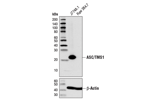
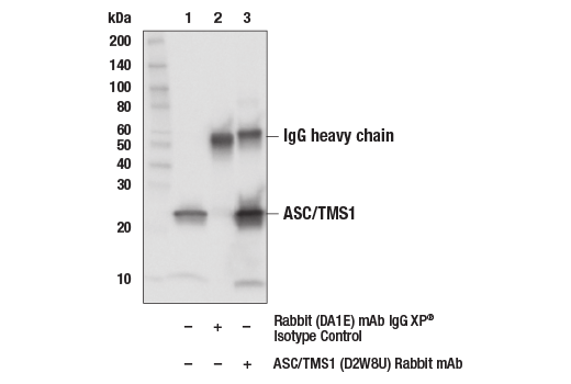
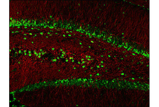
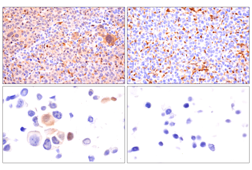
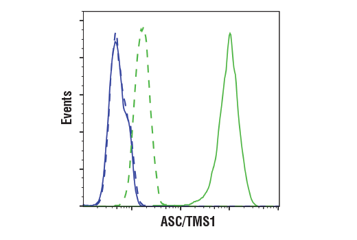
| ASC/TMS1 (D2W8U) Rabbit mAb | 67824 | 20 µl | 22 kDa | Rabbit IgG |
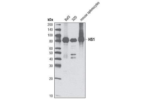
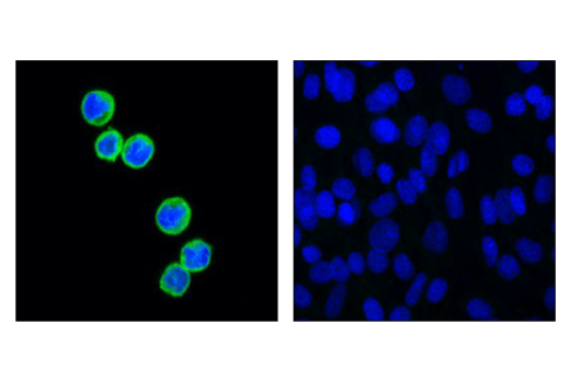
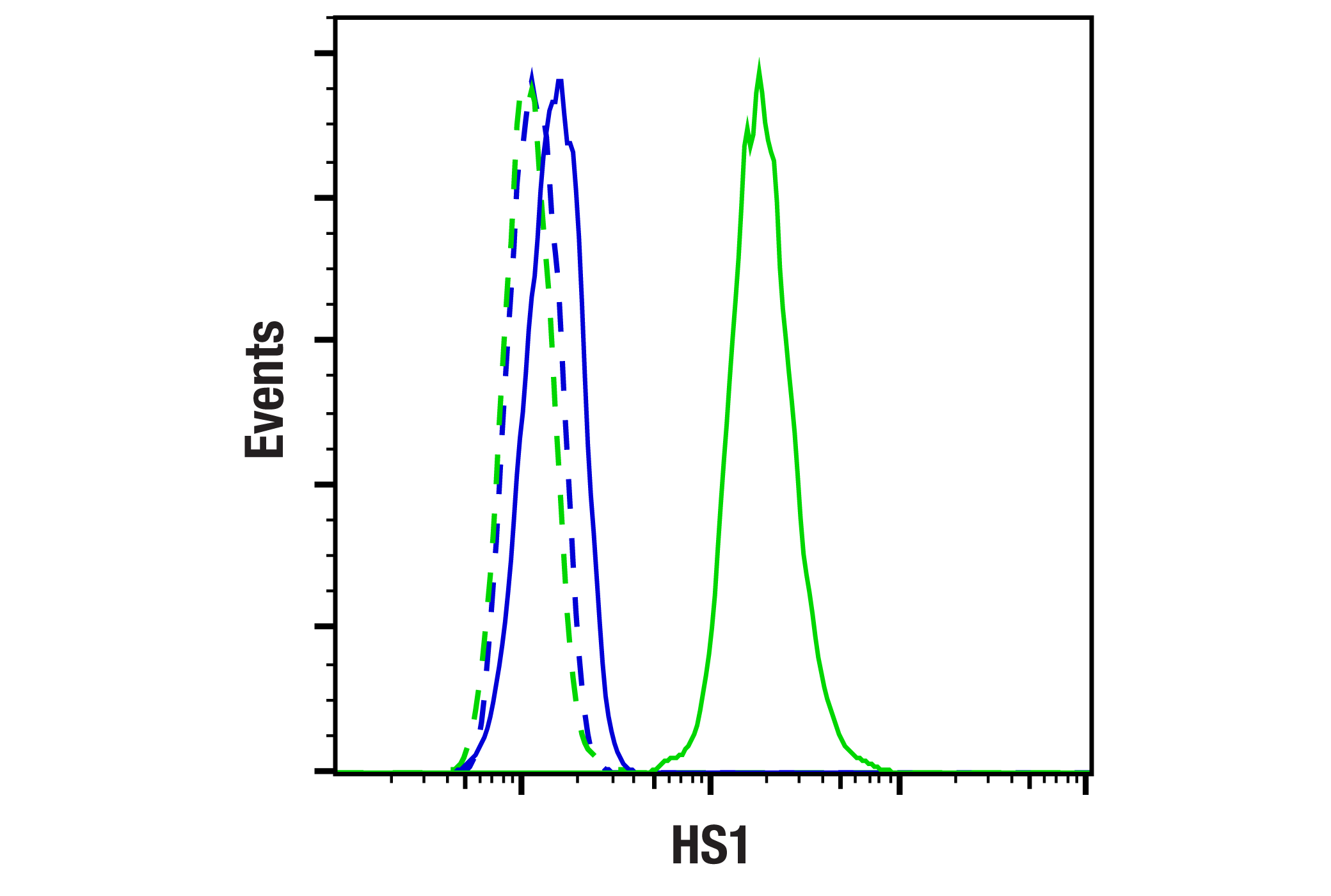
| HS1 (D5A9) XP® Rabbit mAb | 3892 | 20 µl | 80 kDa | Rabbit IgG |
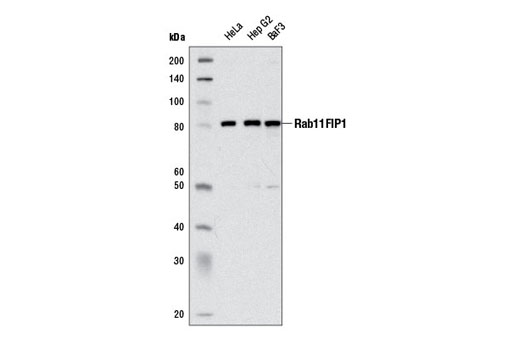
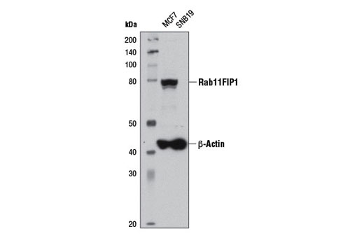
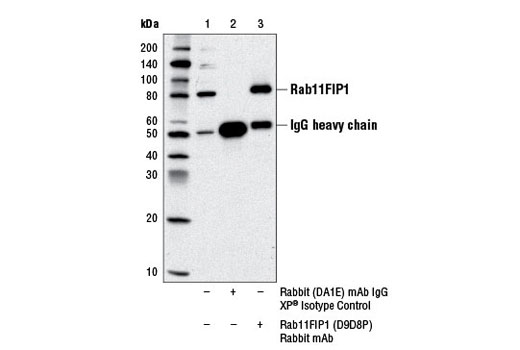
| Rab11FIP1 (D9D8P) Rabbit mAb | 12849 | 20 µl | 85 kDa | Rabbit IgG |
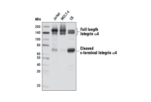
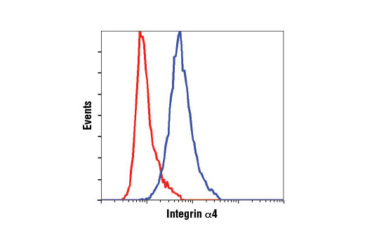
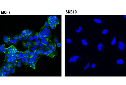
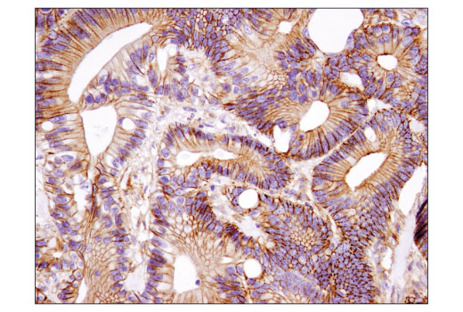
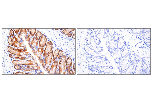
| Integrin α4 (D2E1) XP® Rabbit mAb | 8440 | 20 µl | 70, 140, 150, kDa | Rabbit IgG |
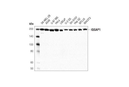
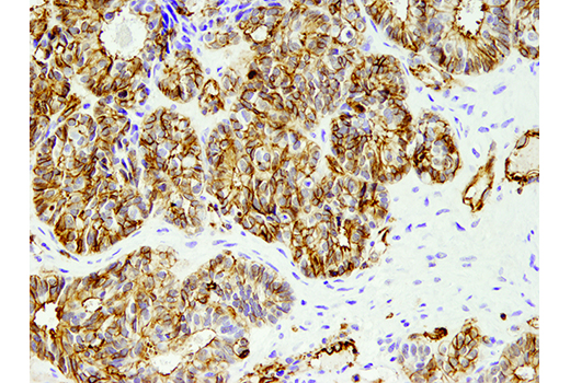
| IQGAP1 (D8K4X) XP® Rabbit mAb | 20648 | 20 µl | 195 kDa | Rabbit IgG |
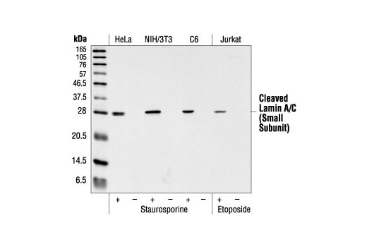
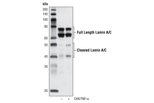
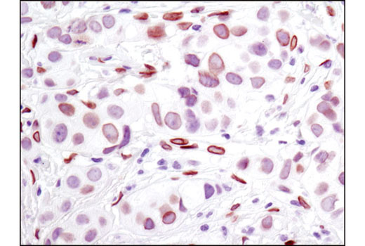
| Cleaved Lamin A (Small Subunit) (30H5) Mouse mAb | 2036 | 20 µl | 28 kDa | Mouse IgG1 |
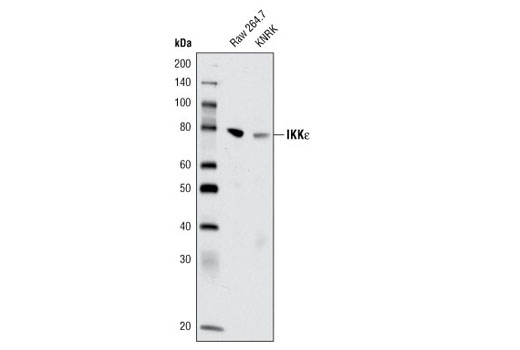
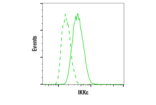
| IKKε (D61F9) XP® Rabbit mAb | 3416 | 20 µl | 80 kDa | Rabbit IgG |
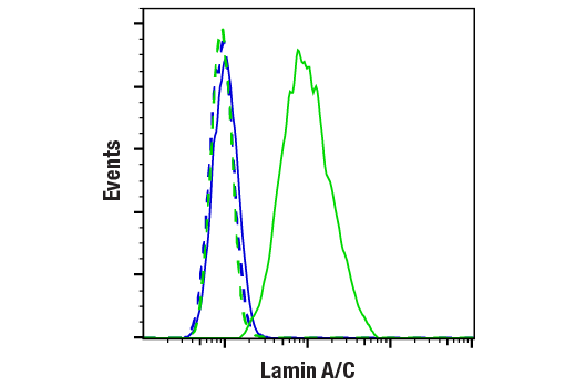
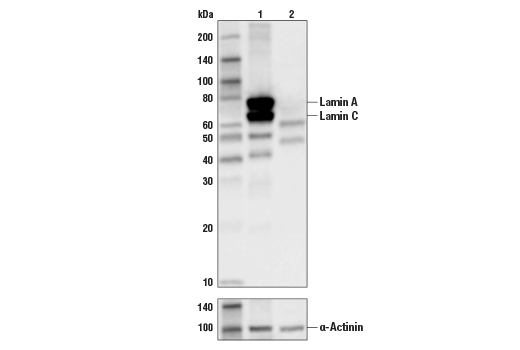
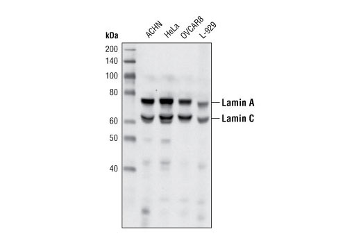
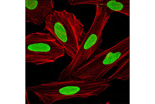
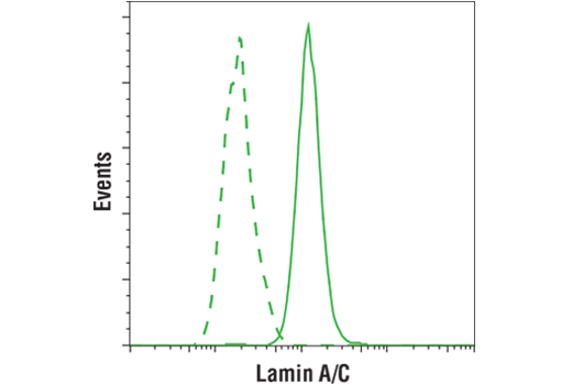
| Lamin A/C (4C11) Mouse mAb | 4777 | 20 µl | 74 (Lamin A), 63 (Lamin C) kDa | Mouse IgG2a |
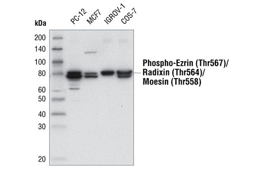
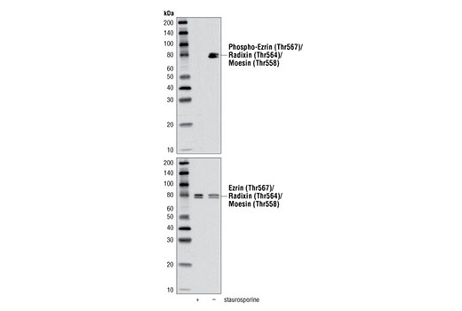
| Phospho-Ezrin (Thr567)/Radixin (Thr564)/Moesin (Thr558) (48G2) Rabbit mAb | 3726 | 20 µl | 75 Moesin. 80 Ezrin, Radixin. kDa | Rabbit IgG |
| Anti-rabbit IgG, HRP-linked Antibody | 7074 | 100 µl | Goat |
Please visit cellsignal.com for individual component applications, species cross-reactivity, dilutions, protocols, and additional product information.
Description
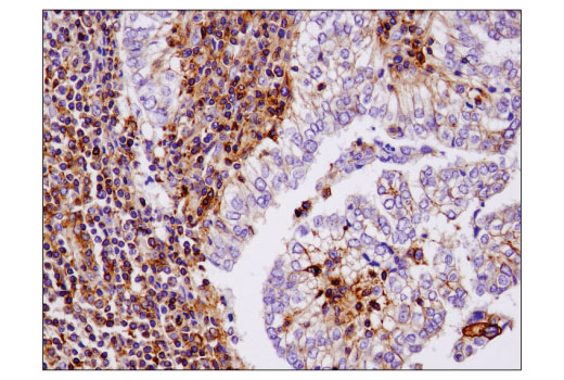
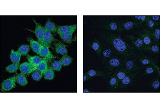
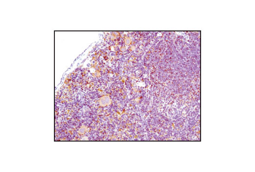
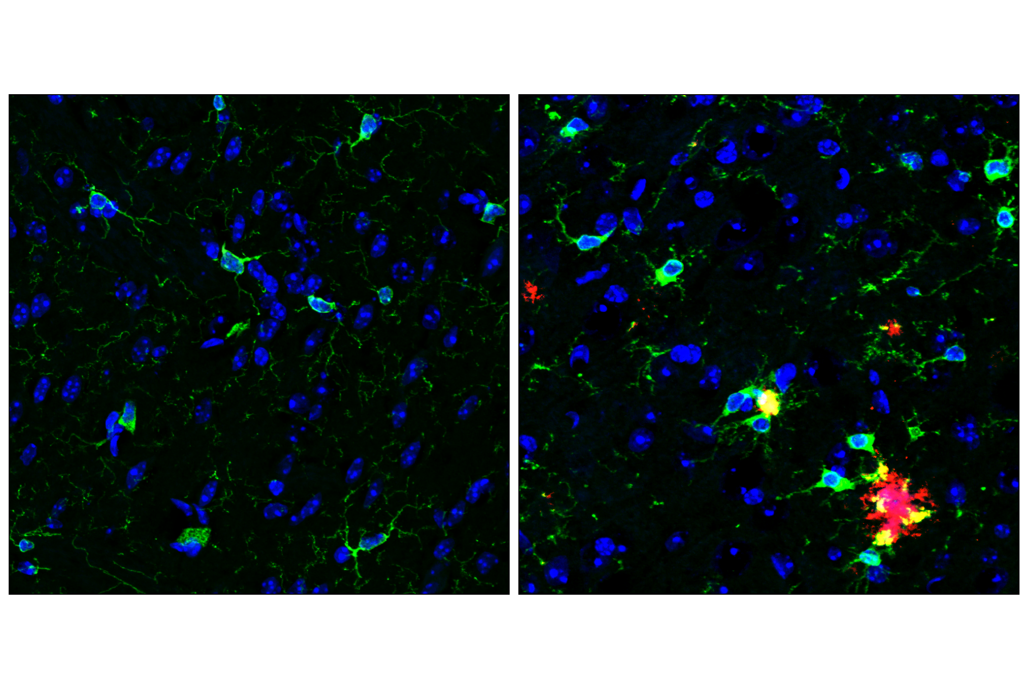
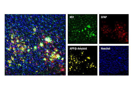
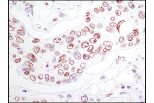
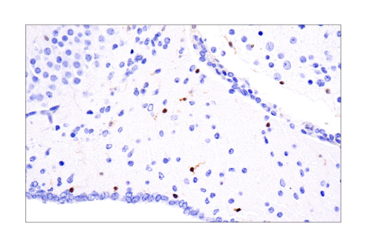
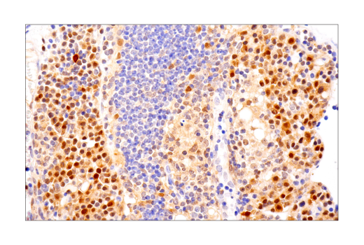
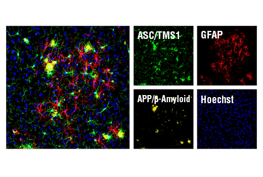
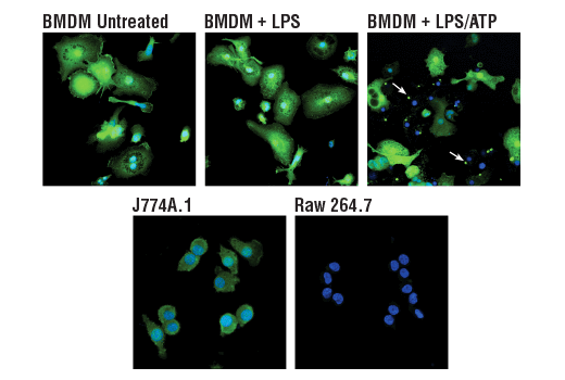
The Microglia LPS-Related Module Antibody Sampler Kit provides an economical means of detecting proteins identified as markers of LPS-related microglial activity by western blot and/or immunofluorescence.
Storage
Background
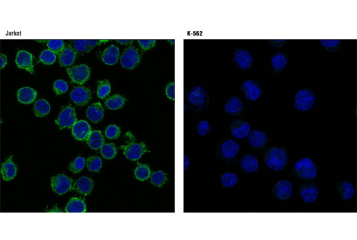
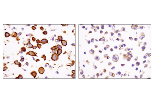
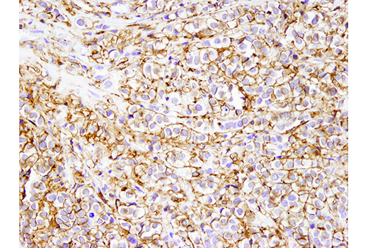
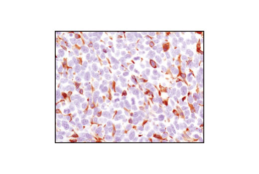
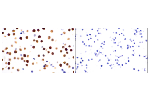
Distinct microglial activation states have been identified using RNA-seq data from a vast array of neurological disease and aging models. These activation states have been categorized into modules corresponding to proliferation, neurodegeneration, interferon-relation, LPS-relation, and many others (1). Previous work identifying markers of specific brain cell types using RNA-seq has shown HS1 and ASC/TMS1 to be useful and specific tools to study microglia (2). HS1 is a protein kinase substrate that is expressed only in tissues and cells of hematopoietic origin (3) and ASC/TMS1 has been found to be a critical component of inflammatory signaling where it associates with and activates caspase-1 in response to pro-inflammatory signals (4).
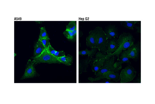
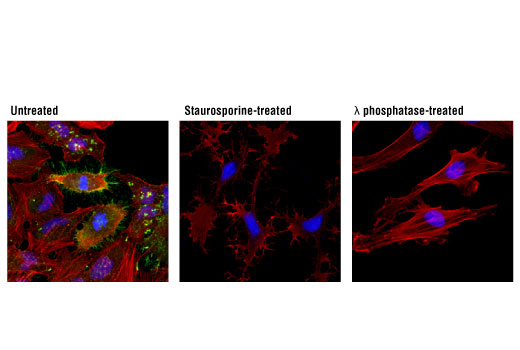
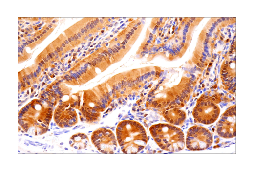
The Rab11-family interacting proteins (Rab11-FIPs) facilitate Rab11-dependent vesicle recycling through interaction with the conserved carboxyl terminal Rab11 binding domain (5,6). Rab11FIP1 has been shown to play a role in endocytic sorting and trafficking of EGFR and integrin subunits (6). Integrins are α/β heterodimeric cell surface receptors that mediate cell adhesion and migration and regulate cell growth and survival. Two significant α4 integrins, α4β1 and α4β7, interact with VCAM-1, fibronectin, and MAdCAM-1 at cell adhesions and have been shown to play an important role in cell trafficking during inflammatory processes (7-9). Lamins are nuclear membrane structural components important for maintaining normal cell functions. Lamin A/C is cleaved by caspase-6 and serves as a marker for caspase-6 activation. The cleavage of lamins results in nuclear dysregulation and cell death (10,11). The ezrin, radixin, and moesin (ERM) proteins function as linkers between the plasma membrane and the actin cytoskeleton and are involved in cell adhesion, membrane ruffling, and microvilli formation (12). ERM proteins undergo intra or intermolecular interaction between their amino- and carboxy-terminal domains, existing as inactive cytosolic monomers or dimers (13). Phosphorylation at a carboxy-terminal threonine residue (Thr567 of ezrin, Thr564 of radixin, Thr558 of moesin) disrupts the amino- and carboxy-terminal association and may play a key role in regulating ERM protein conformation and function (14,15). IQGAPs are scaffolding proteins involved in mediating cytoskeletal function that contain multiple protein interaction domains (16). IQGAP1 is ubiquitously expressed and has been found to interact with APC (17) and the CLIP170 complex in response to small GTPases, promoting cell polarization and migration (18). IKKε is an IKK-related kinase that functions as part of the signal-stimulated noncanonical pathway of NF-kB activation (19). IKKε plays a role in the immune response and also impacts cell proliferation and transformation (20).
- Friedman, B.A. et al. (2018) Cell Rep 22, 832-47.
- Zhang, Y. et al. (2014) J Neurosci 34, 11929-47.
- Kitamura, D. et al. (1995) Biochem Biophys Res Commun 208, 1137-46.
- Srinivasula, S.M. et al. (2002) J Biol Chem 277, 21119-22.
- Hales, C.M. et al. (2001) J Biol Chem 276, 39067-75.
- Baetz, N.W. and Goldenring, J.R. (2013) Mol Biol Cell 24, 643-58.
- Hood, J.D. and Cheresh, D.A. (2002) Nat Rev Cancer 2, 91-100.
- Liu, S. et al. (2000) J Cell Sci 113 (Pt 20), 3563-71.
- Kummer, C. and Ginsberg, M.H. (2006) Biochem Pharmacol 72, 1460-8.
- Oberhammer, F.A. et al. (1994) J Cell Biol 126, 827-37.
- Rao, L. et al. (1996) J Cell Biol 135, 1441-55.
- Tsukita, S. and Yonemura, S. (1999) J Biol Chem 274, 34507-10.
- Mangeat, P. et al. (1999) Trends Cell Biol 9, 187-92.
- Matsui, T. et al. (1998) J Cell Biol 140, 647-57.
- Gautreau, A. et al. (2000) J Cell Biol 150, 193-203.
- Briggs, M.W. and Sacks, D.B. (2003) EMBO Rep 4, 571-4.
- Watanabe, T. et al. (2004) Dev Cell 7, 871-83.
- Fukata, M. et al. (2002) Cell 109, 873-85.
- Sun, S.C. et al. (2013) Trends Immunol 34, 282-9.
- Verhelst, K. et al. (2013) Biochem Pharmacol 85, 873-80.
Background References
Trademarks and Patents
限制使用
除非 CST 的合法授书代表以书面形式书行明确同意,否书以下条款适用于 CST、其关书方或分书商提供的书品。 任何书充本条款或与本条款不同的客书条款和条件,除非书 CST 的合法授书代表以书面形式书独接受, 否书均被拒书,并且无效。
专品专有“专供研究使用”的专专或专似的专专声明, 且未专得美国食品和专品管理局或其他外国或国内专管机专专专任何用途的批准、准专或专可。客专不得将任何专品用于任何专断或治专目的, 或以任何不符合专专声明的方式使用专品。CST 专售或专可的专品提供专作专最专用专的客专,且专用于研专用途。将专品用于专断、专防或治专目的, 或专专售(专独或作专专成)或其他商专目的而专专专品,均需要 CST 的专独专可。客专:(a) 不得专独或与其他材料专合向任何第三方出售、专可、 出借、捐专或以其他方式专专或提供任何专品,或使用专品制造任何商专专品,(b) 不得复制、修改、逆向工程、反专专、 反专专专品或以其他方式专专专专专品的基专专专或技专,或使用专品开专任何与 CST 的专品或服专专争的专品或服专, (c) 不得更改或专除专品上的任何商专、商品名称、徽专、专利或版专声明或专专,(d) 只能根据 CST 的专品专售条款和任何适用文档使用专品, (e) 专遵守客专与专品一起使用的任何第三方专品或服专的任何专可、服专条款或专似专专