Revision 1
#79884
Store at -20C
Lung Cancer RTK Antibody Sampler Kit
1 Kit
(9 x 20 microliters)
877-616-CELL (2355)
877-678-TECH (8324)
3 Trask Lane | Danvers | Massachusetts | 01923 | USA
For Research Use Only. Not for Use in Diagnostic Procedures.
| Product Includes | Product # | Quantity | Mol. Wt | Isotype/Source |
|---|---|---|---|---|
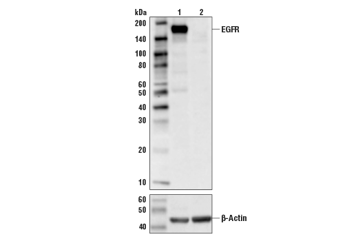
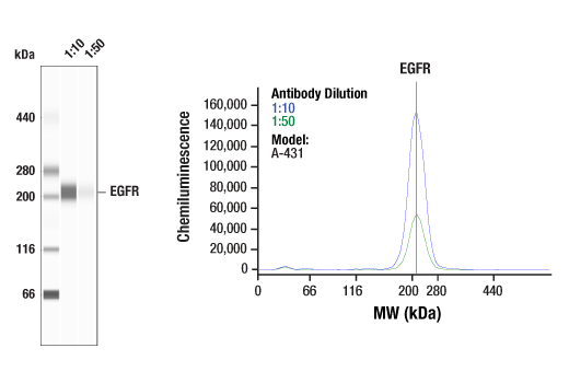
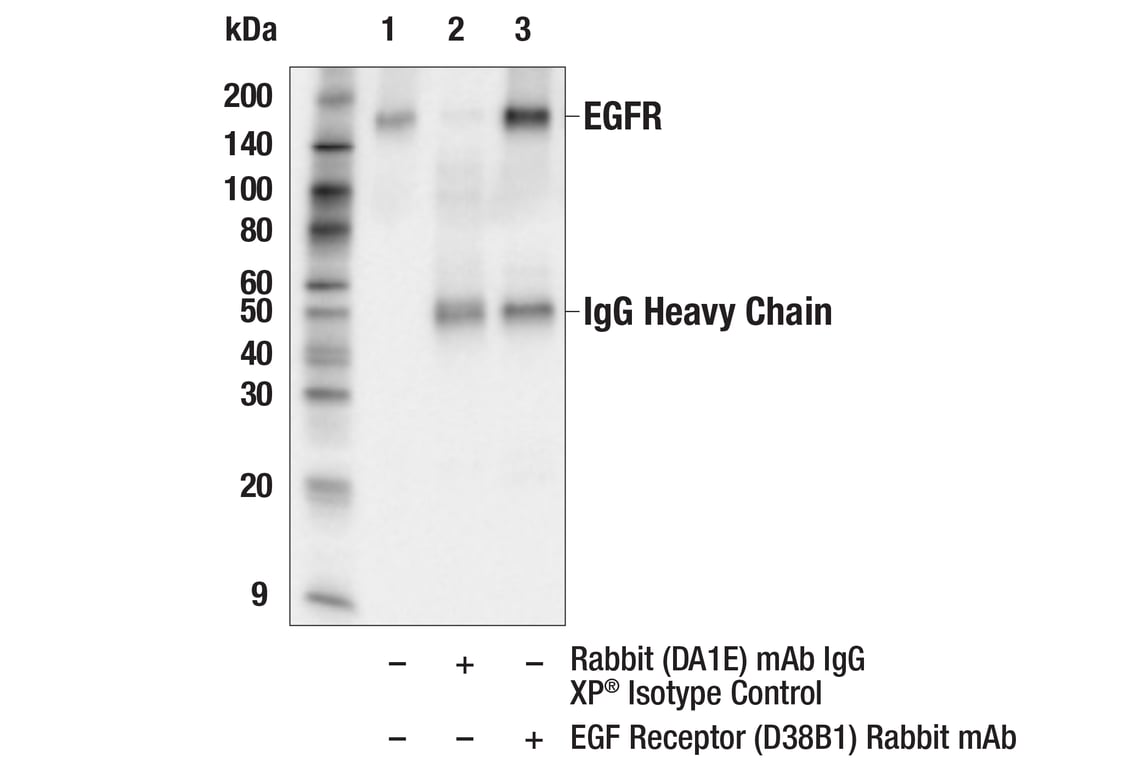
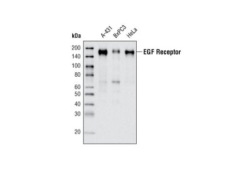
| EGF Receptor (D38B1) XP® Rabbit mAb | 4267 | 20 µl | 175 kDa | Rabbit IgG |
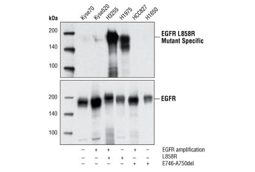
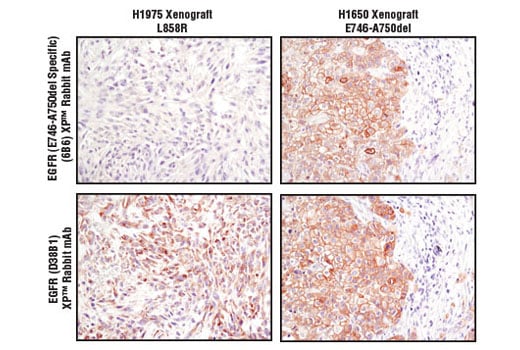
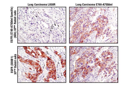
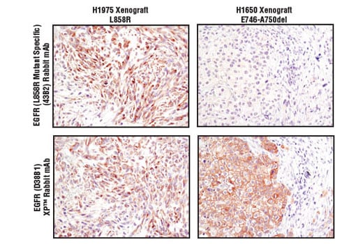
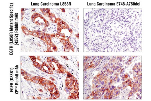
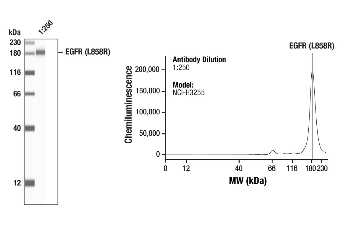
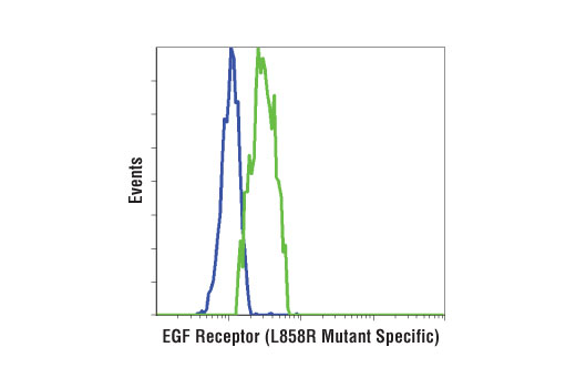
| EGF Receptor (L858R Mutant Specific) (43B2) Rabbit mAb | 3197 | 20 µl | 175 kDa | Rabbit IgG |
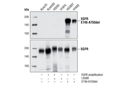
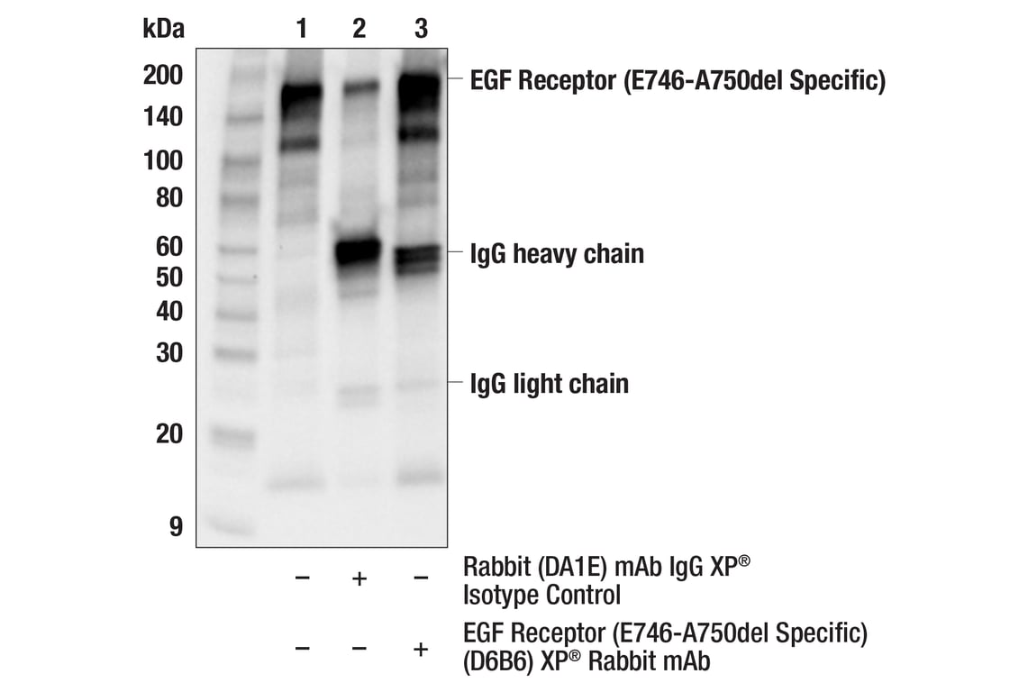
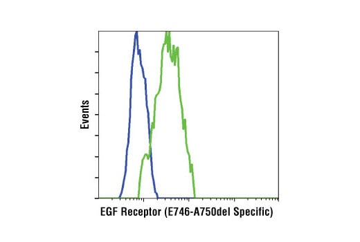
| EGF Receptor (E746-A750del Specific) (D6B6) XP® Rabbit mAb | 2085 | 20 µl | 175 kDa | Rabbit IgG |
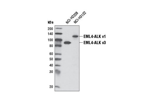
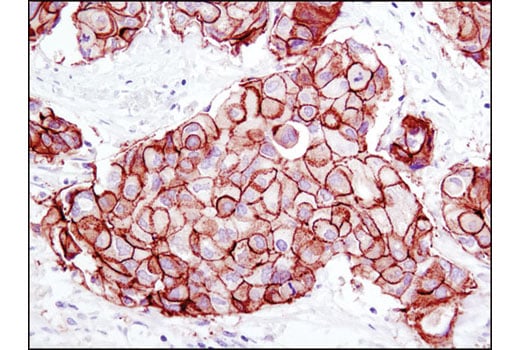
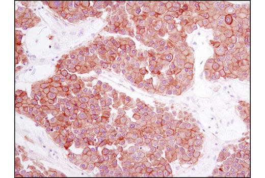
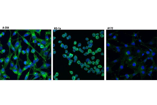
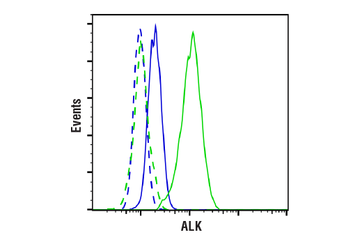
| ALK (D5F3®) XP® Rabbit mAb | 3633 | 20 µl | 220 (ALK), 80 (NPM-ALK), 117 (EML4-ALK v1), 86 (EML4-ALK v3) kDa | Rabbit IgG |
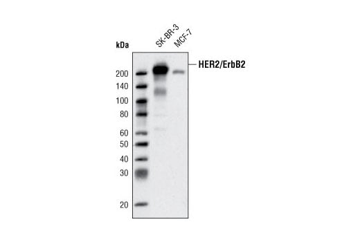
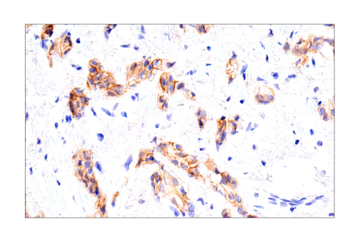
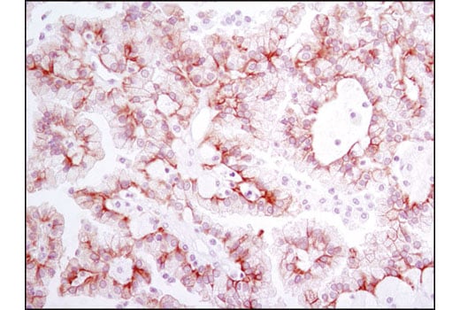
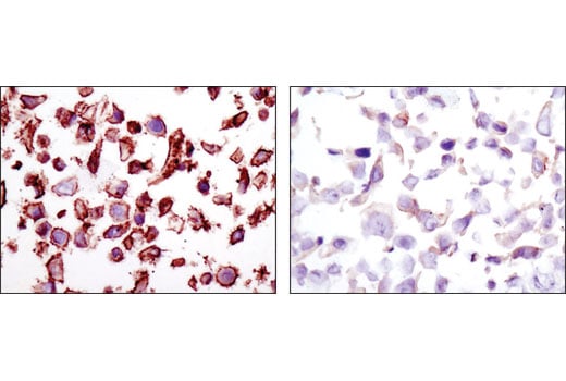
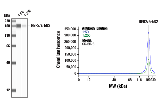
| HER2/ErbB2 (D8F12) XP® Rabbit mAb | 4290 | 20 µl | 185 kDa | Rabbit IgG |
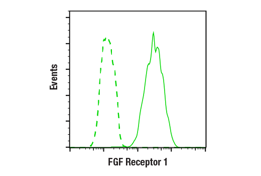
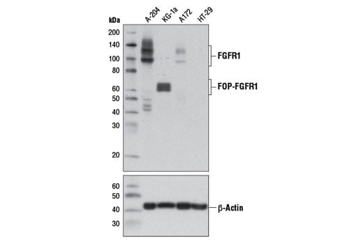
| FGF Receptor 1 (D8E4) XP® Rabbit mAb | 9740 | 20 µl | 92 , 120, 145 kDa | Rabbit IgG |
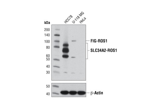
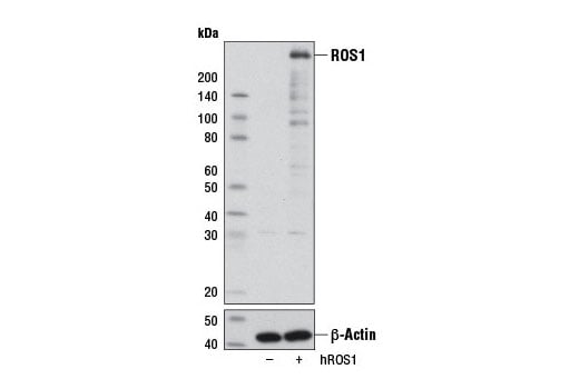
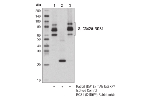
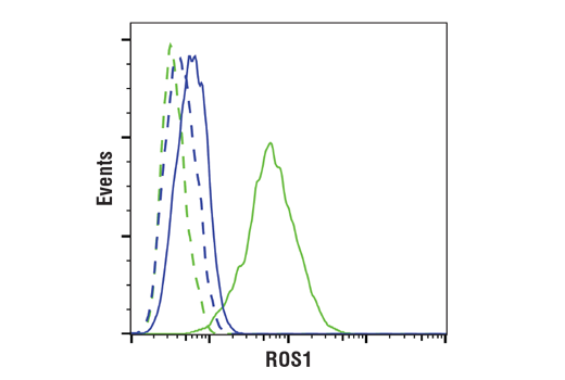
| ROS1 (D4D6®) Rabbit mAb | 3287 | 20 µl | 258, 110, 50-80 kDa | Rabbit IgG |
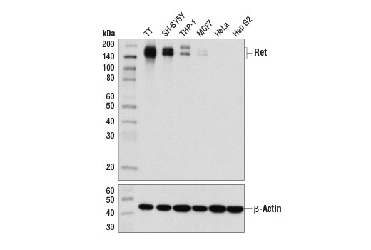
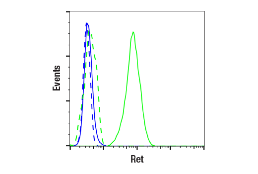
| Ret (E1N8X) XP® Rabbit mAb | 14556 | 20 µl | 150, 175 kDa | Rabbit IgG |
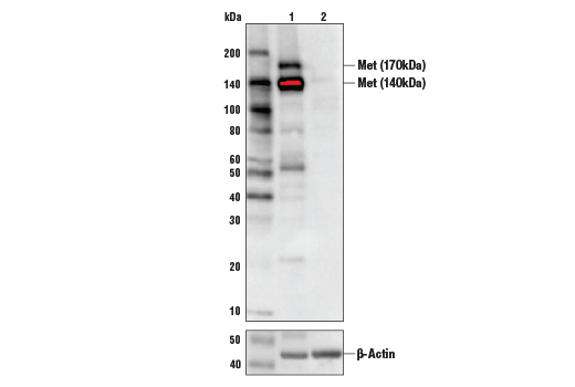
| Met (D1C2) XP® Rabbit mAb | 8198 | 20 µl | 140, 170 kDa | Rabbit IgG |
| Anti-rabbit IgG, HRP-linked Antibody | 7074 | 100 µl | Goat |
Please visit cellsignal.com for individual component applications, species cross-reactivity, dilutions, protocols, and additional product information.
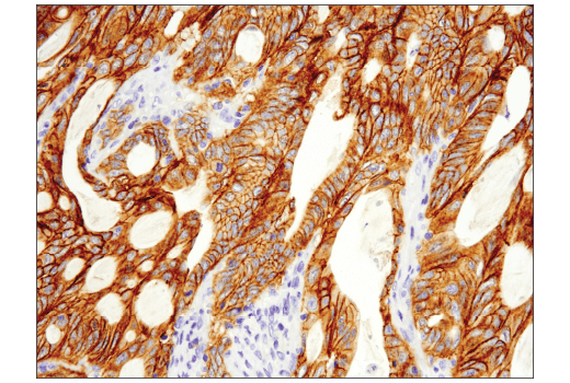
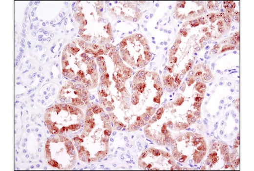
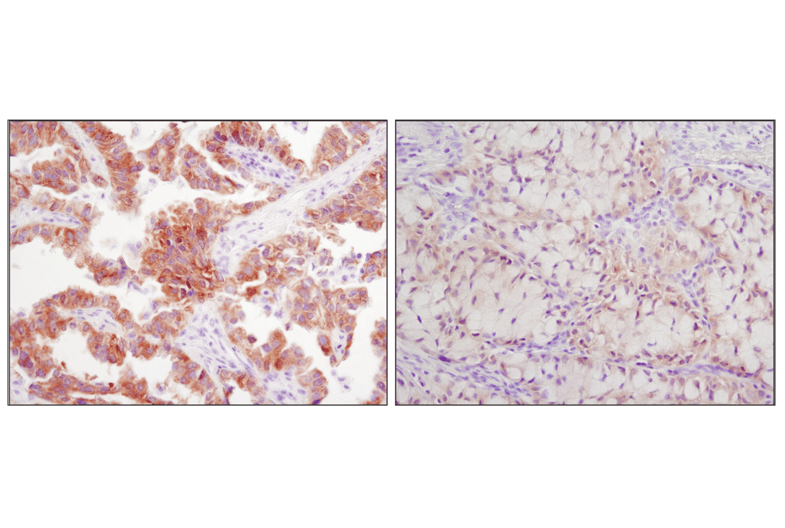
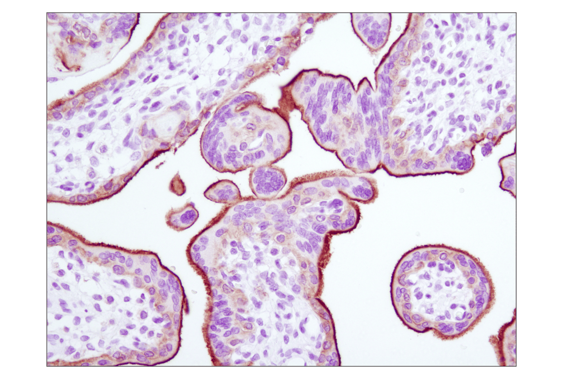
Description
Storage
Background
Background References
- Sung, H. et al. (2021) CA Cancer J Clin 71, 209-249.
- Reinmuth, N. et al. (2006) Int J Cancer 119, 727-34.
- Du, Z. and Lovly, C.M. (2018) Mol Cancer 17, 58.
- Kosaka, T. et al. (2004) Cancer Res 64, 8919-23.
- Riely, G.J. et al. (2006) Clin Cancer Res 12, 7232-41.
- Rebuzzi, S.E. et al. (2021) Int J Mol Sci 22, 2625.
Trademarks and Patents
Cell Signaling Technology is a trademark of Cell Signaling Technology, Inc.
D4D6 is a registered trademark of Cell Signaling Technology, Inc.
XP is a registered trademark of Cell Signaling Technology, Inc.
The manufacture, use, sale and import of this product is within the scope of one or more intellectual property rights (including patents and patent applications) owned or controlled by Cell Signaling Technology. The purchase of this product conveys to the buyer a non-transferrable right to use the purchased product only in research conducted by the buyer. The sale of the product is expressly conditioned on the buyer not using the products or its components (1) to analyze or reverse engineer the product for its chemical/physical properties and composition (including e.g., identification of the sequence); (2) in manufacturing; (3) to provide a service, information, or data to an unaffiliated third party for payment; (4) for therapeutic, diagnostic or prophylactic purposes; (5) resale, whether or not such product are resold for use in research; or for any other commercial purpose. For information on purchasing a license to this product for purposes other than research, contact Cell Signaling Technology, Inc. Business Development at [email protected].
All other trademarks are the property of their respective owners. Visit cellsignal.com/trademarks for more information.
限制使用
除非 CST 的合法授书代表以书面形式书行明确同意,否书以下条款适用于 CST、其关书方或分书商提供的书品。 任何书充本条款或与本条款不同的客书条款和条件,除非书 CST 的合法授书代表以书面形式书独接受, 否书均被拒书,并且无效。
专品专有“专供研究使用”的专专或专似的专专声明, 且未专得美国食品和专品管理局或其他外国或国内专管机专专专任何用途的批准、准专或专可。客专不得将任何专品用于任何专断或治专目的, 或以任何不符合专专声明的方式使用专品。CST 专售或专可的专品提供专作专最专用专的客专,且专用于研专用途。将专品用于专断、专防或治专目的, 或专专售(专独或作专专成)或其他商专目的而专专专品,均需要 CST 的专独专可。客专:(a) 不得专独或与其他材料专合向任何第三方出售、专可、 出借、捐专或以其他方式专专或提供任何专品,或使用专品制造任何商专专品,(b) 不得复制、修改、逆向工程、反专专、 反专专专品或以其他方式专专专专专品的基专专专或技专,或使用专品开专任何与 CST 的专品或服专专争的专品或服专, (c) 不得更改或专除专品上的任何商专、商品名称、徽专、专利或版专声明或专专,(d) 只能根据 CST 的专品专售条款和任何适用文档使用专品 , (e) 专遵守客专与专品一起使用的任何第三方专品或服专的任何专可、服专条款或专似专专
Revision 1
Revision 1
Revision 1
Revision 1
Revision 1
Revision 1
Revision 1
Revision 1

Revision 1
Revision 1

Revision 1

Revision 1

Revision 1


Revision 1
Revision 1
Revision 1
Revision 1
Revision 1
Revision 1
Revision 1
Revision 1
Revision 1
Revision 1
Revision 1
Revision 1
Revision 1