WB, FC-FP, E-P
H M Mk
Endogenous
15
Rabbit
#P05161
9636
Product Information
Product Usage Information
| Application | Dilution |
|---|---|
| Western Blotting | 1:1000 |
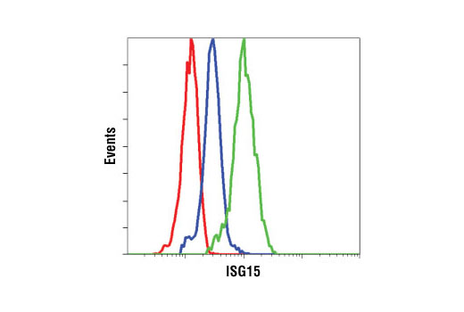
| Flow Cytometry (Fixed/Permeabilized) | 1:200 |
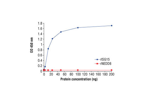
| Peptide ELISA (DELFIA) | 1:100 |
Storage
Specificity / Sensitivity
Species Reactivity:
Human, Mouse, Monkey
Source / Purification
Polyclonal antibodies are produced by immunizing animals with a synthetic peptide corresponding to amino acids from human ISG15 protein. Antibodies are purified by protein A and peptide affinity chromatography.
Background
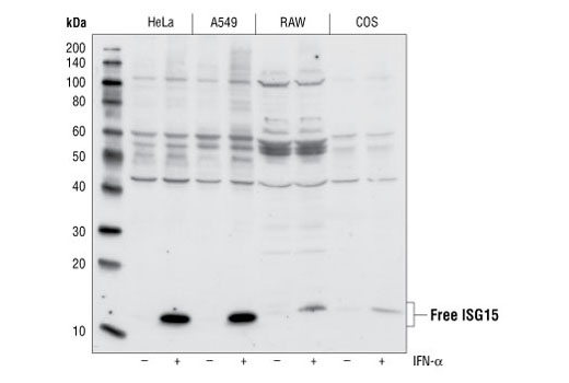
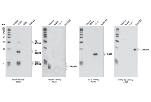
Interferon-stimulated 15 kDa protein (ISG15), also known as ubiquitin cross-reactive protein (UCRP), is a member of the ubiquitin-like protein family and functions in various biological pathways from pregnancy to innate immune responses (1). Expression of ISG15 is stimulated by cellular exposure to type 1 interferons α and β, in addition to infection with viruses such as influenza B (2,3). After exposure to type I interferons, both lymphocytes and monocytes, in addition to some fibroblasts and epithelial cells, release ISG15 into culture medium (1,4). ISG15 has been shown to function as a cytokine, stimulating interferon γ secretion by monocytes and macrophages, proliferation of natural killer cells, and chemotactic responses in neutrophils (4,5). ISG15 has also been shown to function intracellularly, being covalently conjugated to other proteins by E1 (Ube1L), E2 (UbcH8) and E3 ligases via a multi-step process analogous to ubiquitination (6,7). ISG15 is removed from proteins by the ubiquitin processing protease Ubp43 (8). ISG15-protein conjugation (ISGylation) is induced by type 1 interferons, and target proteins include the serine protease inhibitor Serpin 2A, PLCγ1, ERK1/2, Jak1 and Stat1 (9,10). Unlike ubiquitination, ISGylation does not target proteins for degradation, rather ISGylation increases Jak1 and Stat1 activity, enhancing the cellular response to interferons (11).
- Ritchie, K.J. and Zhang, D.E. (2004) Semin. Cell Dev. Biol. 15, 237-246.
- Korant, B.D. et al. (1984) J. Biol. Chem. 259, 14835-14839.
- Haas, A.L. et al. (1987) J. Biol. Chem. 262, 11315-11323.
- Knight, E. and Cordova, B. (1991) J. Immunol. 146, 2280-2284.
- D'Cunha, J. et al. (1996) Proc. Natl. Acad. Sci. USA 93, 211-215.
- Loeb, K.R. and Haas, A.L. (1992) J. Biol. Chem. 267, 7806-7813.
- Zhao, C. et al. (2005) Proc. Natl. Acad. Sci. USA 102, 10200-10205.
- Malakhov, M.P. et al. (2002) J. Biol. Chem. 277, 9976-9981.
- Malakhov, M.P. et al. (2003) J. Biol. Chem. 278, 16608-16613.
- Hamerman, J.A. et al. (2002) J. Immunol. 168, 2415-2423.
- Malakhova, O.A. et al. (2003) Genes Dev. 17, 455-460.
Species Reactivity
Species reactivity is determined by testing in at least one approved application (e.g., western blot).
Western Blot Buffer
IMPORTANT: For western blots, incubate membrane with diluted primary antibody in 5% w/v BSA, 1X TBS, 0.1% Tween® 20 at 4°C with gentle shaking, overnight.
Applications Key
WB: Western Blotting FC-FP: Flow Cytometry (Fixed/Permeabilized) E-P: Peptide ELISA (DELFIA)
Cross-Reactivity Key
H: human M: mouse R: rat Hm: hamster Mk: monkey Vir: virus Mi: mink C: chicken Dm: D. melanogaster X: Xenopus Z: zebrafish B: bovine Dg: dog Pg: pig Sc: S. cerevisiae Ce: C. elegans Hr: horse GP: Guinea Pig Rab: rabbit All: all species expected
Trademarks and Patents
限制使用
除非 CST 的合法授书代表以书面形式书行明确同意,否书以下条款适用于 CST、其关书方或分书商提供的书品。 任何书充本条款或与本条款不同的客书条款和条件,除非书 CST 的合法授书代表以书面形式书独接受, 否书均被拒书,并且无效。
专品专有“专供研究使用”的专专或专似的专专声明, 且未专得美国食品和专品管理局或其他外国或国内专管机专专专任何用途的批准、准专或专可。客专不得将任何专品用于任何专断或治专目的, 或以任何不符合专专声明的方式使用专品。CST 专售或专可的专品提供专作专最专用专的客专,且专用于研专用途。将专品用于专断、专防或治专目的, 或专专售(专独或作专专成)或其他商专目的而专专专品,均需要 CST 的专独专可。客专:(a) 不得专独或与其他材料专合向任何第三方出售、专可、 出借、捐专或以其他方式专专或提供任何专品,或使用专品制造任何商专专品,(b) 不得复制、修改、逆向工程、反专专、 反专专专品或以其他方式专专专专专品的基专专专或技专,或使用专品开专任何与 CST 的专品或服专专争的专品或服专, (c) 不得更改或专除专品上的任何商专、商品名称、徽专、专利或版专声明或专专,(d) 只能根据 CST 的专品专售条款和任何适用文档使用专品, (e) 专遵守客专与专品一起使用的任何第三方专品或服专的任何专可、服专条款或专似专专