Revision 6
#11904
Store at -20C
877-616-CELL (2355)
877-678-TECH (8324)
3 Trask Lane | Danvers | Massachusetts | 01923 | USA
For Research Use Only. Not for Use in Diagnostic Procedures.
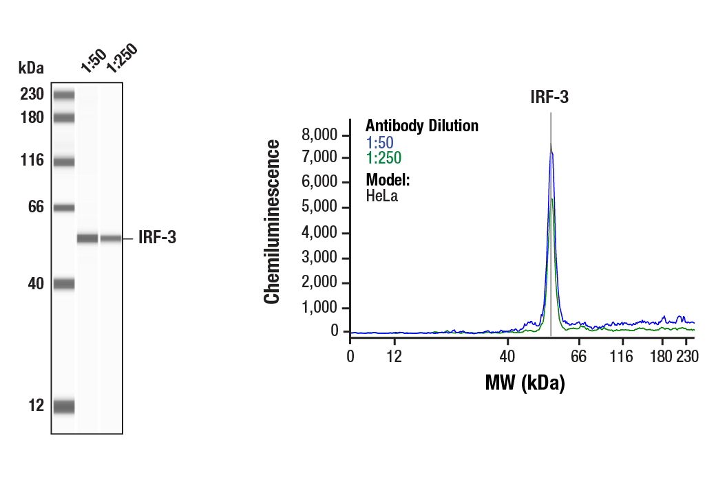
Applications:
W, W-S, IP, IF-IC
Reactivity:
H Mk
Sensitivity:
Endogenous
MW (kDa):
50-55
Source/Isotype:
Rabbit IgG
UniProt ID:
#Q14653
Entrez-Gene Id:
3661
Product Usage Information
| Application | Dilution |
|---|---|
| Western Blotting | 1:1000 |
| Simple Western™ | 1:50 - 1:250 |
| Immunoprecipitation | 1:50 |
| Immunofluorescence (Immunocytochemistry) | 1:200 - 1:800 |
Storage
For a carrier free (BSA and azide free) version of this product see product #62664.
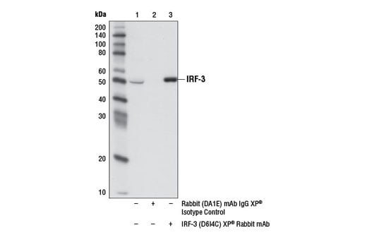
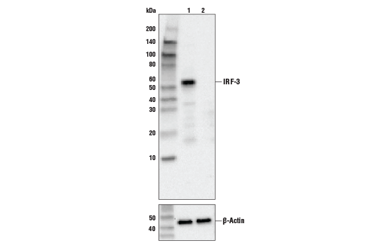
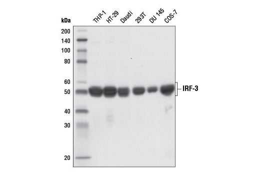
Specificity/Sensitivity
Source / Purification
Background
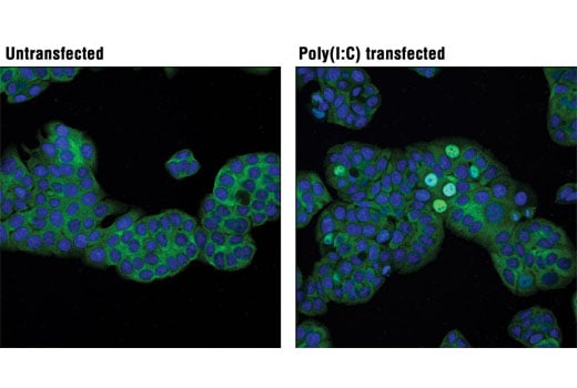
IRF-3 can inhibit cell growth and plays a critical role in controlling the expression of genes in the innate immune response (1-4). In unstimulated cells, IRF-3 is present in the cytoplasm. Viral infection results in phosphorylation of IRF-3 and leads to its translocation to the nucleus where it activates promoters containing IRF-3-binding sites. Phosphorylation of IRF-3 occurs at a cluster of C-terminal Ser and Thr residues (between 385 and 405), leading to its association with the p300/CBP coactivator protein that promotes DNA binding and transcriptional activity (5). During infection, IRF-3 is likely activated through a pathway that includes activation of Toll-like receptors and a kinase complex that includes IKKε and TBK1 (6,7). IRF-3 is phosphorylated at Ser396 following viral infection, expression of viral nucleocapsid, and double-stranded RNA treatment. These events likely play a role in activation of IRF-3 (8).
Background References
- Taniguchi, T. et al. (2001) Annu Rev Immunol 19, 623-55.
- Honda, K. and Taniguchi, T. (2006) Nat Rev Immunol 6, 644-58.
- Hiscott, J. et al. (1999) J Interferon Cytokine Res 19, 1-13.
- Kim, T.Y. et al. (2003) J Biol Chem 278, 15272-8.
- Yoneyama, M. et al. (2002) J Interferon Cytokine Res 22, 73-6.
- Fitzgerald, K.A. et al. (2003) Nat Immunol 4, 491-6.
- Kopp, E. and Medzhitov, R. (2003) Curr Opin Immunol 15, 396-401.
- Servant, M.J. et al. (2003) J Biol Chem 278, 9441-7.
Species Reactivity
Species reactivity is determined by testing in at least one approved application (e.g., western blot).
Western Blot Buffer
IMPORTANT: For western blots, incubate membrane with diluted primary antibody in 5% w/v BSA, 1X TBS, 0.1% Tween® 20 at 4°C with gentle shaking, overnight.
Applications Key
W: Western Blotting W-S: Simple Western™ IP: Immunoprecipitation IF-IC: Immunofluorescence (Immunocytochemistry)
Cross-Reactivity Key
H: Human Mk: Monkey
Trademarks and Patents
Cell Signaling Technology is a trademark of Cell Signaling Technology, Inc.
All other trademarks are the property of their respective owners. Visit cellsignal.com/trademarks for more information.
Limited Uses
Except as otherwise expressly agreed in a writing signed by a legally authorized representative of CST, the following terms apply to Products provided by CST, its affiliates or its distributors. Any Customer's terms and conditions that are in addition to, or different from, those contained herein, unless separately accepted in writing by a legally authorized representative of CST, are rejected and are of no force or effect.
Products are labeled with For Research Use Only or a similar labeling statement and have not been approved, cleared, or licensed by the FDA or other regulatory foreign or domestic entity, for any purpose. Customer shall not use any Product for any diagnostic or therapeutic purpose, or otherwise in any manner that conflicts with its labeling statement. Products sold or licensed by CST are provided for Customer as the end-user and solely for research and development uses. Any use of Product for diagnostic, prophylactic or therapeutic purposes, or any purchase of Product for resale (alone or as a component) or other commercial purpose, requires a separate license from CST. Customer shall (a) not sell, license, loan, donate or otherwise transfer or make available any Product to any third party, whether alone or in combination with other materials, or use the Products to manufacture any commercial products, (b) not copy, modify, reverse engineer, decompile, disassemble or otherwise attempt to discover the underlying structure or technology of the Products, or use the Products for the purpose of developing any products or services that would compete with CST products or services, (c) not alter or remove from the Products any trademarks, trade names, logos, patent or copyright notices or markings, (d) use the Products solely in accordance with CST Product Terms of Sale and any applicable documentation, and (e) comply with any license, terms of service or similar agreement with respect to any third party products or services used by Customer in connection with the Products.
Revision 6
Revision 6