| Product Includes | Product # | Quantity | Mol. Wt | Isotype/Source |
|---|---|---|---|---|
| Doublecortin (E6O6A) Rabbit mAb | 91954 | 20 µl | 45 kDa | Rabbit IgG |
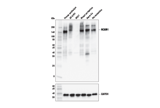
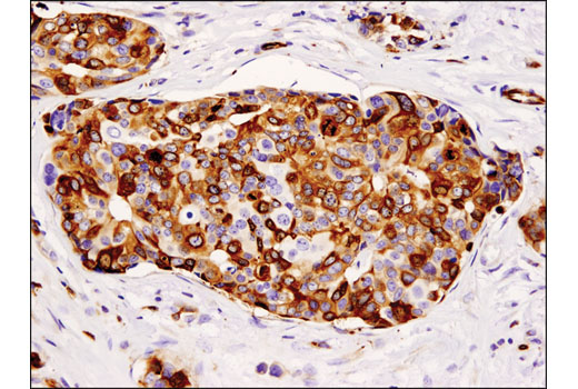
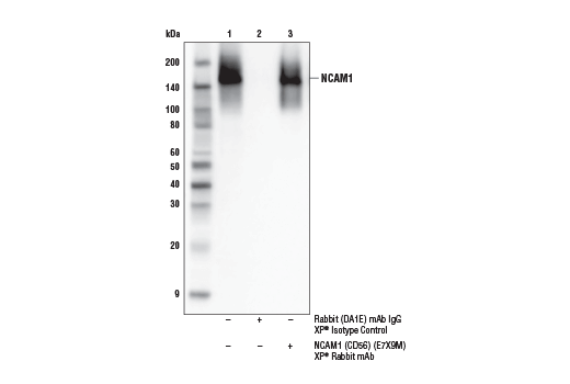
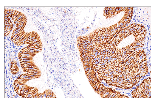
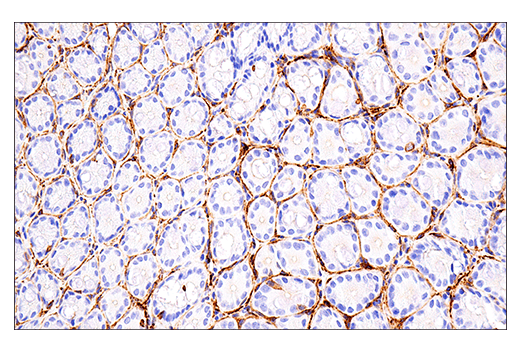
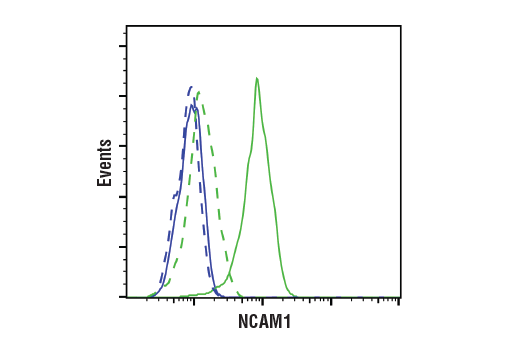
| NCAM1 (CD56) (E7X9M) XP® Rabbit mAb | 99746 | 20 µl | 120 to 220 kDa | Rabbit IgG |
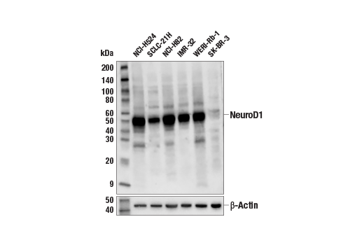
| NeuroD1 (D90G12) Rabbit mAb | 7019 | 20 µl | 49 kDa | Rabbit IgG |
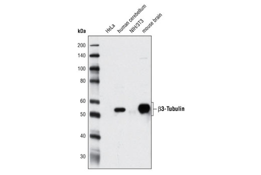
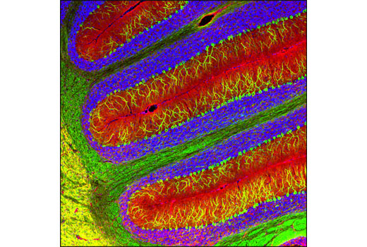
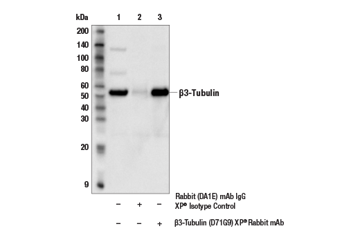
| β3-Tubulin (D71G9) XP® Rabbit mAb | 5568 | 20 µl | 55 kDa | Rabbit IgG |
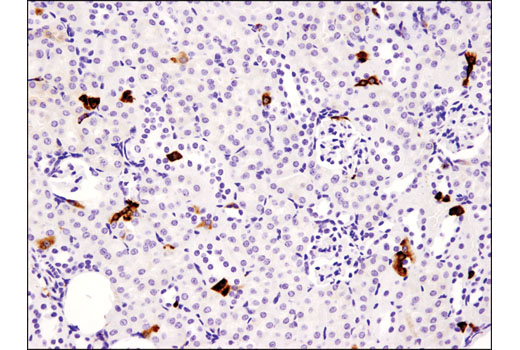
| TBR1 (D6C6X) Rabbit mAb | 49661 | 20 µl | 74 kDa | Rabbit IgG |
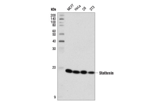
| Stathmin (D1Y5A) Rabbit mAb | 13655 | 20 µl | 19 kDa | Rabbit IgG |
| Anti-rabbit IgG, HRP-linked Antibody | 7074 | 100 µl | Goat |
Please visit cellsignal.com for individual component applications, species cross-reactivity, dilutions, protocols, and additional product information.
Description
The Immature Neuron Marker Antibody Sampler Kit provides an economical means for detecting immature neuron proteins by western blot. The kit includes enough antibodies to perform two western blot experiments with each primary antibody.
Storage
Background
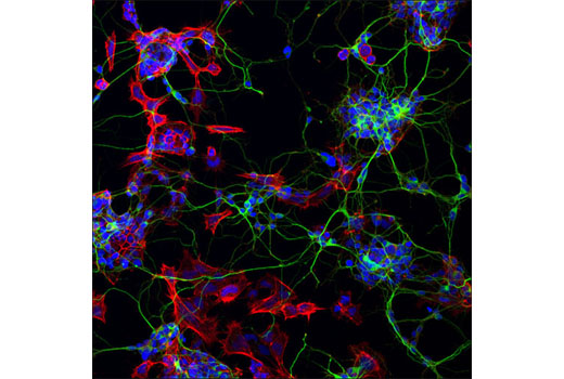
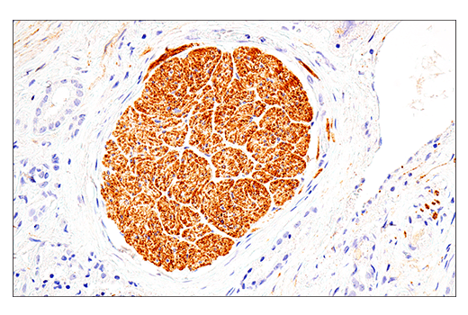
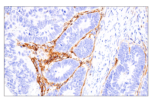
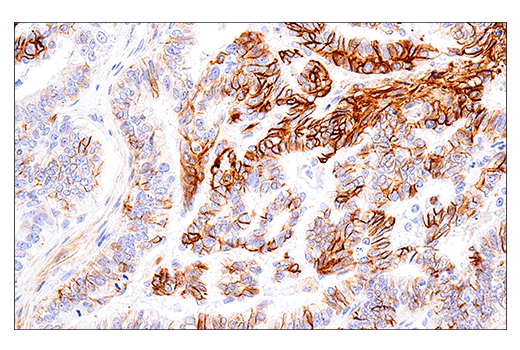
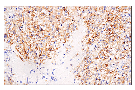
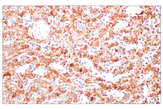
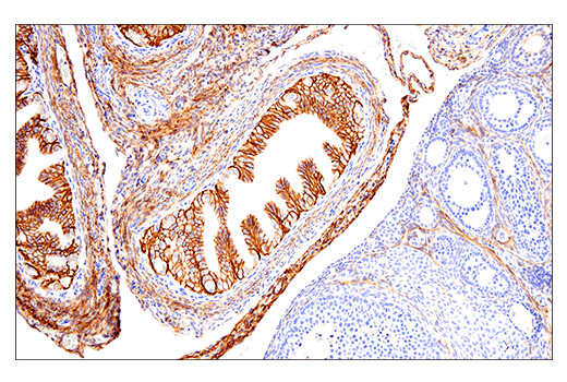
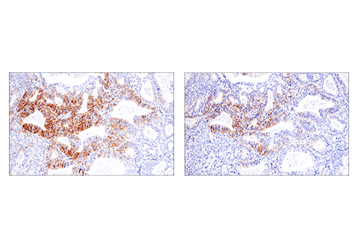
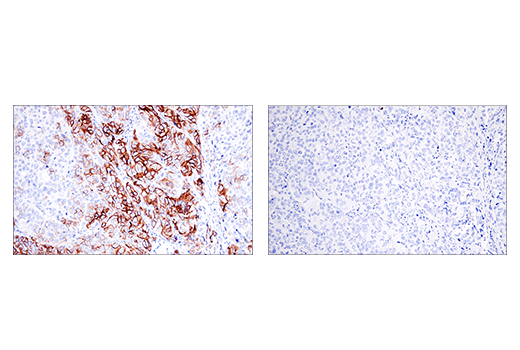
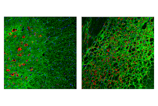
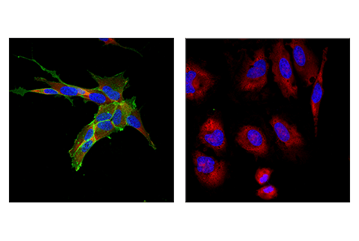
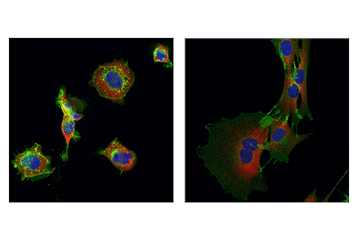
The antibodies in this kit serve to characterize and identify immature neurons. During development, radial glia (RG) cells located in the ventricular zone (VZ) of the brain divide asymmetrically, each producing a neuronal and RG daughter cell. The daughter RG cell is also known as a neural progenitor cell (NPC) or an intermediate progenitor cell (IPC). Newly formed IPCs migrate to the subventricular zone (SVZ) where they divide symmetrically, each giving rise to two post-mitotic neurons that can then migrate to their final destination. In adulthood, NPCs reside within the subgranular zone (SGZ) of the dentate gyrus, and the adult SVZ, which surrounds the lateral ventricles of the cerebral cortex. NPCs within the SGZ and SVZ divide and give rise to immature neurons (1). The cytoskeleton of these cells plays an important role in generating neuronal processes. The cytoskeleton consists of three types of cytosolic fibers: actin microfilaments, intermediate filaments, and microtubules. β3-tubulin is one of six β-tubulin isoforms that make up the building blocks of microtubules (2). Stathmin is a tubulin binding protein that regulates microtubule dynamics in a phosphorylation dependent manner. Stathmin is heavily expressed during neuronal development, mediating differentiation and synaptic plasticity (3,4). Doublecortin is a microtubule-associated protein that facilitates neurite outgrowth and cell migration (5). The dual expression of doublecortin and NCAM (neural cell adhesion molecule, CD56), combined with the lack of expression of mature neuronal markers, is evidence of an immature neuronal phenotype (6). NCAM mediates neuronal attachment, neurite extension, and cell to cell interactions through homo and heterophilic interactions. Polysialic acid (PSA) post-translational modification of NCAM disrupts cell to cell adhesion, promoting axonal growth, cell migration, and synaptic plasticity during neurogenesis (7-9).
Transcription factors also play a key role in immature neuron growth and differentiation. NeuroD1 is a member of the basic helix-loop-helix (bHLH) family of transcription factors. These proteins function by forming heterodimers with E-proteins and binding to the canonical E-box sequence CANNTG (10,11). Neuronal activity results in CaMKII-mediated phosphorylation of NeuroD1 at Ser336, which is necessary for the formation and growth of dendrites (12,13). T-box, brain, 1 (TBR1) is a transcription factor important in vertebrate embryo development. As a member of the T-Box family of transcription factors, TBR1 is expressed in postmitotic glutamatergic projection neurons (14). During cortical neurogenesis, sequential expression of transcription factors Pax6, TBR2, and TBR1 regulates discrete steps in projection neuron differentiation (15).
- Martínez-Cerdeño, V. and Noctor, S.C. (2018) Front Neuroanat 12, 104.
- Jiang, Y.Q. and Oblinger, M.M. (1992) J Cell Sci 103 (Pt 3), 643-51.
- Chauvin, S. and Sobel, A. (2015) Prog Neurobiol 126, 1-18.
- Uchida, S. et al. (2014) Nat Commun 5, 4389.
- Reiner, O. et al. (2004) Cell Cycle 3, 747-51.
- Coviello, S. et al. (2022) Front Neuroanat 16, 851432.
- Seidenfaden, R. et al. (2003) Mol Cell Biol 23, 5908-18.
- Bonfanti, L. and Seki, T. (2021) Cells 10, 2542.
- Wędzony, K. et al. (2013) Pharmacol Rep 65, 1471-8.
- Schonhoff, S.E. et al. (2004) Endocrinology 145, 2639-44.
- Sharma, A. et al. (1999) Mol Cell Biol 19, 704-13.
- Chae, J.H. et al. (2004) Mol Cells 18, 271-88.
- Gaudillière, B. et al. (2004) Neuron 41, 229-41.
- Hevner, R.F. et al. (2001) Neuron 29, 353-66.
- Englund, C. et al. (2005) J Neurosci 25, 247-51.
Background References
Trademarks and Patents
限制使用
除非 CST 的合法授书代表以书面形式书行明确同意,否书以下条款适用于 CST、其关书方或分书商提供的书品。 任何书充本条款或与本条款不同的客书条款和条件,除非书 CST 的合法授书代表以书面形式书独接受, 否书均被拒书,并且无效。
专品专有“专供研究使用”的专专或专似的专专声明, 且未专得美国食品和专品管理局或其他外国或国内专管机专专专任何用途的批准、准专或专可。客专不得将任何专品用于任何专断或治专目的, 或以任何不符合专专声明的方式使用专品。CST 专售或专可的专品提供专作专最专用专的客专,且专用于研专用途。将专品用于专断、专防或治专目的, 或专专售(专独或作专专成)或其他商专目的而专专专品,均需要 CST 的专独专可。客专:(a) 不得专独或与其他材料专合向任何第三方出售、专可、 出借、捐专或以其他方式专专或提供任何专品,或使用专品制造任何商专专品,(b) 不得复制、修改、逆向工程、反专专、 反专专专品或以其他方式专专专专专品的基专专专或技专,或使用专品开专任何与 CST 的专品或服专专争的专品或服专, (c) 不得更改或专除专品上的任何商专、商品名称、徽专、专利或版专声明或专专,(d) 只能根据 CST 的专品专售条款和任何适用文档使用专品, (e) 专遵守客专与专品一起使用的任何第三方专品或服专的任何专可、服专条款或专似专专