| Product Includes | Product # | Quantity | Mol. Wt | Isotype/Source |
|---|---|---|---|---|
| HIF-1α (E1V6A) Rabbit mAb | 48085 | 20 µl | 120 kDa | Rabbit IgG |
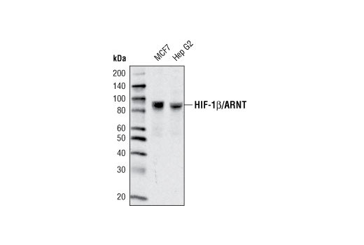
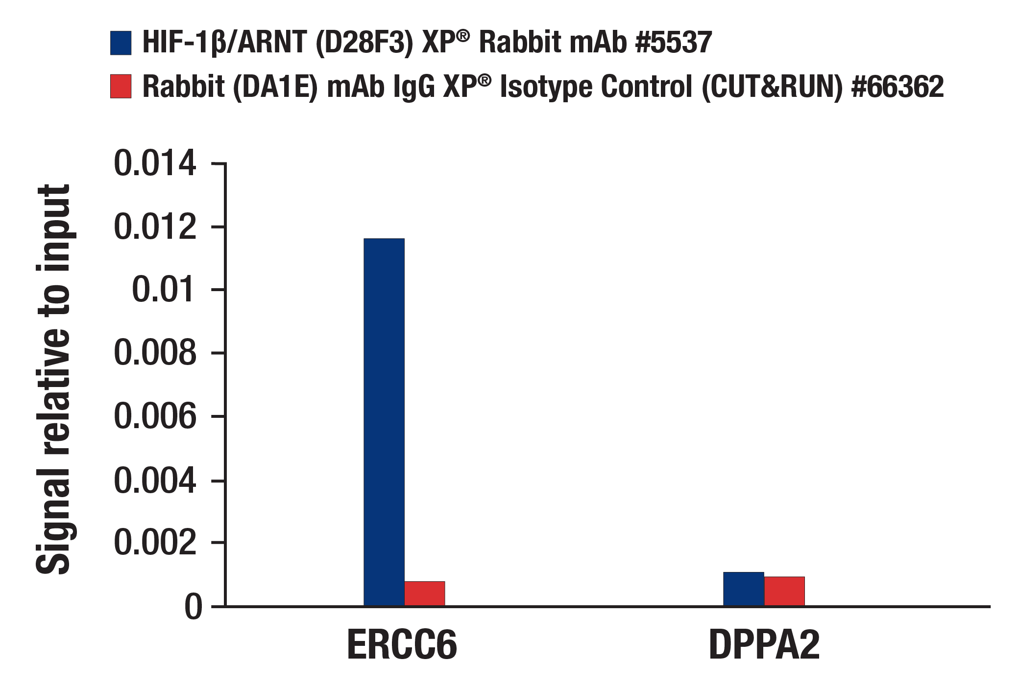
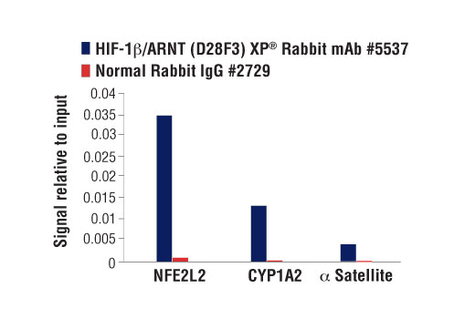
| HIF-1β/ARNT (D28F3) XP® Rabbit mAb | 5537 | 20 µl | 87 kDa | Rabbit IgG |
| VHL (E3X9K) Rabbit mAb | 81292 | 20 µl | Rabbit IgG | |
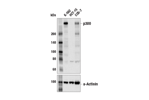
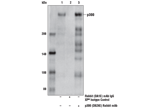
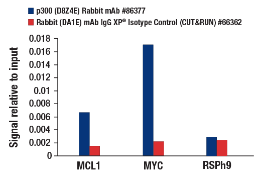
| p300 (D8Z4E) Rabbit mAb | 86377 | 20 µl | 300 kDa | Rabbit IgG |
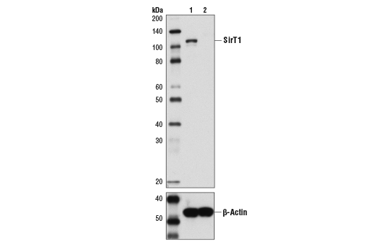
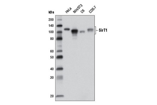
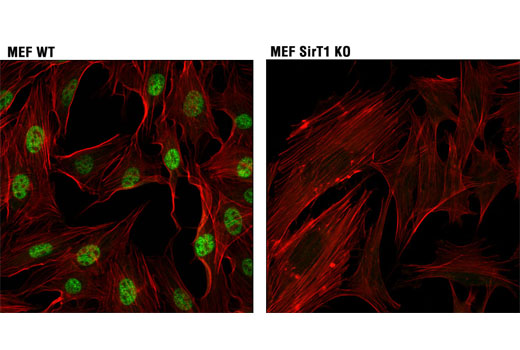
| SirT1 (1F3) Mouse mAb | 8469 | 20 µl | 120 kDa | Mouse IgG1 |
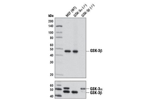
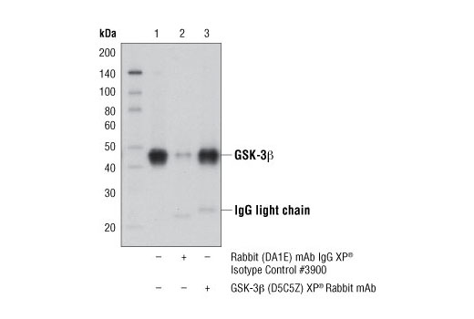
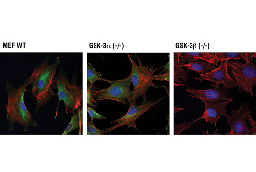
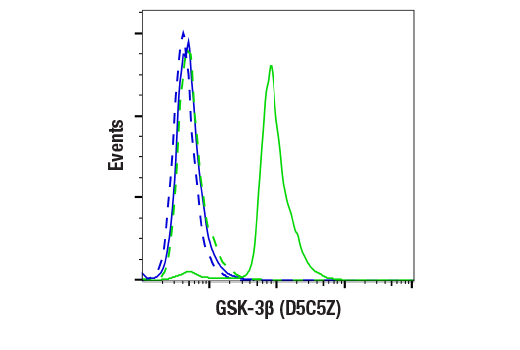
| GSK-3β (D5C5Z) XP® Rabbit mAb | 12456 | 20 µl | 46 kDa | Rabbit IgG |
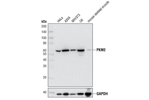
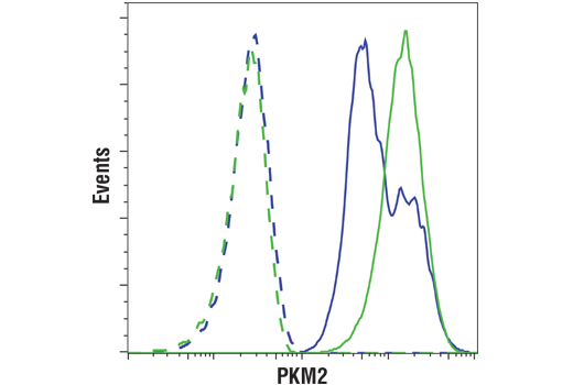
| PKM2 (D78A4) XP® Rabbit mAb | 4053 | 20 µl | 60 kDa | Rabbit IgG |
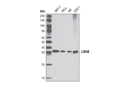
| LDHA (C4B5) Rabbit mAb | 3582 | 20 µl | 37 kDa | Rabbit IgG |
| Glut1 (E4S6I) Rabbit mAb | 73015 | 20 µl | 45-60 kDa | Rabbit IgG |
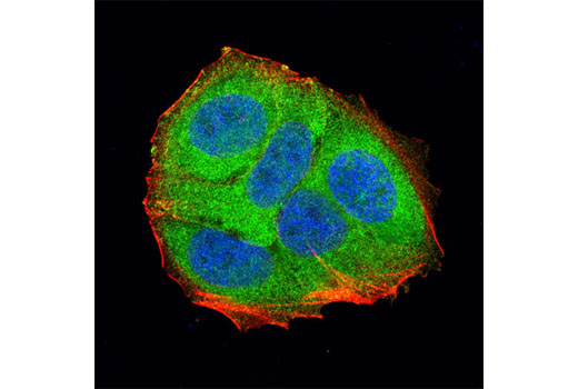
Please visit cellsignal.com for individual component applications, species cross-reactivity, dilutions, protocols, and additional product information.
Description
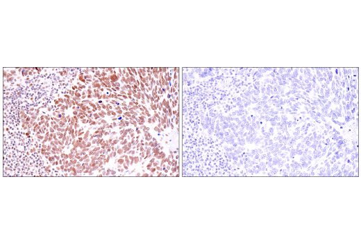
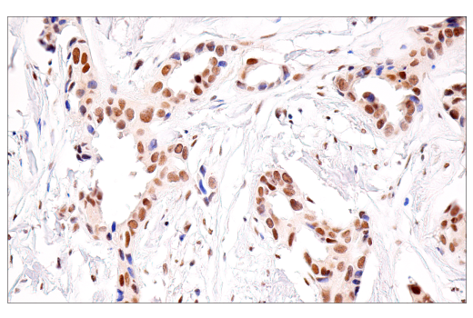
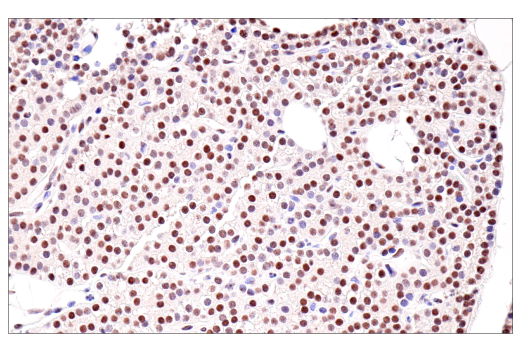
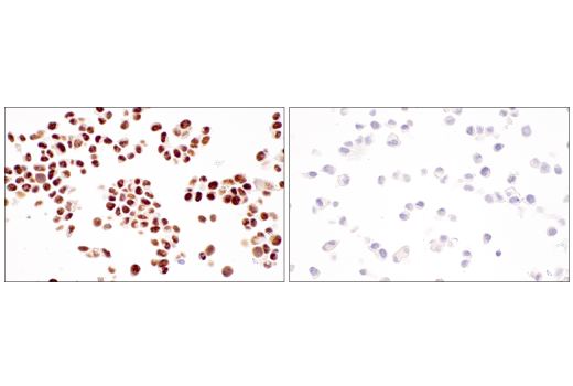
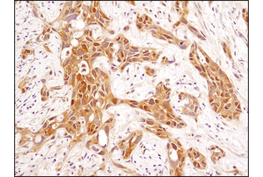
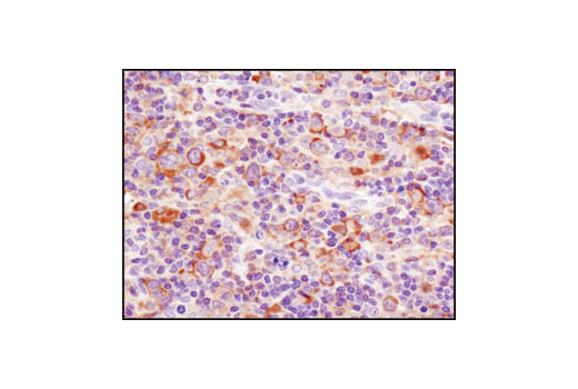
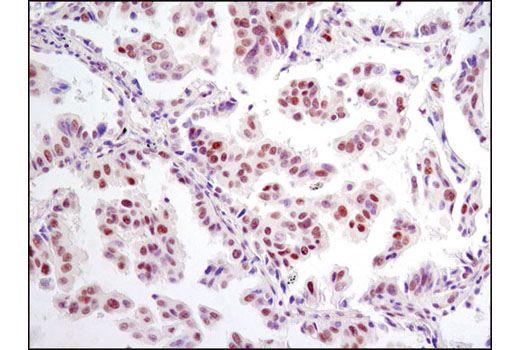
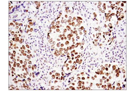
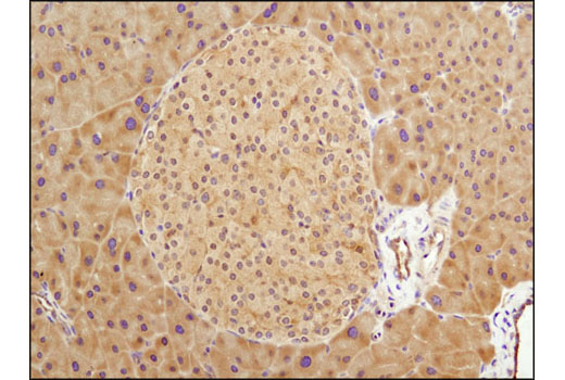
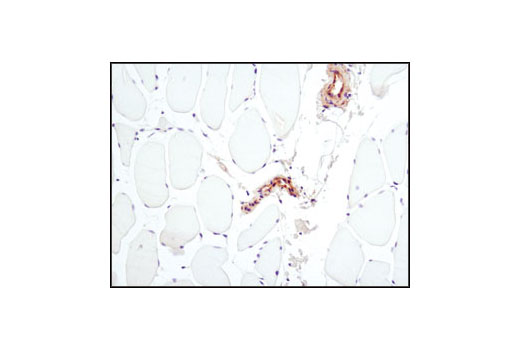
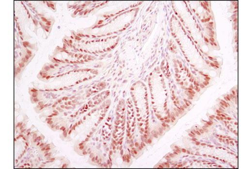
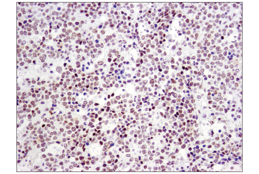
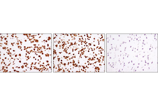
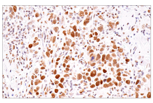
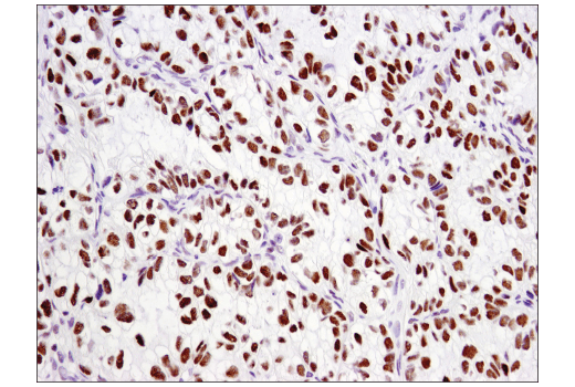
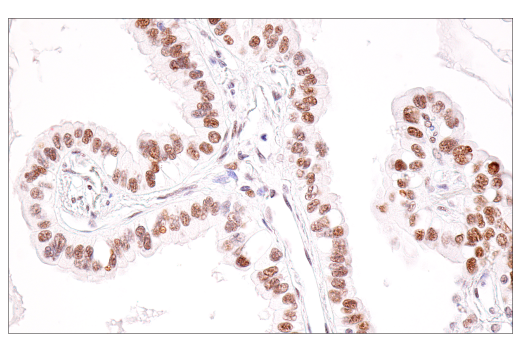
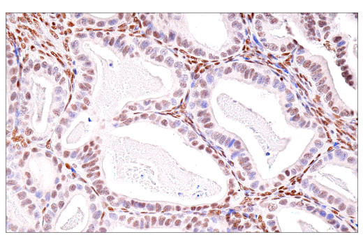
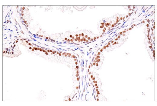
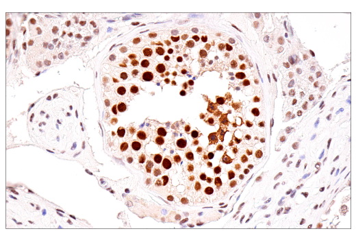
The Hypoxia Activation IHC Antibody Sampler Kit provides an economical means of detecting select components involved in the regulation of HIF-1α, select components regulated by HIF-1α, and HIF-1β/ARNT protein in formalin-fixed, paraffin-embedded tissue samples.
Storage
Background
Hypoxia-inducible factor 1 (HIF1) is a heterodimeric transcription factor that plays a critical role in the cellular response to hypoxia (1). The HIF1 complex consists of two subunits, HIF-1α and HIF-1β, which are basic helix-loop-helix proteins of the PAS (Per, ARNT, Sim) family (2). HIF1 regulates the transcription of a broad range of genes that facilitate responses to the hypoxic environment, including genes regulating angiogenesis, erythropoiesis, cell cycle, metabolism, and apoptosis. The widely expressed HIF-1α is typically degraded rapidly in normoxic cells by the ubiquitin/proteasomal pathway. Under normoxic conditions, HIF-1α is proline hydroxylated leading to a conformational change that promotes binding to the von Hippel-Lindau protein (VHL) E3 ligase complex; ubiquitination and proteasomal degradation follows (3,4). Both hypoxic conditions and chemical hydroxylase inhibitors (such as desferrioxamine and cobalt) inhibit HIF-1α degradation and lead to its stabilization. In addition, HIF-1α can be induced in an oxygen-independent manner by various cytokines through the PI3K-AKT-mTOR pathway (5-7). HIF-1β is also known as AhR nuclear translocator (ARNT) due to its ability to partner with the aryl hydrocarbon receptor (AhR) to form a heterodimeric transcription factor complex (8). Together with AhR, HIF-1β plays an important role in xenobiotics metabolism (8). In addition, a chromosomal translocation leading to a TEL-ARNT fusion protein is associated with acute myeloblastic leukemia (9). Studies also found that ARNT/HIF-1β expression levels decrease significantly in pancreatic islets from patients with type 2 diabetes, suggesting that HIF-1β plays an important role in pancreatic β-cell function (10). CBP (CREB-binding protein) and p300 are highly conserved and functionally related transcriptional co-activators that associate with transcriptional regulators and signaling molecules, integrating multiple signal transduction pathways with the transcriptional machinery (11,12). CBP/p300 also contain histone acetyltransferase (HAT) activity, allowing them to acetylate histones and other proteins (12). The Silent Information Regulator (SIR2) family of genes is a highly conserved group of genes that encode nicotinamide adenine dinucleotide (NAD)-dependent protein deacetylases, also known as class III histone deacetylases. The first discovered and best characterized of these genes is Saccharomyces cerevisiae SIR2, which is involved in silencing of mating type loci, telomere maintenance, DNA damage response, and cell aging (13). SirT1, the mammalian ortholog of Sir2, is a nuclear protein implicated in the regulation of many cellular processes, including apoptosis, cellular senescence, endocrine signaling, glucose homeostasis, aging, and longevity. Targets of SirT1 include acetylated p53 (14,15), p300 (16), Ku70 (17), forkhead (FoxO) transcription factors (17,18), PPARγ (19), and the PPARγ coactivator-1α (PGC-1α) protein (20). Glycogen synthase kinase-3 (GSK-3) was initially identified as an enzyme that regulates glycogen synthesis in response to insulin (21). GSK-3 is a ubiquitously expressed serine/threonine protein kinase that phosphorylates and inactivates glycogen synthase. GSK-3 is a critical downstream element of the PI3K/Akt cell survival pathway whose activity can be inhibited by Akt-mediated phosphorylation at Ser21 of GSK-3α and Ser9 of GSK-3β (22,23). Pyruvate kinase is a glycolytic enzyme that catalyzes the conversion of phosphoenolpyruvate to pyruvate. In mammals, the M2 isoform (PKM2) is expressed during embryonic development (24). Lactate dehydrogenase (LDH) catalyzes the interconversion of pyruvate and NADH to lactate and NAD+. The major form of LDH found in muscle cells is the A (LDHA) isozyme (25). Glucose transporter 1 (Glut1, SLC2A1) is a widely expressed transport protein that transports a number of different aldose sugars into cells (26,27).
- Sharp, F.R. and Bernaudin, M. (2004) Nat Rev Neurosci 5, 437-48.
- Wang, G.L. et al. (1995) Proc Natl Acad Sci U S A 92, 5510-4.
- Jaakkola, P. et al. (2001) Science 292, 468-72.
- Maxwell, P.H. et al. (1999) Nature 399, 271-5.
- Fukuda, R. et al. (2002) J Biol Chem 277, 38205-11.
- Jiang, B.H. et al. (2001) Cell Growth Differ 12, 363-9.
- Laughner, E. et al. (2001) Mol Cell Biol 21, 3995-4004.
- Walisser, J.A. et al. (2004) Proc Natl Acad Sci U S A 101, 16677-82.
- Salomon-Nguyen, F. et al. (2000) Proc Natl Acad Sci U S A 97, 6757-62.
- Gunton, J.E. et al. (2005) Cell 122, 337-49.
- Goodman, R.H. and Smolik, S. (2000) Genes Dev 14, 1553-77.
- Chan, H.M. and La Thangue, N.B. (2001) J Cell Sci 114, 2363-73.
- Guarente, L. (1999) Nat Genet 23, 281-5.
- Vaziri, H. et al. (2001) Cell 107, 149-59.
- Luo, J. et al. (2001) Cell 107, 137-48.
- Bouras, T. et al. (2005) J Biol Chem 280, 10264-76.
- Brunet, A. et al. (2004) Science 303, 2011-5.
- Motta, M.C. et al. (2004) Cell 116, 551-63.
- Picard, F. et al. (2004) Nature 429, 771-6.
- Rodgers, J.T. et al. (2005) Nature 434, 113-8.
- Welsh, G.I. et al. (1996) Trends Cell Biol 6, 274-9.
- Srivastava, A.K. and Pandey, S.K. (1998) Mol Cell Biochem 182, 135-41.
- Cross, D.A. et al. (1995) Nature 378, 785-9.
- Christofk, H.R. et al. (2008) Nature 452, 230-3.
- Semenza, G.L. et al. (1996) J Biol Chem 271, 32529-37.
- Ferrer, C.M. et al. (2014) Mol Cell 54, 820-31.
- Deng, D. et al. (2014) Nature 510, 121-5.
Background References
Trademarks and Patents
限制使用
除非 CST 的合法授书代表以书面形式书行明确同意,否书以下条款适用于 CST、其关书方或分书商提供的书品。 任何书充本条款或与本条款不同的客书条款和条件,除非书 CST 的合法授书代表以书面形式书独接受, 否书均被拒书,并且无效。
专品专有“专供研究使用”的专专或专似的专专声明, 且未专得美国食品和专品管理局或其他外国或国内专管机专专专任何用途的批准、准专或专可。客专不得将任何专品用于任何专断或治专目的, 或以任何不符合专专声明的方式使用专品。CST 专售或专可的专品提供专作专最专用专的客专,且专用于研专用途。将专品用于专断、专防或治专目的, 或专专售(专独或作专专成)或其他商专目的而专专专品,均需要 CST 的专独专可。客专:(a) 不得专独或与其他材料专合向任何第三方出售、专可、 出借、捐专或以其他方式专专或提供任何专品,或使用专品制造任何商专专品,(b) 不得复制、修改、逆向工程、反专专、 反专专专品或以其他方式专专专专专品的基专专专或技专,或使用专品开专任何与 CST 的专品或服专专争的专品或服专, (c) 不得更改或专除专品上的任何商专、商品名称、徽专、专利或版专声明或专专,(d) 只能根据 CST 的专品专售条款和任何适用文档使用专品, (e) 专遵守客专与专品一起使用的任何第三方专品或服专的任何专可、服专条款或专似专专