| Product Includes | Product # | Quantity | Mol. Wt | Isotype/Source |
|---|---|---|---|---|
| CD3ε (D7A6E™) XP® Rabbit mAb | 85061 | 20 µl | 23 kDa | Rabbit IgG |
| CD8α (C8/144B) Mouse mAb | 70306 | 20 µl | Mouse IgG1 | |
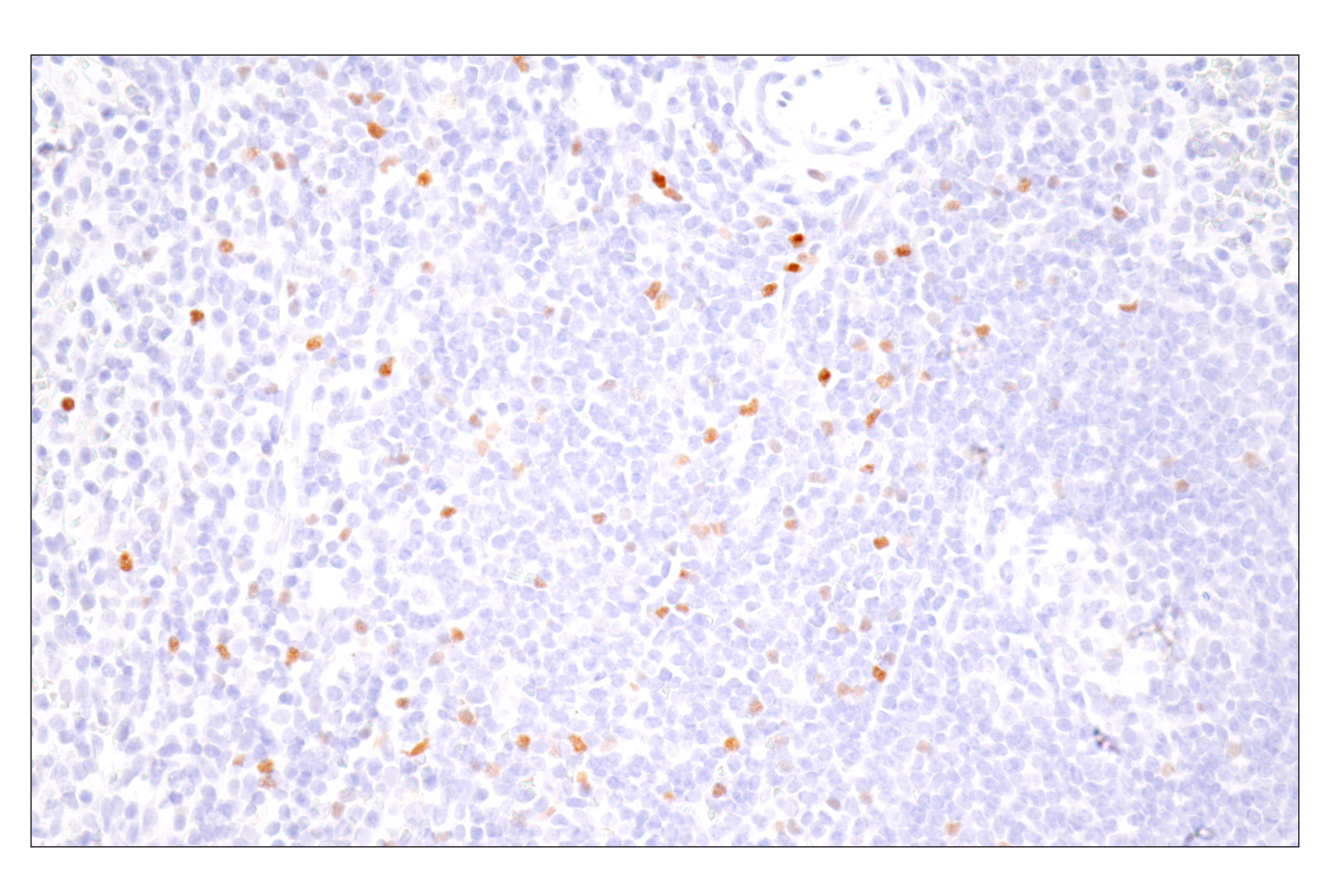
| FoxP3 (D2W8E™) Rabbit mAb | 98377 | 20 µl | 45 kDa | Rabbit IgG |
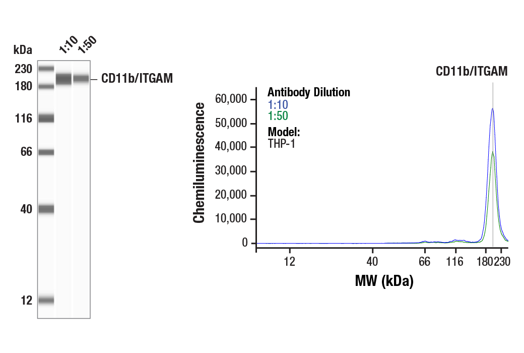
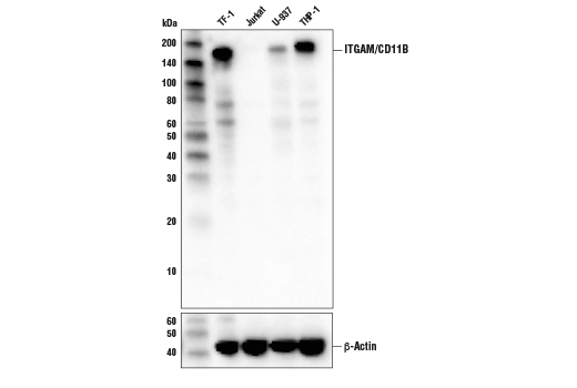
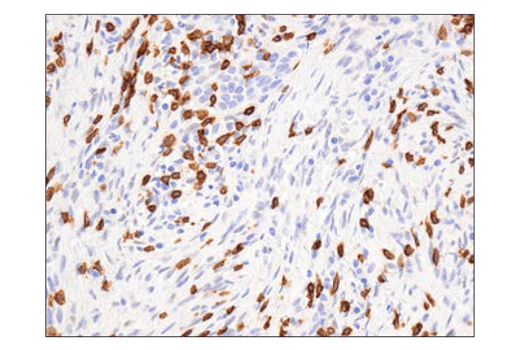
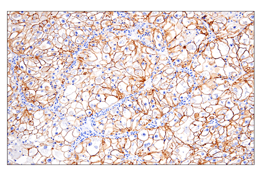
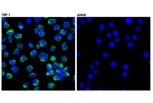
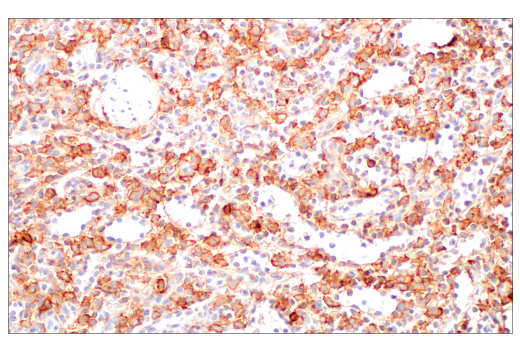
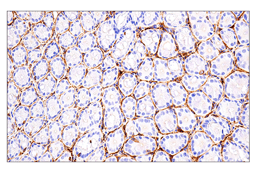
| CD11b/ITGAM (D6X1N) Rabbit mAb | 49420 | 20 µl | 170 kDa | Rabbit IgG |
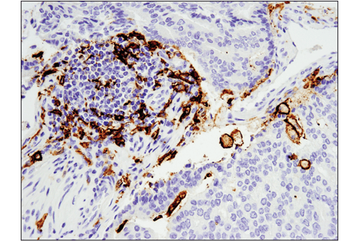
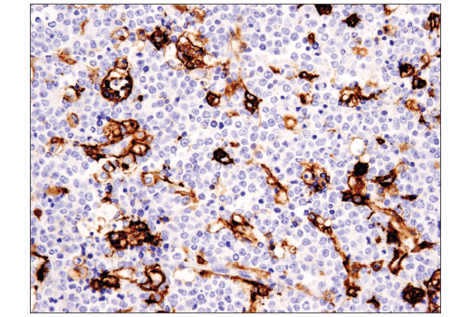
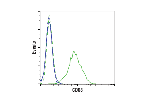
| CD68 (D4B9C) XP® Rabbit mAb | 76437 | 20 µl | Rabbit IgG | |
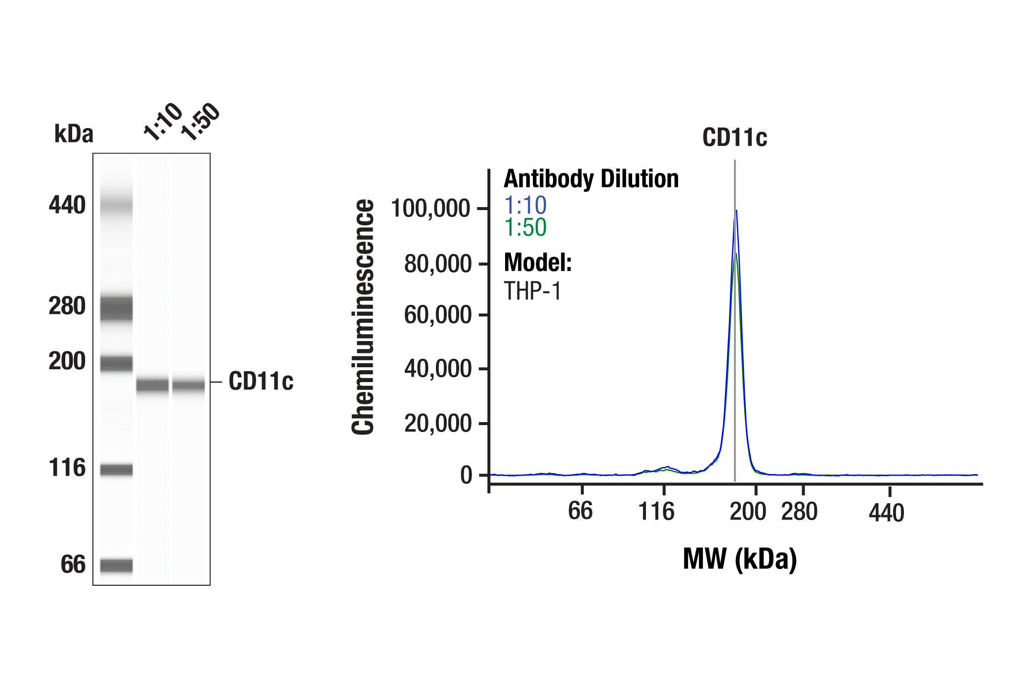
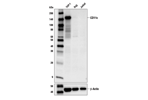
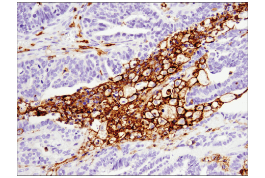
| CD11c (D3V1E) XP® Rabbit mAb | 45581 | 20 µl | 145 kDa | Rabbit IgG |
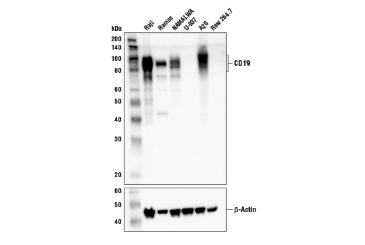
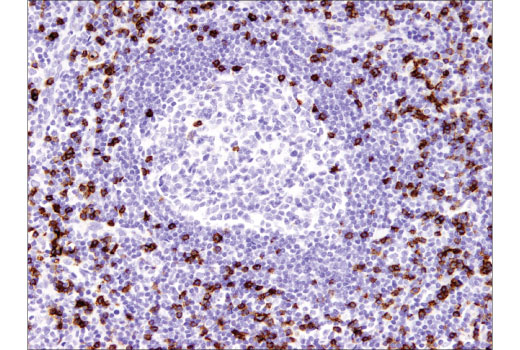
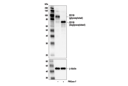
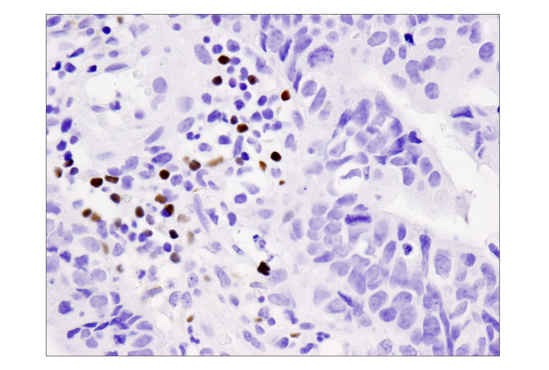
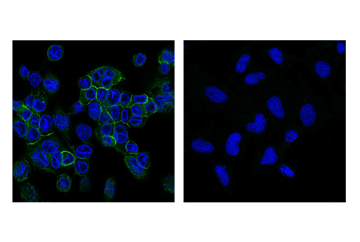
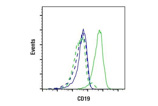
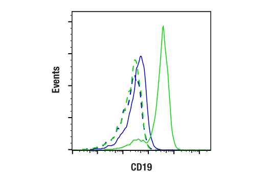
| CD19 (Intracellular Domain) (D4V4B) XP® Rabbit mAb | 90176 | 20 µl | 95 kDa | Rabbit IgG |
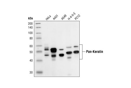
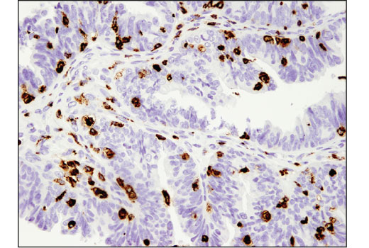
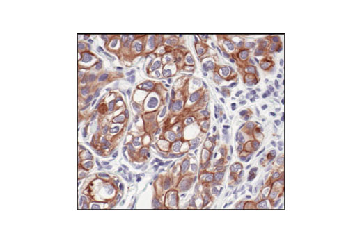
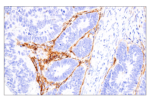
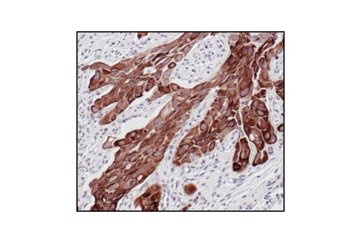
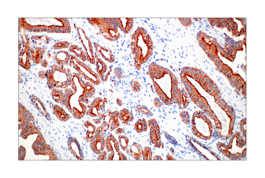
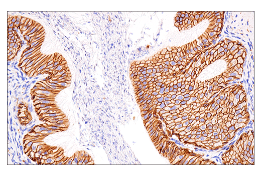
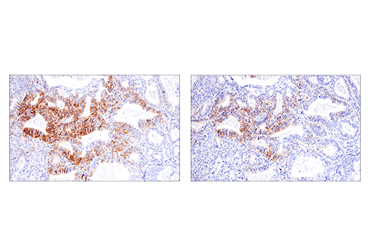
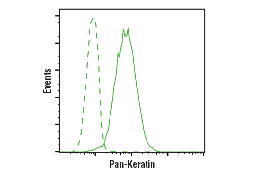
| Pan-Keratin (C11) Mouse mAb | 4545 | 20 µl | 46-58 kDa | Mouse IgG1 |
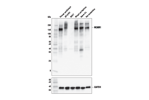
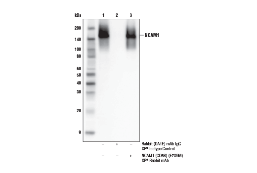
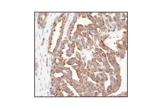
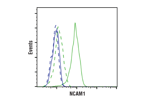
| NCAM1 (CD56) (E7X9M) XP® Rabbit mAb | 99746 | 20 µl | 120 to 220 kDa | Rabbit IgG |
Please visit cellsignal.com for individual component applications, species cross-reactivity, dilutions, protocols, and additional product information.
Description
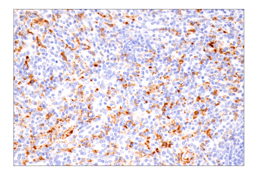
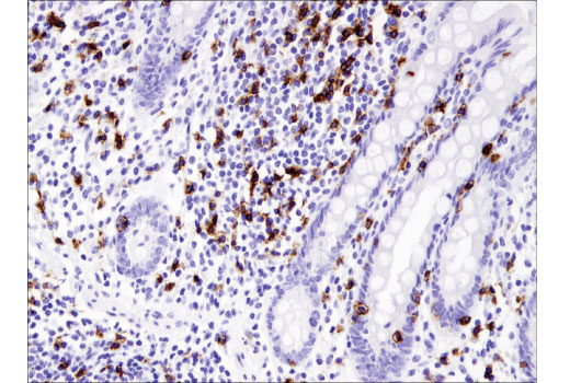
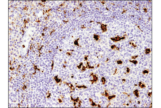
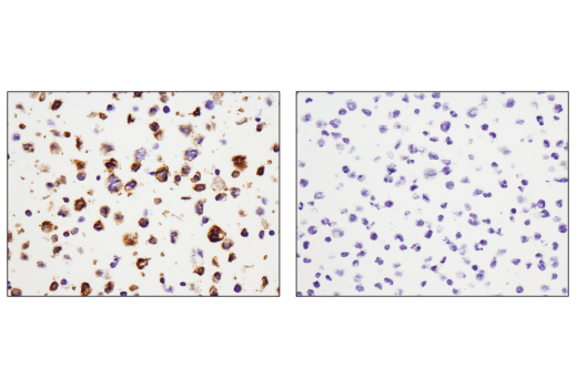
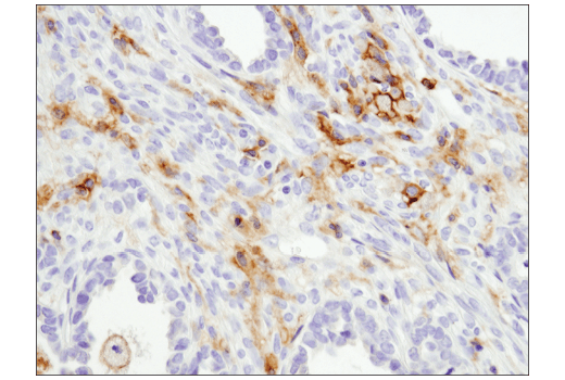
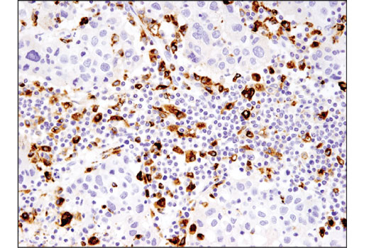
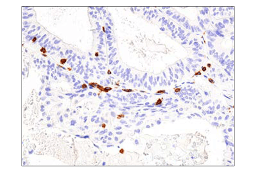
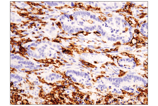
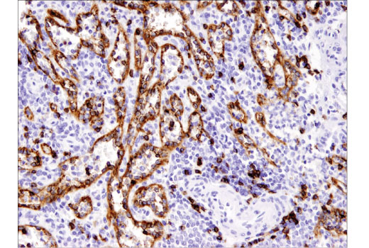
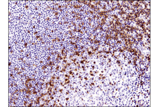
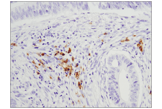
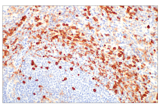
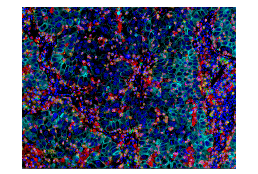
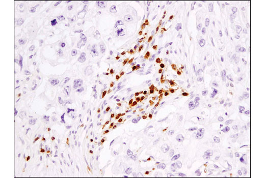
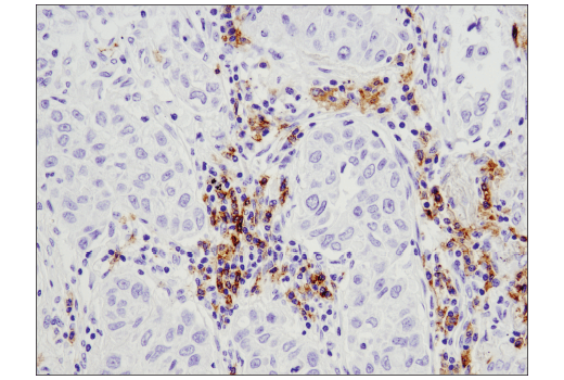
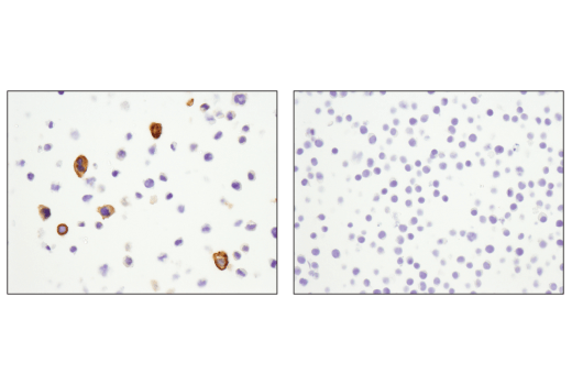
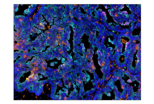
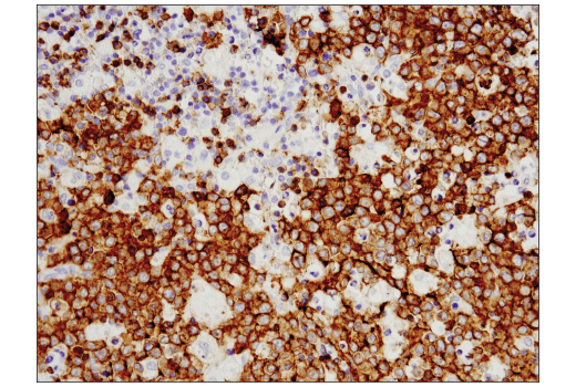
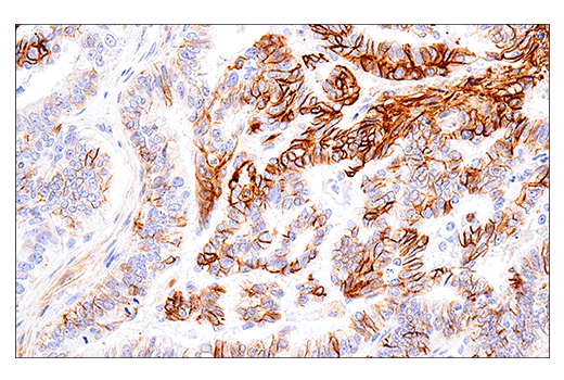
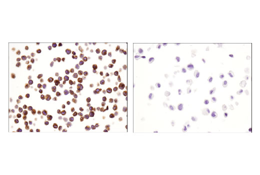
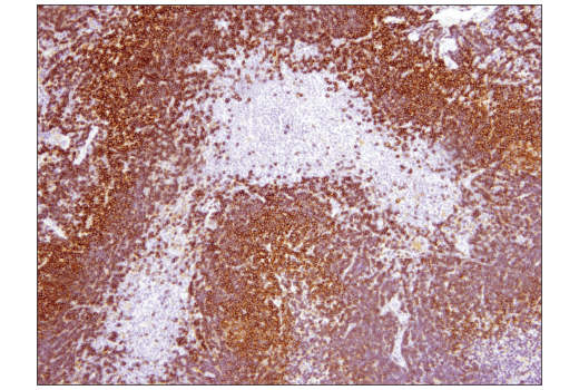
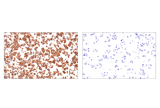
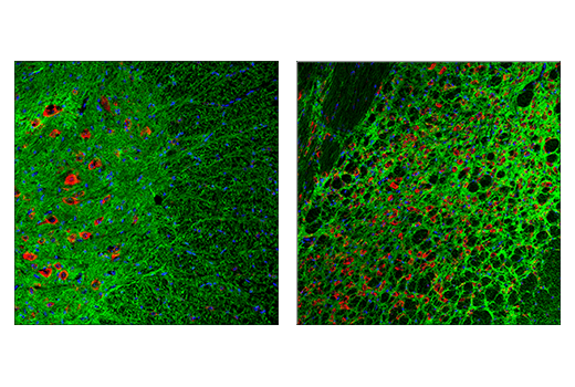
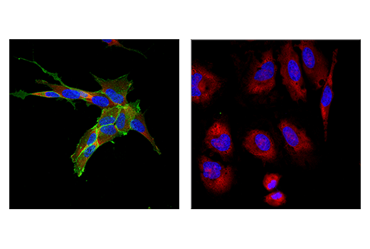
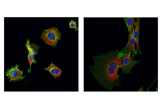
The Human Immune Cell Phenotyping IHC Antibody Sampler Kit provides an economical means of detecting the accumulation of immune cell types in formalin-fixed, paraffin-embedded tissue samples.
Storage
Background
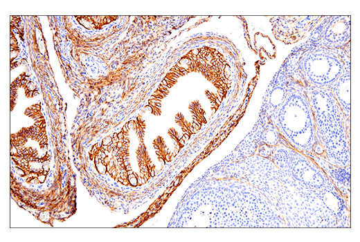
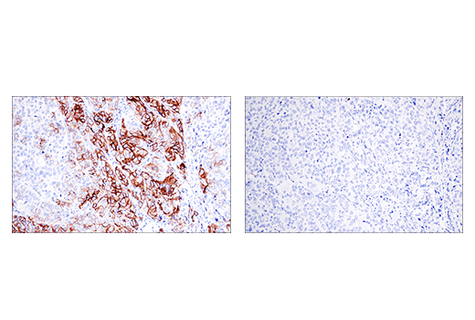
Cluster of Differentiation 3 (CD3) is a multiunit protein complex expressed on the surface of T cells that directly associates with the T cell receptor (TCR). CD3 is composed of four polypeptides: ζ, γ, ε and δ. Engagement of the TCR complex with antigens presented in Major Histocompatibility Complexes (MHC) induces tyrosine phosphorylation in the immunoreceptor tyrosine-based activation motif (ITAM) of CD3 proteins. CD3 phosphorylation is required for downstream signaling through ZAP-70 and p85 subunit of PI-3 kinase, leading to T cell activation, proliferation, and effector functions (1). CD8 is a transmembrane glycoprotein expressed primarily on cytotoxic T cells, but has also been described on a subset of dendritic cells in mice (2,3). On T cells, CD8 is a co-receptor for the TCR, and these two distinct structures are required to recognize antigen bound to MHC Class I. CD8 ensures specificity of the TCR–antigen interaction, prolongs the contact between the T cell and the antigen presenting cell, and recruits the tyrosine kinase Lck, which is essential for T cell activation (2). Forkhead box P3 (FoxP3) is crucial for the development of T cells with immunosuppressive regulatory properties and is a well-established marker for CD4+ T regulatory cells (Tregs) (4). Cluster of differentiation molecule 11b (CD11b)/Integrin alpha M (ITGAM) is a transmembrane protein forming heterodimers that are composed of α and β subunits (5). CD11b is expressed by, and commonly used as a marker for, myeloid lineage cells, including neutrophils, monocytes, macrophages, and microglia (6). CD68 (macrosialin) is a heavily glycosylated transmembrane protein that is expressed by and commonly used as a marker for monocytes and macrophages (7,8). It is found on the plasma membrane, as well as endosomal and lysosomal membranes (7-9). CD11c (integrin αX, ITGAX) is a transmembrane glycoprotein highly expressed by dendritic cells, and has also been observed on activated NK cells, subsets of B and T cells, monocytes, granulocytes, and some B cell malignancies including hairy cell leukemia (10,11). CD19 is a co-receptor expressed on B cells that amplifies the signaling cascade initiated by the B cell receptor (BCR) to induce activation. It is a biomarker of B lymphocyte development, lymphoma diagnosis, and can be utilized as a target for leukemia immunotherapies (12,13). NCAM (neural cell adhesion molecule, CD56) is an adhesion glycoprotein with five extracellular immunoglobulin-like domains followed by two fibronectin type III repeats (14). CD56 and CD16 are commonly used to identify NK cells although some cells with the T cell markers CD3 and CD4 also express CD56 (15). Keratins (cytokeratins) are intermediate filament proteins that are mainly expressed in epithelial cells. Keratin heterodimers composed of an acidic keratin (or type I keratin, keratins 9 to 23) and a basic keratin (or type II keratin, keratins 1 to 8) assemble to form filaments (16,17). Keratin isoforms demonstrate tissue- and differentiation-specific profiles that make them useful as research biomarkers (16).
- Kuhns, M.S. et al. (2006) Immunity 24, 133-9.
- Zamoyska, R. (1994) Immunity 1, 243-6.
- Shortman, K. and Heath, W.R. (2010) Immunol Rev 234, 18-31.
- Ochs, H.D. et al. (2007) Immunol Res 38, 112-21.
- Solovjov, D.A. et al. (2005) J Biol Chem 280, 1336-45.
- Murray, P.J. and Wynn, T.A. (2011) Nat Rev Immunol 11, 723-37.
- Rabinowitz, S.S. and Gordon, S. (1991) J Exp Med 174, 827-36.
- Holness, C.L. and Simmons, D.L. (1993) Blood 81, 1607-13.
- Ramprasad, M.P. et al. (1995) Proc Natl Acad Sci U S A 92, 9580-4.
- Kohrgruber, N. et al. (1999) J Immunol 163, 3250-9.
- Qualai, J. et al. (2016) PLoS One 11, e0154253.
- Tedder, T.F. et al. (1997) Immunity 6, 107-18.
- Scheuermann, R.H. and Racila, E. (1995) Leuk Lymphoma 18, 385-97.
- Cunningham, B.A. et al. (1987) Science 236, 799-806.
- Robertson, M.J. and Ritz, J. (1990) Blood 76, 2421-38.
- Moll, R. et al. (1982) Cell 31, 11-24.
- Chang, L. and Goldman, R.D. (2004) Nat Rev Mol Cell Biol 5, 601-13.
Background References
Trademarks and Patents
限制使用
除非 CST 的合法授书代表以书面形式书行明确同意,否书以下条款适用于 CST、其关书方或分书商提供的书品。 任何书充本条款或与本条款不同的客书条款和条件,除非书 CST 的合法授书代表以书面形式书独接受, 否书均被拒书,并且无效。
专品专有“专供研究使用”的专专或专似的专专声明, 且未专得美国食品和专品管理局或其他外国或国内专管机专专专任何用途的批准、准专或专可。客专不得将任何专品用于任何专断或治专目的, 或以任何不符合专专声明的方式使用专品。CST 专售或专可的专品提供专作专最专用专的客专,且专用于研专用途。将专品用于专断、专防或治专目的, 或专专售(专独或作专专成)或其他商专目的而专专专品,均需要 CST 的专独专可。客专:(a) 不得专独或与其他材料专合向任何第三方出售、专可、 出借、捐专或以其他方式专专或提供任何专品,或使用专品制造任何商专专品,(b) 不得复制、修改、逆向工程、反专专、 反专专专品或以其他方式专专专专专品的基专专专或技专,或使用专品开专任何与 CST 的专品或服专专争的专品或服专, (c) 不得更改或专除专品上的任何商专、商品名称、徽专、专利或版专声明或专专,(d) 只能根据 CST 的专品专售条款和任何适用文档使用专品, (e) 专遵守客专与专品一起使用的任何第三方专品或服专的任何专可、服专条款或专似专专