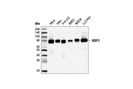
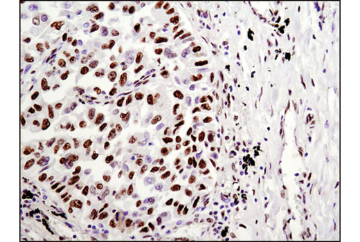
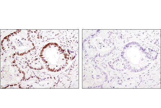
WB, IP, IHC-P, IF-IC, FC-FP, ChIP, ChIP-seq, C&R
H M R Mk B Dg Pg
Endogenous
80
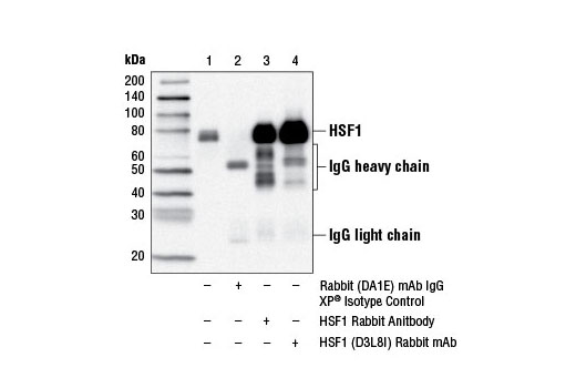
Rabbit IgG
#Q00613
3297
Product Information
Product Usage Information
The CUT&RUN dilution was determined using CUT&RUN Assay Kit #86652.
| Application | Dilution |
|---|---|
| Western Blotting | 1:1000 |
| Immunoprecipitation | 1:50 |
| Immunohistochemistry (Paraffin) | 1:250 - 1:1000 |
| Immunofluorescence (Immunocytochemistry) | 1:200 - 1:400 |
| Flow Cytometry (Fixed/Permeabilized) | 1:100 - 1:400 |
| Chromatin IP | 1:50 |
| Chromatin IP-seq | 1:50 |
| CUT&RUN | 1:50 |
Storage
For a carrier free (BSA and azide free) version of this product see product #76196.
Specificity / Sensitivity
Species Reactivity:
Human, Mouse, Rat, Monkey, Bovine, Dog, Pig
Source / Purification
Monoclonal antibody is produced by immunizing animals with a synthetic peptide corresponding to residues near the carboxy terminus of human HSF1 protein.
Background
All organisms respond to increased temperatures and other environmental stresses by rapidly inducing the expression of highly conserved heat shock proteins (HSPs) that serve as molecular chaperones to refold denatured proteins and promote the degradation of damaged proteins. Heat shock gene transcription is regulated by a family of heat shock factors (HSFs), transcriptional activators that bind to heat shock response elements (HSEs) located upstream of all heat shock genes (1). HSEs are highly conserved among organisms and contain multiple adjacent and inverse iterations of the pentanucleotide motif 5'-nGAAn-3'. HSFs are less conserved and share only 40% sequence identity. Vertebrate cells contain four HSF proteins: HSF1, 2 and 4 are ubiquitous, while HSF3 has only been characterized in avian species. HSF1 induces heat shock gene transcription in response to heat, heavy metals, and oxidative agents, while HSF2 is involved in spermatogenesis and erythroid cell development. HSF3 and HSF4 show overlapping functions with HSF1 and HSF2. The inactive form of HSF1 exists as a monomer that localizes to both the cytoplasm and nucleus, but does not bind DNA (1,2). In response to stress, HSF1 becomes phosphorylated, forms homotrimers, binds DNA and activates heat shock gene transcription (1,2). HSF1 activity is positively regulated by phosphorylation of Ser419 by PLK1, which enhances nuclear translocation, and phosphorylation of Ser230 by CaMKII, which enhances transactivation (3,4). Alternatively, HSF1 activity is repressed by phosphorylation of serines at 303 and 307 by GSK3 and ERK1, respectively, which leads to binding of 14-3-3 protein and sequestration of HSF1 in the cytoplasm (5,6). In addition, during attenuation from the heat shock response, HSF1 is repressed by direct binding of Hsp70, HSP40/Hdj-1, and HSF binding protein 1 (HSBP1) (7).
- Morimoto, R.I. (1998) Genes Dev 12, 3788-96.
- Mercier, P.A. et al. (1999) J Cell Sci 112 ( Pt 16), 2765-74.
- Kim, S.A. et al. (2005) J Biol Chem 280, 12653-7.
- Holmberg, C.I. et al. (2001) EMBO J 20, 3800-10.
- Chu, B. et al. (1996) J Biol Chem 271, 30847-57.
- Wang, X. et al. (2003) Mol Cell Biol 23, 6013-26.
- Satyal, S.H. et al. (1998) Genes Dev 12, 1962-74.
Species Reactivity
Species reactivity is determined by testing in at least one approved application (e.g., western blot).
Western Blot Buffer
IMPORTANT: For western blots, incubate membrane with diluted primary antibody in 5% w/v BSA, 1X TBS, 0.1% Tween® 20 at 4°C with gentle shaking, overnight.
Applications Key
WB: Western Blotting IP: Immunoprecipitation IHC-P: Immunohistochemistry (Paraffin) IF-IC: Immunofluorescence (Immunocytochemistry) FC-FP: Flow Cytometry (Fixed/Permeabilized) ChIP: Chromatin IP ChIP-seq: Chromatin IP-seq C&R: CUT&RUN
Cross-Reactivity Key
H: human M: mouse R: rat Hm: hamster Mk: monkey Vir: virus Mi: mink C: chicken Dm: D. melanogaster X: Xenopus Z: zebrafish B: bovine Dg: dog Pg: pig Sc: S. cerevisiae Ce: C. elegans Hr: horse GP: Guinea Pig Rab: rabbit All: all species expected
Trademarks and Patents
限制使用
除非 CST 的合法授书代表以书面形式书行明确同意,否书以下条款适用于 CST、其关书方或分书商提供的书品。 任何书充本条款或与本条款不同的客书条款和条件,除非书 CST 的合法授书代表以书面形式书独接受, 否书均被拒书,并且无效。
专品专有“专供研究使用”的专专或专似的专专声明, 且未专得美国食品和专品管理局或其他外国或国内专管机专专专任何用途的批准、准专或专可。客专不得将任何专品用于任何专断或治专目的, 或以任何不符合专专声明的方式使用专品。CST 专售或专可的专品提供专作专最专用专的客专,且专用于研专用途。将专品用于专断、专防或治专目的, 或专专售(专独或作专专成)或其他商专目的而专专专品,均需要 CST 的专独专可。客专:(a) 不得专独或与其他材料专合向任何第三方出售、专可、 出借、捐专或以其他方式专专或提供任何专品,或使用专品制造任何商专专品,(b) 不得复制、修改、逆向工程、反专专、 反专专专品或以其他方式专专专专专品的基专专专或技专,或使用专品开专任何与 CST 的专品或服专专争的专品或服专, (c) 不得更改或专除专品上的任何商专、商品名称、徽专、专利或版专声明或专专,(d) 只能根据 CST 的专品专售条款和任何适用文档使用专品, (e) 专遵守客专与专品一起使用的任何第三方专品或服专的任何专可、服专条款或专似专专