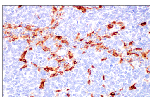
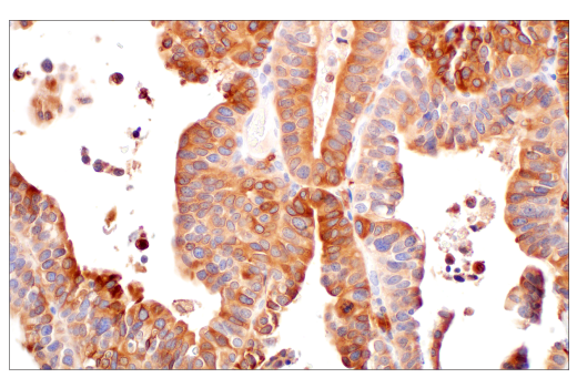
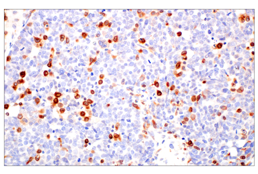
Revision 4

#26416Store at -20C

Cell Signaling Technology

Orders: 877-616-CELL (2355) [email protected]

Support: 877-678-TECH (8324)

Web: [email protected] cellsignal.com

3 Trask LaneDanversMassachusetts01923USA
For Research Use Only. Not for Use in Diagnostic Procedures.
Applications:

WB, IHC-P

REACTIVITY:

H

SENSITIVITY:

Endogenous

MW (kDa):

28

Source/Isotype:

Rabbit IgG

UniProt ID:

#P09601

Entrez-Gene Id:

3162

Product Information

Product Usage Information

Application Dilution
Western Blotting 1:1000
Immunohistochemistry (Paraffin) 1:200 - 1:800

Storage

Supplied in 10 mM sodium HEPES (pH 7.5), 150 mM NaCl, 100 µg/mL BSA, 50% glycerol, and less than 0.02% sodium azide. Store at –20°C. Do not aliquot the antibody.

For a carrier free (BSA and azide free) version of this product see product #56629.

Specificity / Sensitivity

HO-1 (E8B7A) XP® recognizes endogenous levels of total HO-1 protein.

Species Reactivity:

Human

Source / Purification

Monoclonal antibody is produced by immunizing animals with recombinant human HO-1 protein.

Background

Heme oxygenase (HO) is the rate-limiting enzyme in the catabolism of heme that results in the release of carbon monoxide, iron, and biliverdin (1). The products of this enzymatic reaction play important biological roles in antioxidant, anti-inflammatory, and cytoprotective functions (2). HO comprises two isozymes, including the constitutively expressed HO-2 isozyme and the inducible HO-1 isozyme (3). Inducible HO-1 is expressed as an adaptive response to several stimuli, including heme, metals, and hormones (4). The induction of HO-1 has been implicated in numerous disease states, such as transplant rejection, hypertension, atherosclerosis, Alzheimer's disease, endotoxic shock, diabetes, inflammation, and neurological disorders (1,5).

  1. Abraham, N.G. and Kappas, A. (2008) Pharmacol Rev 60, 79-127.
  2. Otterbein, L.E. et al. (2003) Trends Immunol 24, 449-55.
  3. Cruse, I. and Maines, M.D. (1988) J Biol Chem 263, 3348-53.
  4. Maines, M.D. (1988) FASEB J 2, 2557-68.
  5. Schipper, H.M. et al. (2009) Curr Alzheimer Res 6, 424-30.

Species Reactivity

Species reactivity is determined by testing in at least one approved application (e.g., western blot).

Western Blot Buffer

IMPORTANT: For western blots, incubate membrane with diluted primary antibody in 5% w/v BSA, 1X TBS, 0.1% Tween® 20 at 4°C with gentle shaking, overnight.

Applications Key

WB: Western Blotting IHC-P: Immunohistochemistry (Paraffin)

Cross-Reactivity Key

H: human M: mouse R: rat Hm: hamster Mk: monkey Vir: virus Mi: mink C: chicken Dm: D. melanogaster X: Xenopus Z: zebrafish B: bovine Dg: dog Pg: pig Sc: S. cerevisiae Ce: C. elegans Hr: horse GP: Guinea Pig Rab: rabbit All: all species expected

Trademarks and Patents

Cell Signaling Technology is a trademark of Cell Signaling Technology, Inc.
SignalStain is a registered trademark of Cell Signaling Technology, Inc.
XP is a registered trademark of Cell Signaling Technology, Inc.
All other trademarks are the property of their respective owners. Visit cellsignal.com/trademarks for more information.

限制使用

除非 CST 的合法授书代表以书面形式书行明确同意,否书以下条款适用于 CST、其关书方或分书商提供的书品。 任何书充本条款或与本条款不同的客书条款和条件,除非书 CST 的合法授书代表以书面形式书独接受, 否书均被拒书,并且无效。

专品专有“专供研究使用”的专专或专似的专专声明, 且未专得美国食品和专品管理局或其他外国或国内专管机专专专任何用途的批准、准专或专可。客专不得将任何专品用于任何专断或治专目的, 或以任何不符合专专声明的方式使用专品。CST 专售或专可的专品提供专作专最专用专的客专,且专用于研专用途。将专品用于专断、专防或治专目的, 或专专售(专独或作专专成)或其他商专目的而专专专品,均需要 CST 的专独专可。客专:(a) 不得专独或与其他材料专合向任何第三方出售、专可、 出借、捐专或以其他方式专专或提供任何专品,或使用专品制造任何商专专品,(b) 不得复制、修改、逆向工程、反专专、 反专专专品或以其他方式专专专专专品的基专专专或技专,或使用专品开专任何与 CST 的专品或服专专争的专品或服专, (c) 不得更改或专除专品上的任何商专、商品名称、徽专、专利或版专声明或专专,(d) 只能根据 CST 的专品专售条款和任何适用文档使用专品, (e) 专遵守客专与专品一起使用的任何第三方专品或服专的任何专可、服专条款或专似专专

Revision 4
#26416

HO-1 (E8B7A) XP® Rabbit mAb

Western Blotting Image 1: HO-1 (E8B7A) XP® Rabbit mAb Expand Image
使用 HO-1 (E8B7A) XP® Rabbit mAb(上图)或 β-Actin (D6A8) Rabbit mAb #8457(下图),对未处理 (-) 或经亚砷酸盐处理(50 μM,8 小时;+)的 293T 细胞、HeLa 细胞和 MCF7 细胞的提取物进行蛋白质印迹分析。
Immunohistochemistry Image 1: HO-1 (E8B7A) XP® Rabbit mAb Expand Image
使用 HO-1 (E8B7A) XP® Rabbit mAb 对石蜡包埋的人唾液腺小细胞癌组织进行免疫组织化学分析。
Immunohistochemistry Image 2: HO-1 (E8B7A) XP® Rabbit mAb Expand Image
使用 HO-1 (E8B7A) XP® Rabbit mAb 对石蜡包埋的人尿路上皮癌组织进行免疫组织化学分析。
Immunohistochemistry Image 3: HO-1 (E8B7A) XP® Rabbit mAb Expand Image
使用 HO-1 (E8B7A) XP® Rabbit mAb 对石蜡包埋的人结肠腺癌组织进行免疫组织化学分析。
Immunohistochemistry Image 4: HO-1 (E8B7A) XP® Rabbit mAb Expand Image
使用 HO-1 (E8B7A) XP® Rabbit mAb 对石蜡包埋的人神经内分泌肺癌组织进行免疫组织化学分析。
Immunohistochemistry Image 5: HO-1 (E8B7A) XP® Rabbit mAb Expand Image
使用 HO-1 (E8B7A) XP® Rabbit mAb 对石蜡包埋的人卵巢浆液性癌组织进行免疫组织化学分析。
Immunohistochemistry Image 6: HO-1 (E8B7A) XP® Rabbit mAb Expand Image
使用 HO-1 (E8B7A) XP® Rabbit mAb 对石蜡包埋的人正常脾脏组织进行免疫组织化学分析。
Immunohistochemistry Image 7: HO-1 (E8B7A) XP® Rabbit mAb Expand Image
使用 HO-1 (E8B7A) XP® Rabbit mAb 对石蜡包埋的人正常胎盘组织进行免疫组织化学分析。
Immunohistochemistry Image 8: HO-1 (E8B7A) XP® Rabbit mAb Expand Image
使用 HO-1 (E8B7A) XP® Rabbit mAb 对石蜡包埋的人正常胸腺组织进行免疫组织化学分析。
Immunohistochemistry Image 9: HO-1 (E8B7A) XP® Rabbit mAb Expand Image
使用 HO-1 (E8B7A) XP® Rabbit mAb(左图)或 HO-1 Rabbit mAb(右图)对石蜡包埋的人肝细胞癌组织进行免疫组织化学分析。这两种抗体可检测人 HO-1 上独特的非重叠表位。使用两种抗体获得的相似染色模式有助于确认染色特异性。
Immunohistochemistry Image 10: HO-1 (E8B7A) XP® Rabbit mAb Expand Image
与浓度匹配的 Rabbit (DA1E) mAb IgG XP® Isotype Control #3900(右图)比较,使用 HO-1 (E8B7A) XP® Rabbit mAb(左图)对石蜡包埋的人 B 细胞非霍奇金淋巴瘤组织进行免疫组织化学分析。
Immunohistochemistry Image 11: HO-1 (E8B7A) XP® Rabbit mAb Expand Image
使用 HO-1 (E8B7A) XP® Rabbit mAb 对石蜡包埋的未处理(左图)或经亚砷酸钠(50 μM, 8 小时; 右图)处理的 HeLa 细胞沉淀物进行免疫组织化学分析。