| Product Includes | Product # | Quantity | Mol. Wt | Isotype/Source |
|---|---|---|---|---|
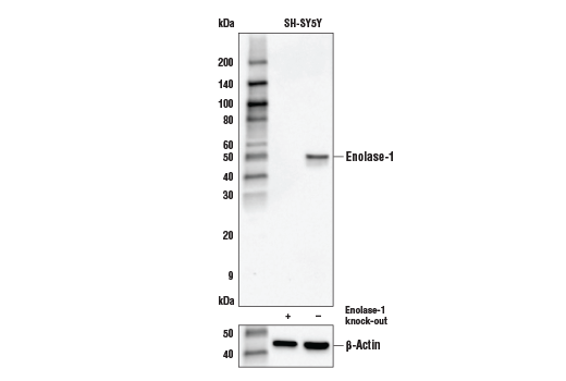
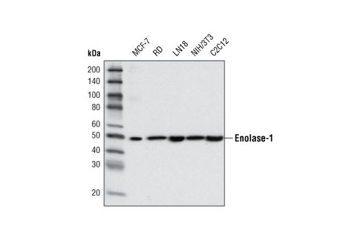
| Enolase-1 Antibody | 3810 | 20 µl | 47 kDa | Rabbit |
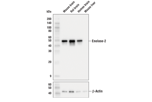
| Enolase-2 (E2H9X) XP® Rabbit mAb | 24330 | 20 µl | 47 kDa | Rabbit IgG |
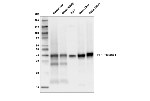
| FBP1/FBPase 1 (D2T7F) Rabbit mAb | 59172 | 20 µl | 39 kDa | Rabbit IgG |
| GPI (E2Q8J) XP® Rabbit mAb | 94068 | 20 µl | 60 kDa | Rabbit IgG |
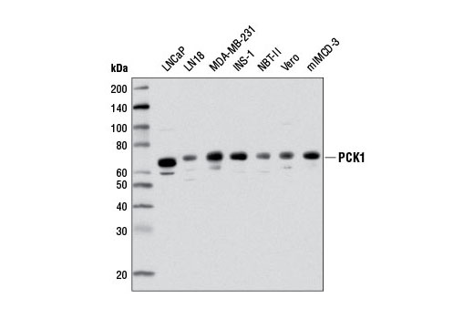
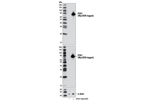
| PCK1 (D12F5) Rabbit mAb | 12940 | 20 µl | 63 kDa | Rabbit IgG |
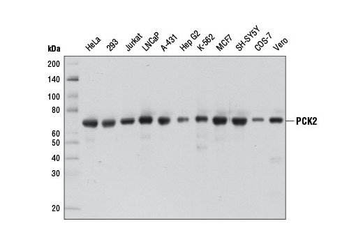
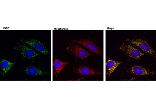
| PCK2 (D3E11) Rabbit mAb | 8565 | 20 µl | 71 kDa | Rabbit IgG |
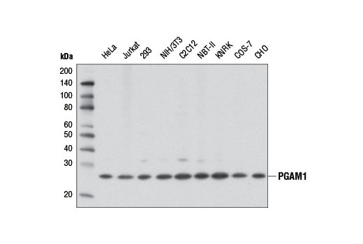
| PGAM1 (D3J9T) Rabbit mAb | 12098 | 20 µl | 28 kDa | Rabbit IgG |
| PGK1 Antibody | 68540 | 20 µl | 43 kDa | Rabbit |
| Pyruvate Carboxylase Antibody | 66470 | 20 µl | 130 kDa | Rabbit |
| Anti-rabbit IgG, HRP-linked Antibody | 7074 | 100 µl | Goat |
Please visit cellsignal.com for individual component applications, species cross-reactivity, dilutions, protocols, and additional product information.
Description
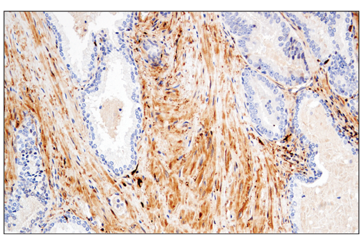
The Gluconeogenesis Antibody Sampler Kit provides an economical means of detecting select components involved in the gluconeogenesis metabolism pathway. The kit includes enough antibodies to perform two western blot experiments with each primary antibody.
Storage
Background
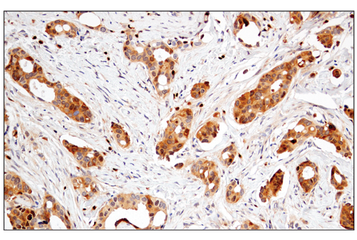
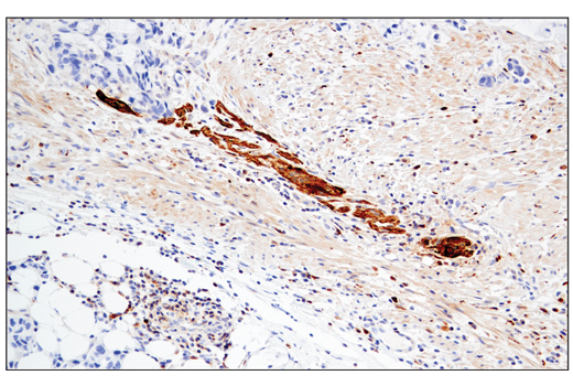
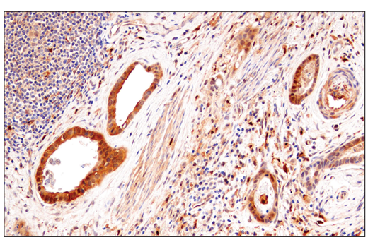
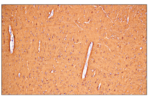
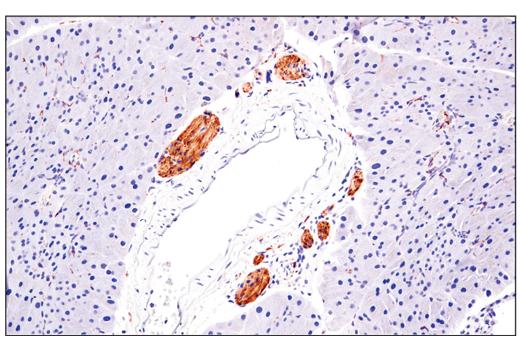
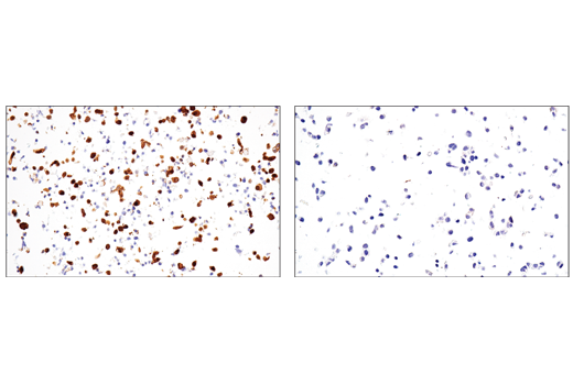
Enolase is an important glycolytic enzyme involved in the interconversion of 2-phosphoglycerate to phosphoenolpyruvate. Mammalian enolase exists as three subunits: enolase-1 (α-enolase), enolase-2 (γ-enolase), and enolase-3 (β-enolase). Expression of the enolase isoforms differs in a tissue specific manner (1). Enolase-1 plays a key role in anaerobic metabolism under hypoxic conditions and may act as a cell surface plasminogen receptor during tissue invasion (2,3). Abnormal expression of enolase-1 is associated with tumor progression in some cases of breast and lung cancer (4-7). Alternatively, an enolase-1 splice variant (MBP-1) binds the c-myc promoter p2 and may function as a tumor suppressor. For this reason, enolase-1 is considered as a potential therapeutic target in the treatment of some forms of cancer (8). Research studies have shown elevated levels of neuron-specific enolase-2 in neuroblastoma (1) and small-cell lung cancer (9,10). Fructose-1,6-bisphosphatase 1 (FBP1 or FBPase 1), a rate-limiting enzyme in gluconeogenesis, catalyzes the conversion of fructose-1,6-bisphosphate to fructose-6-phosphate (11). Inhibition of FBP1 expression in basal-like breast cancer (BLBC) cells leads to metabolic reprogramming, including enhanced glycolysis, which leads to increased glucose uptake, biosynthesis of macromolecules, and activation of PKM2 (11). This metabolic reprogramming endows tumor cells with cancer stem cell (CSC)-like properties, thereby increasing their tumorigenicity (11). Depletion of FBP1 was also reported in more than 600 clear cell renal cell carcinoma (ccRCC) tumors, suggesting that FBP1 may inhibit ccRCC tumor progression (12). Glucose-6-phosphate isomerase (GPI) is a multi-functional protein belonging to the glucose phosphate isomerase family (13,14). As an intracellular metabolic enzyme, GPI plays a pivotal role in glycolysis and gluconeogenesis by catalyzing the interconversion of D-glucose-6-phosphate and D-fructose-6-phosphate (15). GPI is also secreted, where it functions as a cytokine (referred to as Autocrine Motility Factor, AMF), acting via the E3-ubiquitin-protein ligase AMFR/gp78 (16). In normal tissues, GPI/AMF has been shown to promote both immune cell maturation and neuronal cell survival (17,18). It is also secreted in abundance by some tumor cells (19), where it has been shown to promote tumor cell migration and metastasis (20,21). Phosphoenolpyruvate carboxykinase 1 (PCK1, PEPCK1, or PEPCK-C) is a cytosolic enzyme responsible for the conversion of oxaloacetate to phosphoenolpyruvate (22). PCK1 and PCK2 are involved in controlling the rate-limiting step of gluconeogenesis in the liver, which generates glucose from non-carbohydrate substrates, such as lactate and glycerol (23, 24). PCK2 (PEPCK2 or PEPCK-M) encodes an isoform of phosphoenolpyruvate carboxykinase (PEPCK) that is found in the mitochondria of renal and hepatic tissues (22). Phosphoglycerate mutase (PGAM1) catalyzes the conversion of 3-phosphoglycerate to 2-phosphoglycerate during glycolysis (25-29). Research studies have shown increased PGAM1 expression in cancer (25-28) and mental disease (29). PGK1 (phosphoglycerate kinase) is an essential enzyme in the glycolysis pathway (30). It catalyzes the reversible phospho-transfer reaction from 1,3-diphosphoglycerate to ADP to form ATP and 3-phosphoglycerate. The expression of PGK1 is upregulated in many cancer types and plays an important role in cancer cell proliferation and metastasis (31-34). Pyruvate carboxylase (PC) catalyzes the carboxylation of pyruvate to oxaloacetate to replenish TCA cycle intermediates. It is also critical in regulating gluconeogenesis in the liver (35).
- Pancholi, V. (2001) Cell Mol Life Sci 58, 902-20.
- Redlitz, A. et al. (1995) Eur J Biochem 227, 407-15.
- Jiang, B.H. et al. (1997) Cancer Res 57, 5328-35.
- Peebles, K.A. et al. (2003) Carcinogenesis 24, 651-7.
- Zhang, L. et al. (2000) J Surg Res 93, 108-19.
- Wu, W. et al. (2002) Clin Exp Metastasis 19, 319-26.
- Hennipman, A. et al. (1988) Tumour Biol 9, 241-8.
- Feo, S. et al. (2000) FEBS Lett 473, 47-52.
- Stern, P. et al. (2007) Tumour Biol 28, 84-92.
- O'Shea, P. et al. (1995) Ir J Med Sci 164, 31-6.
- Dong, C. et al. (2013) Cancer Cell 23, 316-31.
- Li, B. et al. (2014) Nature 513, 251-5.
- Haga, A. et al. (2000) Biochim Biophys Acta 1480, 235-44.
- Jeffery, C.J. et al. (2000) Biochemistry 39, 955-64.
- Kim, J.W. and Dang, C.V. (2005) Trends Biochem Sci 30, 142-50.
- Fairbank, M. et al. (2009) Mol Biosyst 5, 793-801.
- Gurney, M.E. et al. (1986) Science 234, 574-81.
- Gurney, M.E. et al. (1986) Science 234, 566-74.
- Lucarelli, G. et al. (2015) Medicine (Baltimore) 94, e2117.
- Liotta, L.A. et al. (1986) Proc Natl Acad Sci U S A 83, 3302-6.
- Funasaka, T. and Raz, A. (2007) Cancer Metastasis Rev 26, 725-35.
- Caton, P.W. et al. (2009) Life Sci 84, 738-44.
- Yoon, J.C. et al. (2001) Nature 413, 131-8.
- Fischer, S. et al. (2010) Biol Reprod 83, 859-65.
- Vander Heiden, M.G. et al. (2010) Science 329, 1492-9.
- Jacobowitz, D.M. et al. (2008) Microvasc Res 76, 89-93.
- Ren, F. et al. (2010) Mol Cancer 9, 81.
- Evans, M.J. et al. (2005) Nat Biotechnol 23, 1303-7.
- Martins-de-Souza, D. et al. (2009) BMC Psychiatry 9, 17.
- Beutler, E. (2007) Br J Haematol 136, 3-11.
- Wilson, R.B. et al. (2019) Pleura Peritoneum 4, 20190003.
- Yu, T. et al. (2017) Cancer Res 77, 5782-5794.
- Hu, H. et al. (2017) Hepatology 65, 515-528.
- Cao, H. et al. (2017) Cancer Chemother Pharmacol 79, 985-994.
- Cappel, D.A. et al. (2019) Cell Metab 29, 1291-1305.e8.
Background References
Trademarks and Patents
限制使用
除非 CST 的合法授书代表以书面形式书行明确同意,否书以下条款适用于 CST、其关书方或分书商提供的书品。 任何书充本条款或与本条款不同的客书条款和条件,除非书 CST 的合法授书代表以书面形式书独接受, 否书均被拒书,并且无效。
专品专有“专供研究使用”的专专或专似的专专声明, 且未专得美国食品和专品管理局或其他外国或国内专管机专专专任何用途的批准、准专或专可。客专不得将任何专品用于任何专断或治专目的, 或以任何不符合专专声明的方式使用专品。CST 专售或专可的专品提供专作专最专用专的客专,且专用于研专用途。将专品用于专断、专防或治专目的, 或专专售(专独或作专专成)或其他商专目的而专专专品,均需要 CST 的专独专可。客专:(a) 不得专独或与其他材料专合向任何第三方出售、专可、 出借、捐专或以其他方式专专或提供任何专品,或使用专品制造任何商专专品,(b) 不得复制、修改、逆向工程、反专专、 反专专专品或以其他方式专专专专专品的基专专专或技专,或使用专品开专任何与 CST 的专品或服专专争的专品或服专, (c) 不得更改或专除专品上的任何商专、商品名称、徽专、专利或版专声明或专专,(d) 只能根据 CST 的专品专售条款和任何适用文档使用专品, (e) 专遵守客专与专品一起使用的任何第三方专品或服专的任何专可、服专条款或专似专专