| Product Includes | Product # | Quantity | Mol. Wt | Isotype/Source |
|---|---|---|---|---|
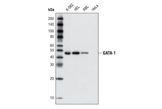
| GATA-1 (D52H6) XP® Rabbit mAb | 3535 | 20 µl | 43 kDa | Rabbit IgG |
| GATA-2 (E9T6F) Rabbit mAb | 79802 | 20 µl | 51 kDa | Rabbit IgG |
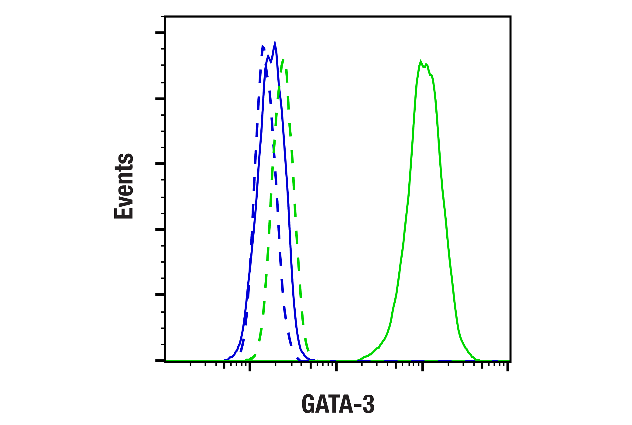
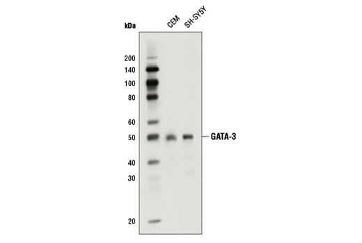
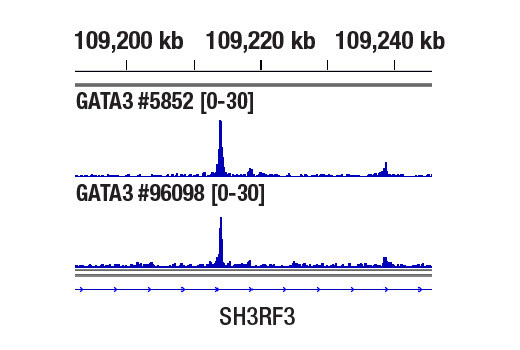
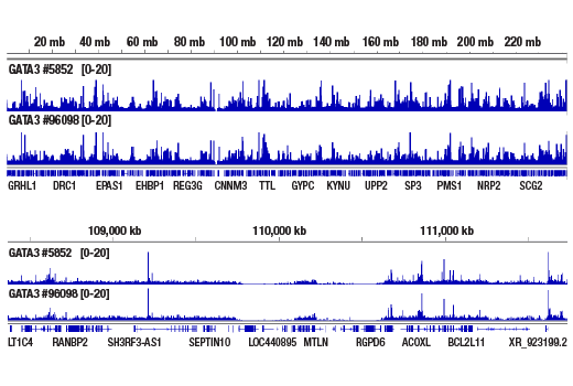
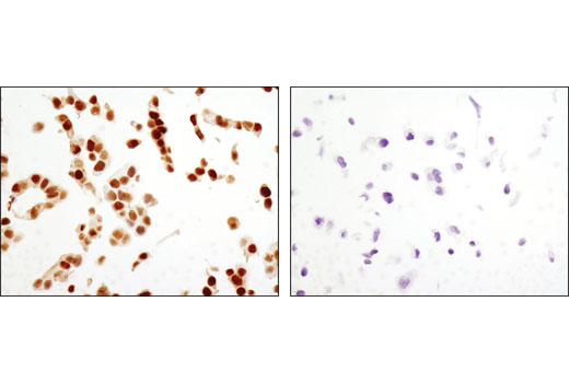
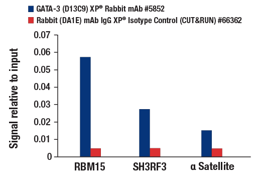
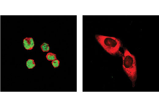
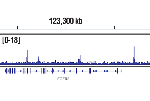
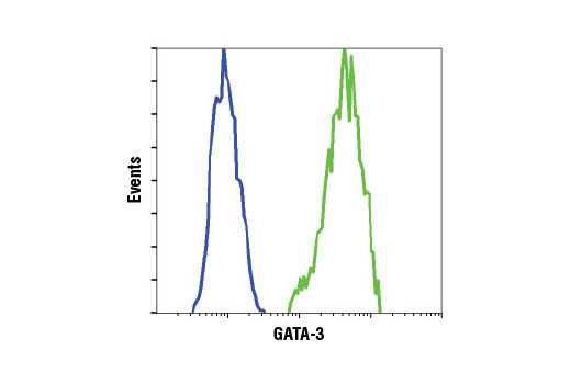
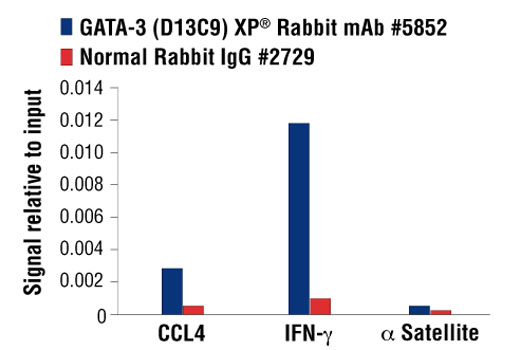
| GATA-3 (D13C9) XP® Rabbit mAb | 5852 | 20 µl | 48 kDa | Rabbit IgG |
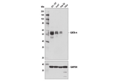
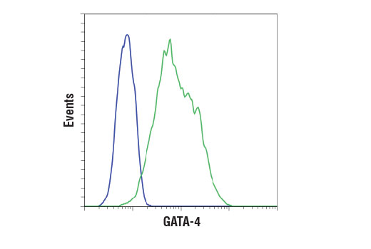
| GATA-4 (D3A3M) Rabbit mAb | 36966 | 20 µl | 55 kDa | Rabbit IgG |
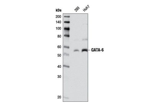
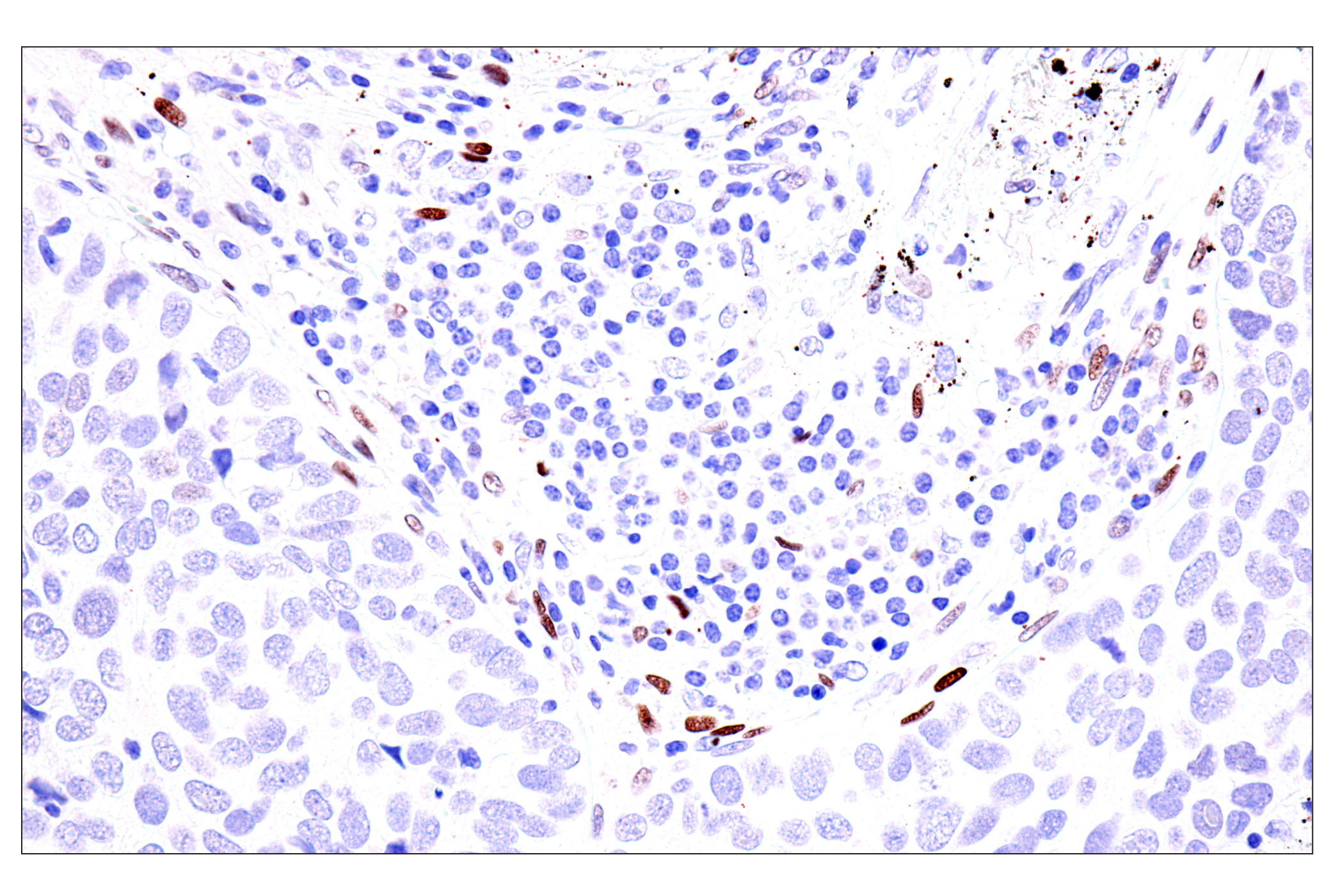
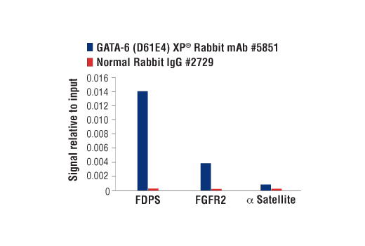
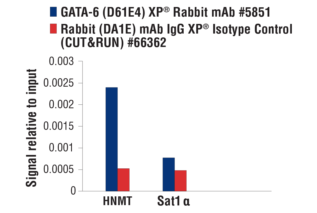
| GATA-6 (D61E4) XP® Rabbit mAb | 5851 | 20 µl | 55 kDa | Rabbit IgG |
| Anti-rabbit IgG, HRP-linked Antibody | 7074 | 100 µl | Goat |
Please visit cellsignal.com for individual component applications, species cross-reactivity, dilutions, protocols, and additional product information.
Description
The GATA Transcription Factor Antibody Sampler Kit provides an economical means of evaluating total levels of GATA family proteins. The kit includes enough antibodies to perform two western blot experiments with each primary antibody.
Storage
Background
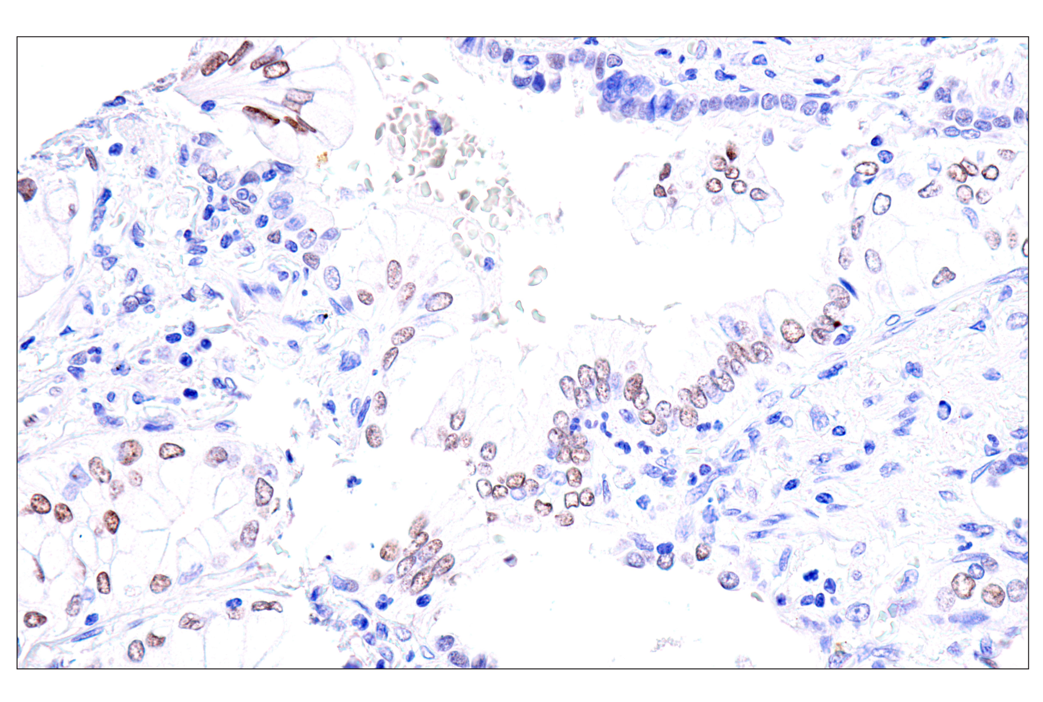
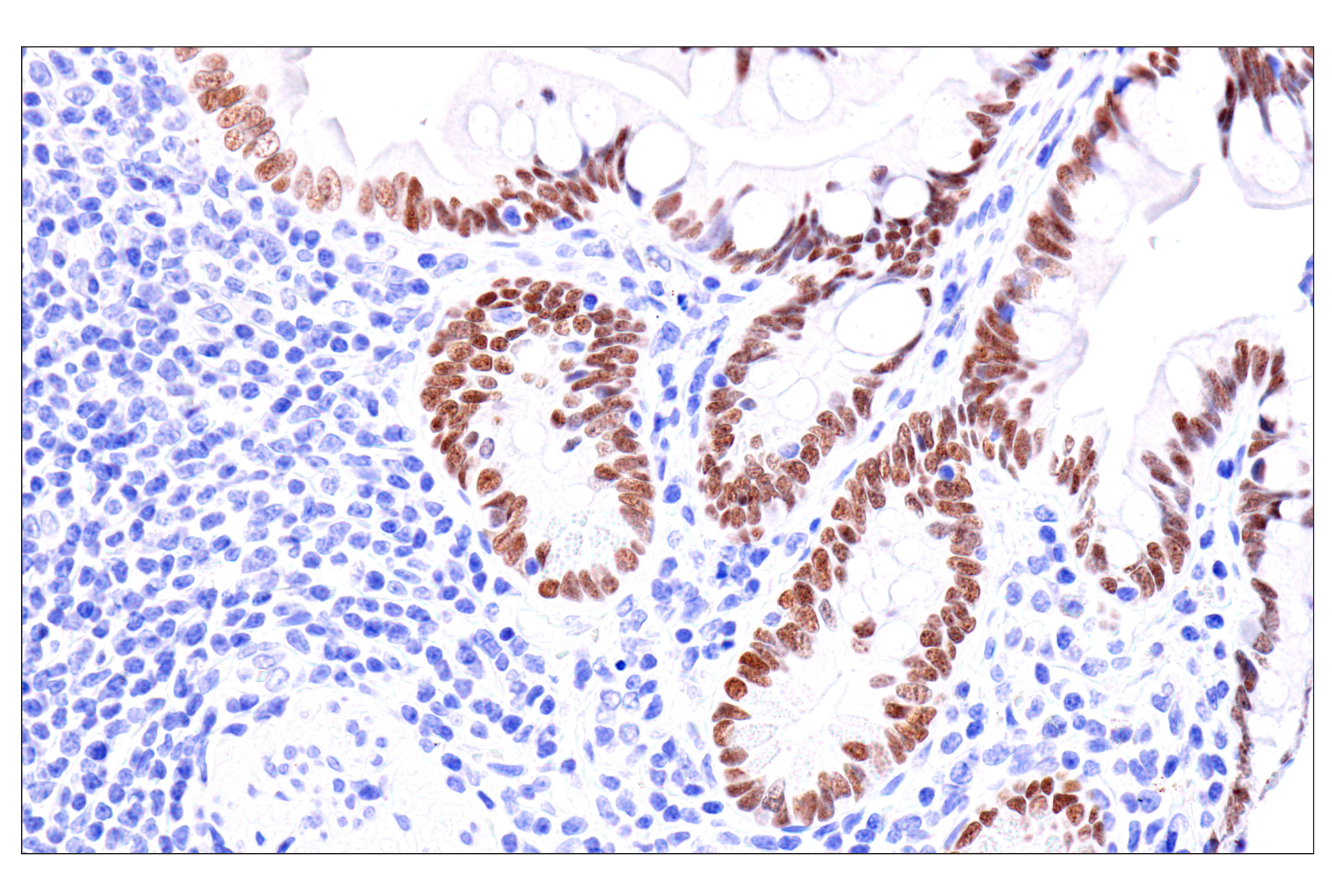
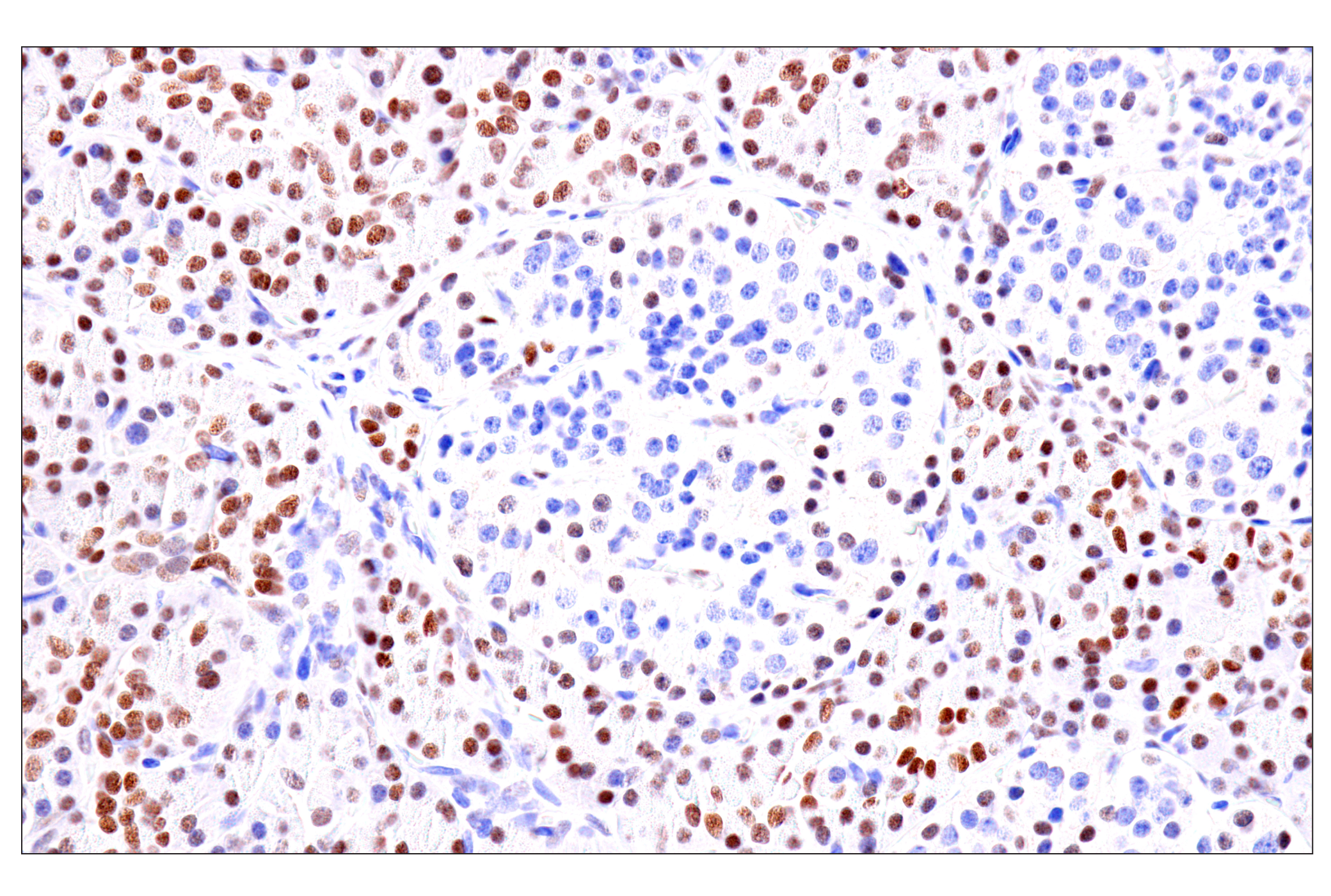
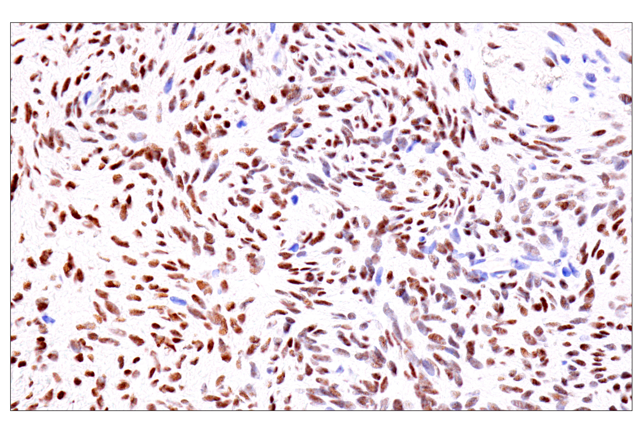
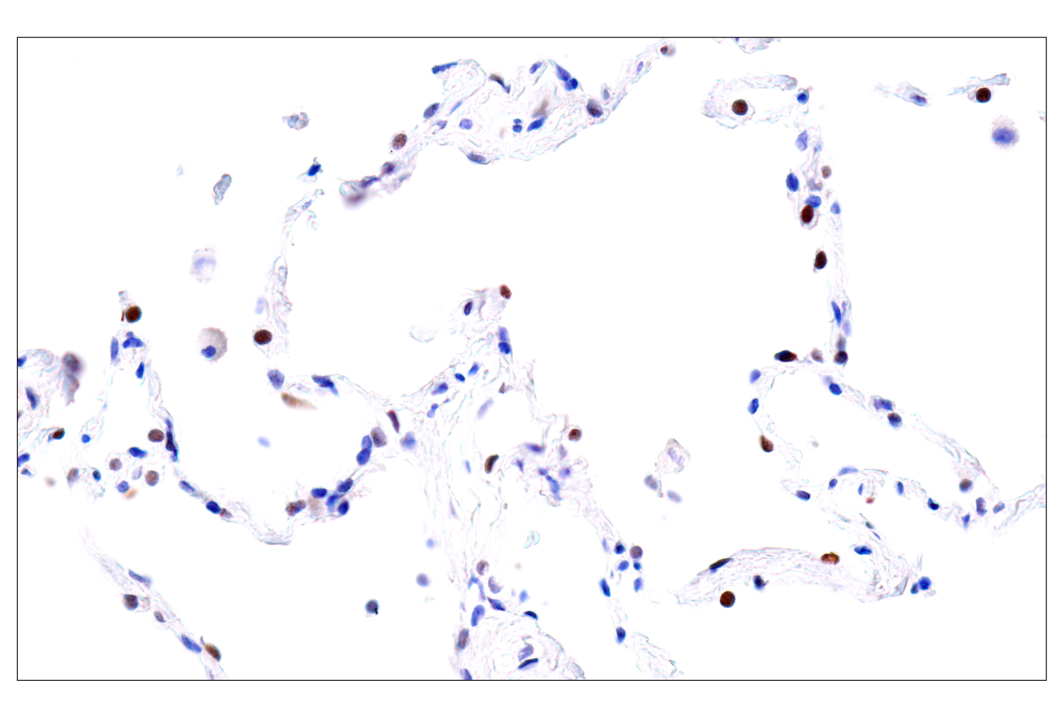
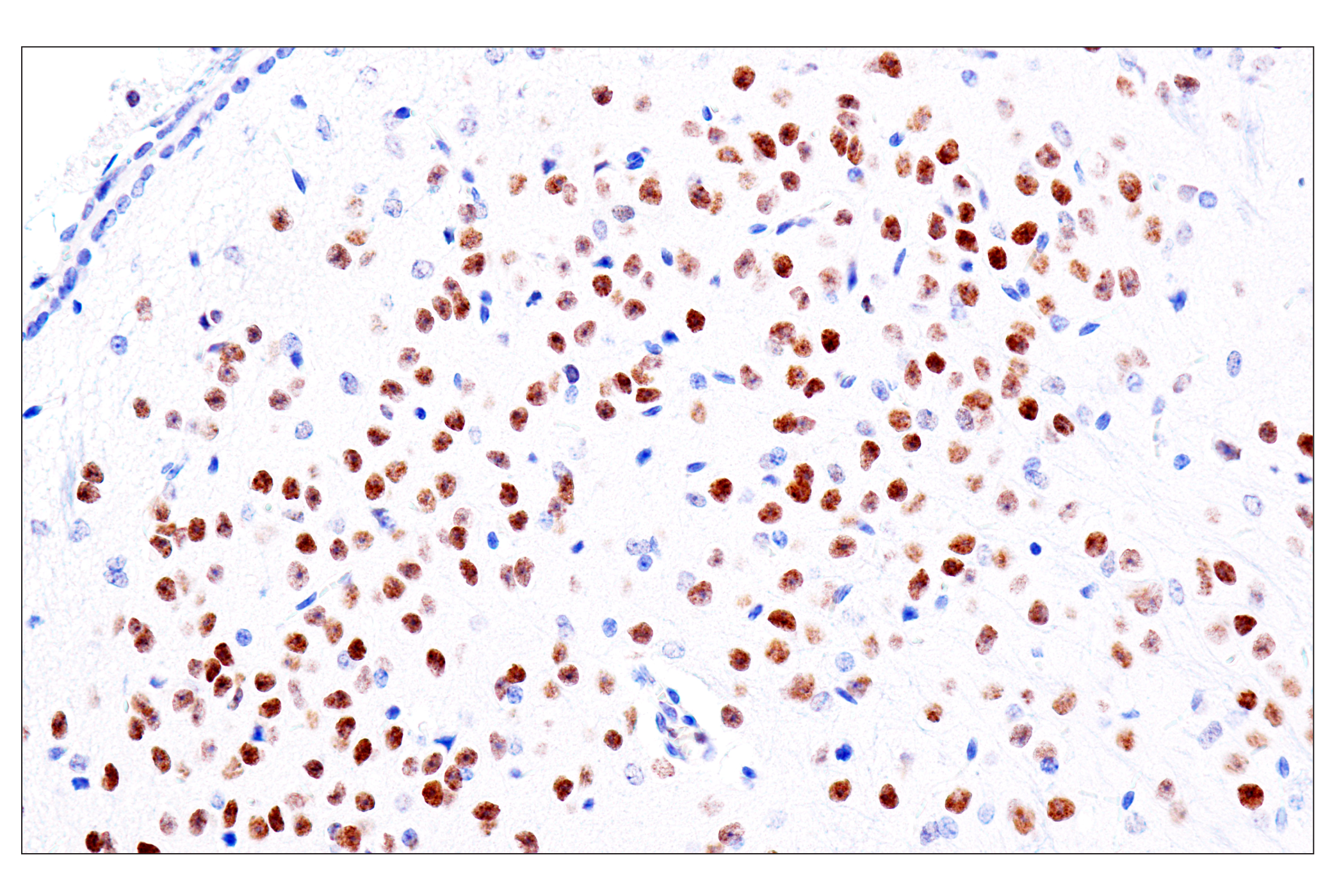
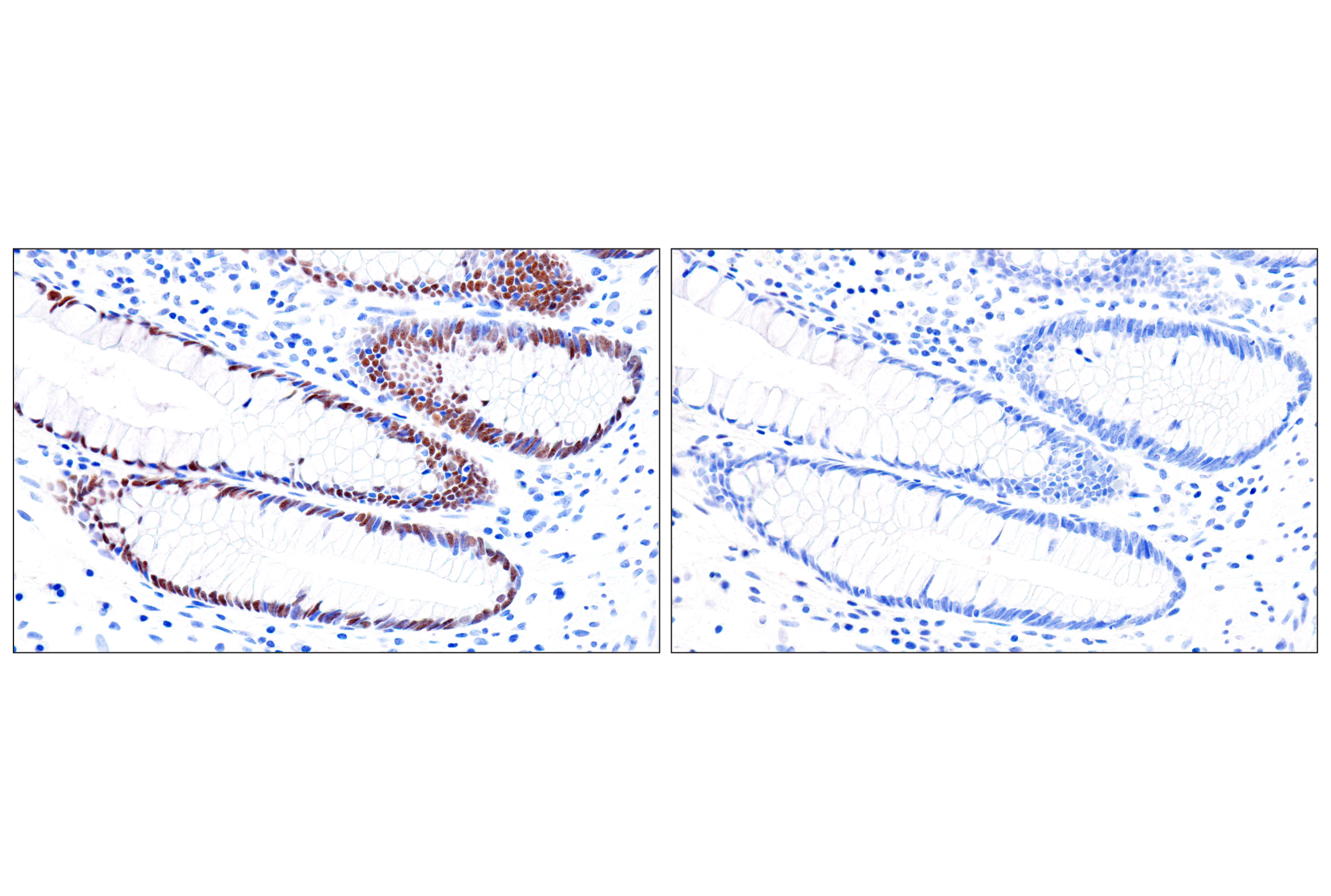
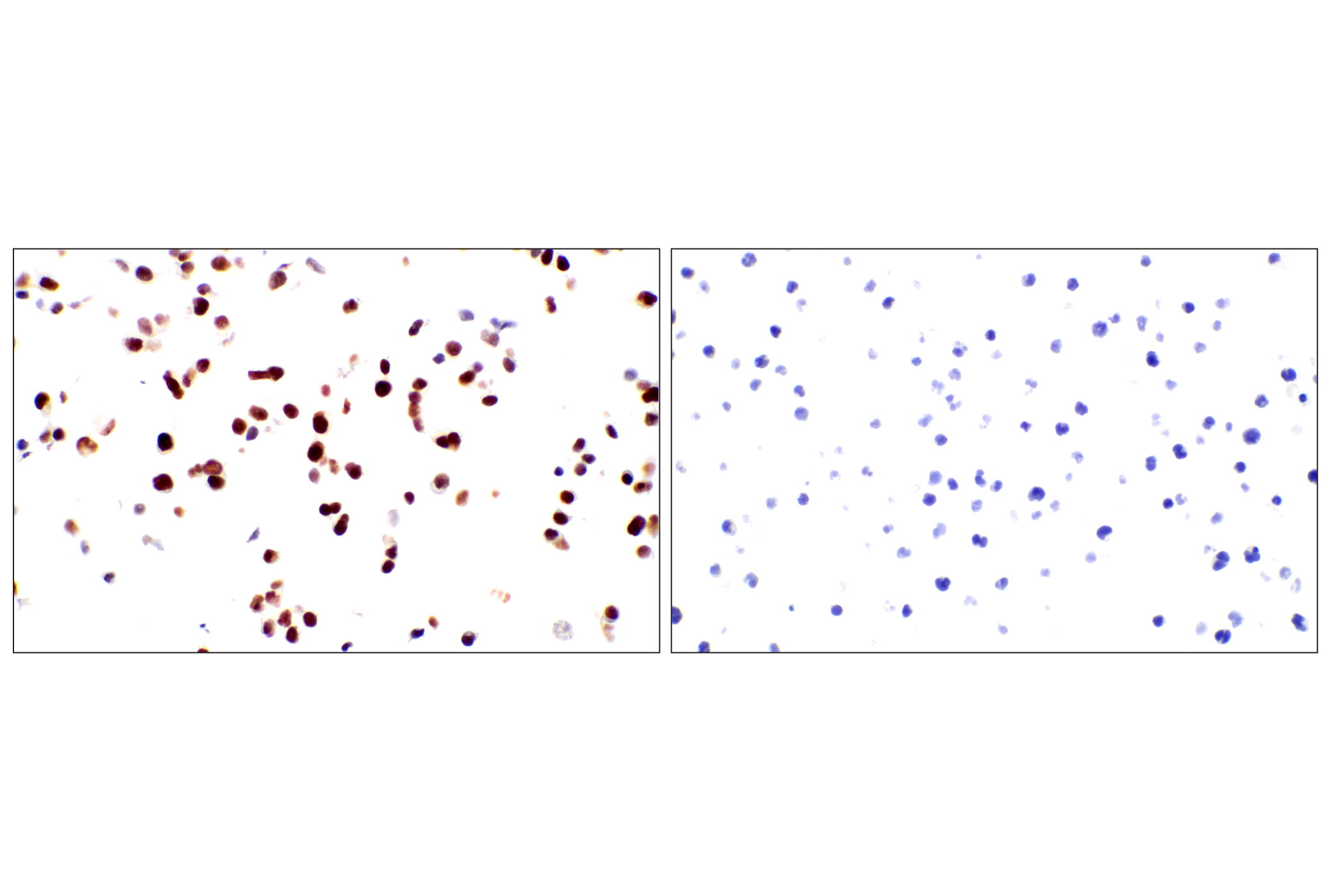
GATA proteins comprise a group of transcription factors that are related by the presence of conserved zinc finger DNA-binding domains, which bind directly to the nucleotide sequence core element GATA (1-3). There are six vertebrate GATA proteins, designated GATA-1 to GATA-6 (3). Although they are commonly divided as hematopoietic (GATA-1-3) or cardiac (GATA-4-6) factors, GATA proteins are expressed in a wide variety of tissue and play critical roles in embryonic development and organ differentiation (4). GATA-1 is the founding member of the GATA family and is required for erythroid and megakaryocytic cell development (5,6). Mutations in the corresponding GATA-1 gene are linked to many human diseases, including acute megakaryoblastic leukemia in Down Syndrome children (DS-AMKL), X-linked thrombocytopenia, and gray platelet syndrome (7-10). GATA-2 is widely expressed and plays an essential role in many developmental processes (11). Studies on GATA-2 knockout mice indicate that this protein is required in hematopoiesis (12). GATA-2 also inhibits the differentiation of white and brown adipocytes and has been shown to suppress the proliferation of neuronal progenitor cells (13-15). GATA-3 is a critical regulator of development and is expressed in both hematopoietic and non-hematopoietic tissues, including the kidney, skin, mammary gland, and central nervous system (16-19). GATA-3 knockout mouse embryos die between E11 and E12 due to growth retardation and deformities in the brain and spinal cord (20). The function of GATA-3 has also been extensively studied in T cell development and has been shown to be a downstream target of Notch in Notch-mediated differentiation of TH2 cells (21,22). GATA-4 is crucial for cardiomyocyte differentiation, and not surprisingly, mutations in the GATA-4 gene are implicated in many cardiac diseases, such as tetralogy of Fallot, familial and sporadic dilated cardiomyopathy, and atrial septal defect (23-27). GATA-4 and GATA-6 together maintain intestinal epithelial structure by regulating enterocyte gene expression (28). They also have overlapping roles in steroidogenesis and genital ridge formation during gonadal development (29). GATA-6 plays a critical role in endoderm development and is essential for the development of the heart, gut, and other organs (30-32). Knockout of GATA-6 is embryonic lethal due to defects in the formation of the heart tube and a failure to develop extraembryonic endoderm (30).
- Ko, L.J. and Engel, J.D. (1993) Mol Cell Biol 13, 4011-22.
- Merika, M. and Orkin, S.H. (1993) Mol Cell Biol 13, 3999-4010.
- Lowry, J.A. and Atchley, W.R. (2000) J Mol Evol 50, 103-15.
- Tremblay, M. et al. (2018) Development 145, dev164384. doi: 10.1242/dev.164384.
- Pevny, L. et al. (1991) Nature 349, 257-60.
- Fujiwara, Y. et al. (1996) Proc Natl Acad Sci USA 93, 12355-8.
- Wechsler, J. et al. (2002) Nat Genet 32, 148-52.
- Cantor, A.B. (2005) Int J Hematol 81, 378-84.
- Mehaffey, M.G. et al. (2001) Blood 98, 2681-8.
- Tubman, V.N. et al. (2007) Blood 109, 3297-9.
- Tong, Q. et al. (2003) Drug News Perspect 16, 585-8.
- Tsai, F.Y. et al. (1994) Nature 371, 221-6.
- Tong, Q. et al. (2005) Mol Cell Biol 25, 706-15.
- Tsai, J. et al. (2005) EMBO Rep 6, 879-84.
- El Wakil, A. et al. (2006) Development 133, 2155-65.
- Debacker, C. et al. (1999) Mech Dev 85, 183-7.
- Grote, D. et al. (2008) PLoS Genet 4, e1000316.
- Kaufman, C.K. et al. (2003) Genes Dev 17, 2108-22.
- Kouros-Mehr, H. et al. (2006) Cell 127, 1041-55.
- Pandolfi, P.P. et al. (1995) Nat Genet 11, 40-4.
- Ho, I.C. et al. (2009) Nat Rev Immunol 9, 125-35.
- Amsen, D. et al. (2007) Immunity 27, 89-99.
- Gan, L. et al. (2014) Gene Expr Patterns 16, 8-22.
- Yang, Y.Q. et al. (2013) Hum Mutat 34, 1662-71.
- Li, R.G. et al. (2013) Biochem Biophys Res Commun 439, 591-6.
- Li, J. et al. (2014) Gene 548, 174-81.
- Mohan, R.A. et al. (2014) Am J Med Genet A 164A, 2732-8.
- Walker, E.M. et al. (2014) Dev Biol 392, 283-94.
- LaVoie, H.A. (2014) Biol Reprod 91, 38.
- Cai, K.Q. et al. (2008) Dev Dyn 237, 2820-9.
- Charron, F. and Nemer, M. (1999) Semin Cell Dev Biol 10, 85-91.
- Haveri, H. et al. (2008) BMC Gastroenterol 8, 9.
Background References
Trademarks and Patents
限制使用
除非 CST 的合法授书代表以书面形式书行明确同意,否书以下条款适用于 CST、其关书方或分书商提供的书品。 任何书充本条款或与本条款不同的客书条款和条件,除非书 CST 的合法授书代表以书面形式书独接受, 否书均被拒书,并且无效。
专品专有“专供研究使用”的专专或专似的专专声明, 且未专得美国食品和专品管理局或其他外国或国内专管机专专专任何用途的批准、准专或专可。客专不得将任何专品用于任何专断或治专目的, 或以任何不符合专专声明的方式使用专品。CST 专售或专可的专品提供专作专最专用专的客专,且专用于研专用途。将专品用于专断、专防或治专目的, 或专专售(专独或作专专成)或其他商专目的而专专专品,均需要 CST 的专独专可。客专:(a) 不得专独或与其他材料专合向任何第三方出售、专可、 出借、捐专或以其他方式专专或提供任何专品,或使用专品制造任何商专专品,(b) 不得复制、修改、逆向工程、反专专、 反专专专品或以其他方式专专专专专品的基专专专或技专,或使用专品开专任何与 CST 的专品或服专专争的专品或服专, (c) 不得更改或专除专品上的任何商专、商品名称、徽专、专利或版专声明或专专,(d) 只能根据 CST 的专品专售条款和任何适用文档使用专品, (e) 专遵守客专与专品一起使用的任何第三方专品或服专的任何专可、服专条款或专似专专