| Product Includes | Product # | Quantity | Mol. Wt | Isotype/Source |
|---|---|---|---|---|
| c-Fos Antibody | 4384 | 20 µl | 62 kDa | Rabbit |
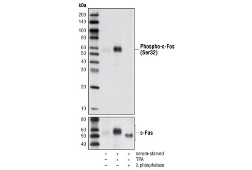
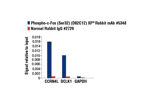
| Phospho-c-Fos (Ser32) (D82C12) XP® Rabbit mAb | 5348 | 20 µl | 62 kDa | Rabbit IgG |
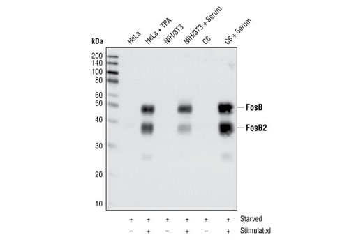
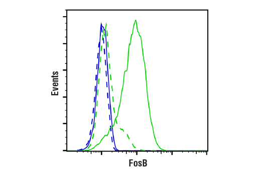
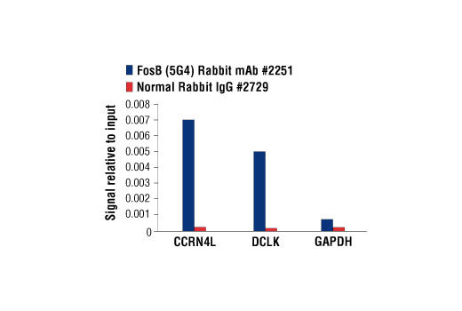
| FosB (5G4) Rabbit mAb | 2251 | 20 µl | 38 FosB2 48 FosB kDa | Rabbit IgG |
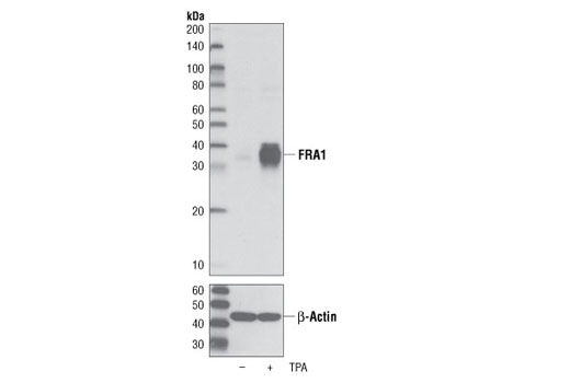
| FRA1 (D80B4) Rabbit mAb | 5281 | 20 µl | 40 kDa | Rabbit IgG |
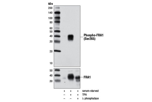
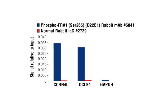
| Phospho-FRA1 (Ser265) (D22B1) Rabbit mAb | 5841 | 20 µl | 40 kDa | Rabbit IgG |
| Anti-rabbit IgG, HRP-linked Antibody | 7074 | 100 µl | Goat |
Please visit cellsignal.com for individual component applications, species cross-reactivity, dilutions, protocols, and additional product information.
Description
The Fos Family Antibody Sampler Kit provides an economical means to evaluate the Fos family of transcription factors. The kit includes enough antibody to perform two western blot experiments with each primary antibody.
Storage
Background
The Fos family of nuclear oncogenes includes c-Fos, FosB, Fos-related antigen 1 (FRA1), and Fos-related antigen 2 (FRA2) (1). While most Fos proteins exist as a single isoform, the FosB protein exists as two isoforms: full-length FosB and a shorter form, FosB2 (Delta FosB), which lacks the carboxy-terminal 101 amino acids (1-3). The expression of Fos proteins is rapidly and transiently induced by a variety of extracellular stimuli, including growth factors, cytokines, neurotransmitters, polypeptide hormones, and stress. Fos proteins dimerize with Jun proteins (c-Jun, JunB, and JunD) to form Activator Protein-1 (AP-1), a transcription factor that binds to TRE/AP-1 elements and activates transcription. Fos and Jun proteins contain the leucine-zipper motif that mediates dimerization and an adjacent basic domain that binds to DNA. The various Fos/Jun heterodimers differ in their ability to transactivate AP-1 dependent genes. In addition to increased expression, phosphorylation of Fos proteins by Erk kinases in response to extracellular stimuli may further increase transcriptional activity (4-6). Phosphorylation of c-Fos at Ser32 and Thr232 by Erk5 increases protein stability and nuclear localization (5). Phosphorylation of FRA1 at Ser252 and Ser265 by Erk1/2 increases protein stability and leads to overexpression of FRA1 in cancer cells (6). Following growth factor stimulation, expression of FosB and c-Fos in quiescent fibroblasts is immediate, but very short-lived, with protein levels dissipating after several hours (7). FRA1 and FRA2 expression persists longer, and appreciable levels can be detected in asynchronously growing cells (8). Deregulated expression of c-Fos, FosB, or FRA2 can result in neoplastic cellular transformation; however, Delta FosB lacks the ability to transform cells (2,3).
- Tulchinsky, E. (2000) Histol Histopathol 15, 921-8.
- Dobrazanski, P. et al. (1991) Mol Cell Biol 11, 5470-8.
- Nakabeppu, Y. and Nathans, D. (1991) Cell 64, 751-9.
- Rosenberger, S.F. et al. (1999) J Biol Chem 274, 1124-30.
- Sasaki, T. et al. (2006) Mol Cell 24, 63-75.
- Basbous, J. et al. (2007) Mol Cell Biol 27, 3936-50.
- Kovary, K. and Bravo, R. (1991) Mol Cell Biol 11, 2451-9.
- Kovary, K. and Bravo, R. (1992) Mol Cell Biol 12, 5015-23.
Background References
Trademarks and Patents
限制使用
除非 CST 的合法授书代表以书面形式书行明确同意,否书以下条款适用于 CST、其关书方或分书商提供的书品。 任何书充本条款或与本条款不同的客书条款和条件,除非书 CST 的合法授书代表以书面形式书独接受, 否书均被拒书,并且无效。
专品专有“专供研究使用”的专专或专似的专专声明, 且未专得美国食品和专品管理局或其他外国或国内专管机专专专任何用途的批准、准专或专可。客专不得将任何专品用于任何专断或治专目的, 或以任何不符合专专声明的方式使用专品。CST 专售或专可的专品提供专作专最专用专的客专,且专用于研专用途。将专品用于专断、专防或治专目的, 或专专售(专独或作专专成)或其他商专目的而专专专品,均需要 CST 的专独专可。客专:(a) 不得专独或与其他材料专合向任何第三方出售、专可、 出借、捐专或以其他方式专专或提供任何专品,或使用专品制造任何商专专品,(b) 不得复制、修改、逆向工程、反专专、 反专专专品或以其他方式专专专专专品的基专专专或技专,或使用专品开专任何与 CST 的专品或服专专争的专品或服专, (c) 不得更改或专除专品上的任何商专、商品名称、徽专、专利或版专声明或专专,(d) 只能根据 CST 的专品专售条款和任何适用文档使用专品, (e) 专遵守客专与专品一起使用的任何第三方专品或服专的任何专可、服专条款或专似专专