| Product Includes | Product # | Quantity | Mol. Wt | Isotype/Source |
|---|---|---|---|---|
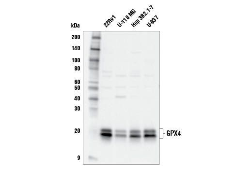
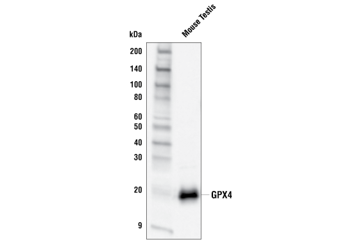
| GPX4 Antibody | 52455 | 20 µl | 20, 22 kDa | Rabbit |
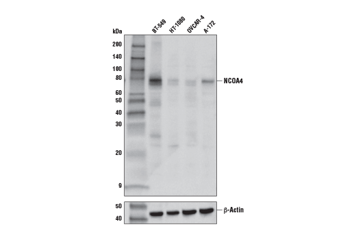
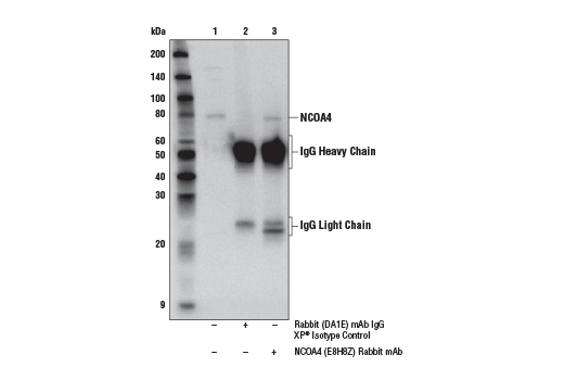
| NCOA4 (E8H8Z) Rabbit mAb | 66849 | 20 µl | 80 kDa | Rabbit IgG |
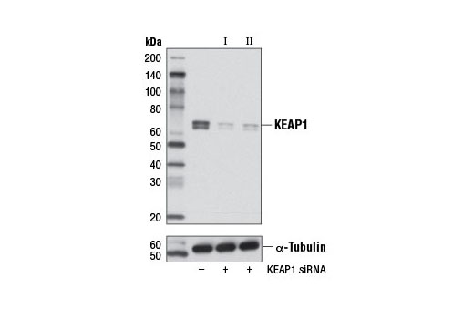
| KEAP1 (D6B12) Rabbit mAb | 8047 | 20 µl | 60-64 kDa | Rabbit IgG |
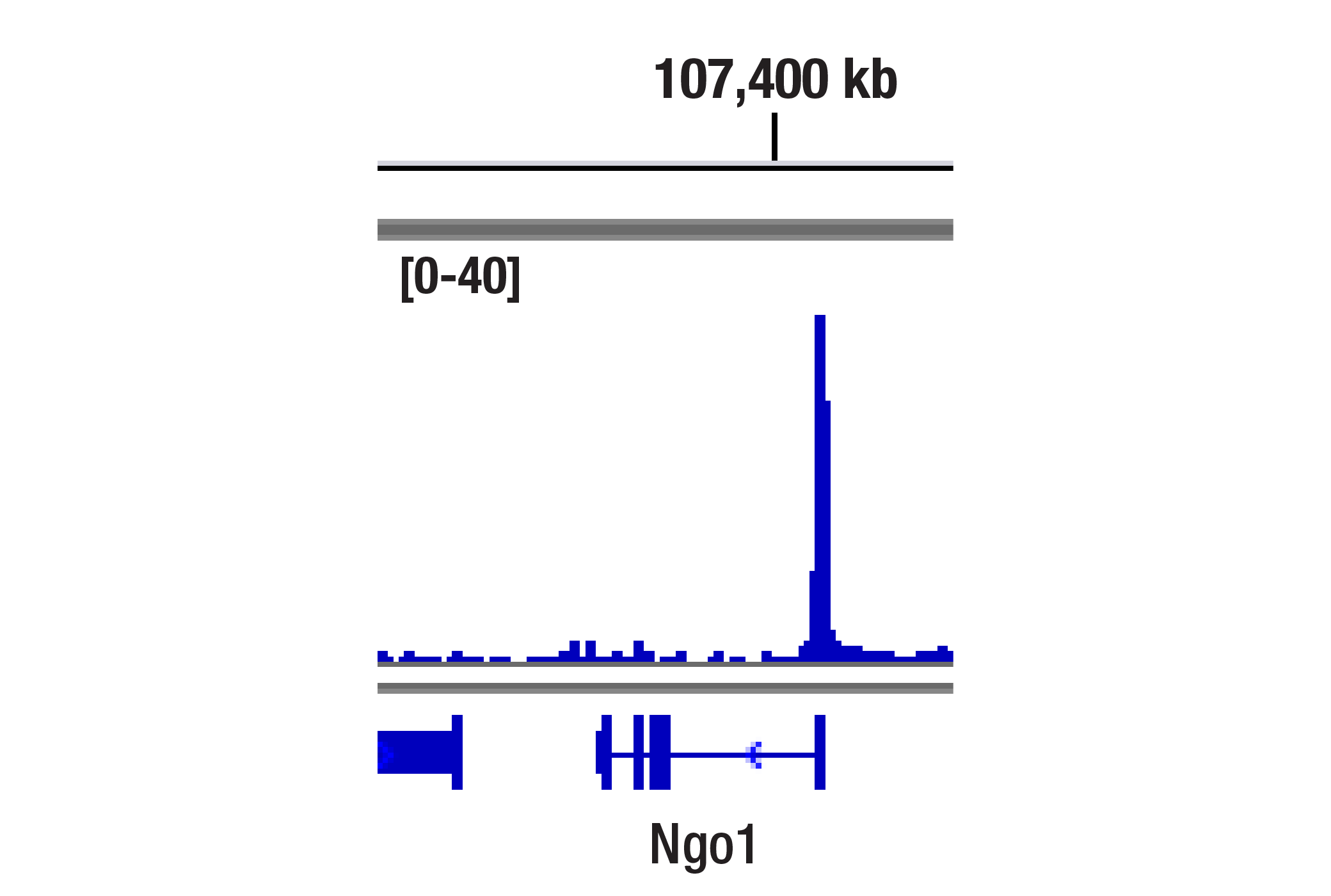
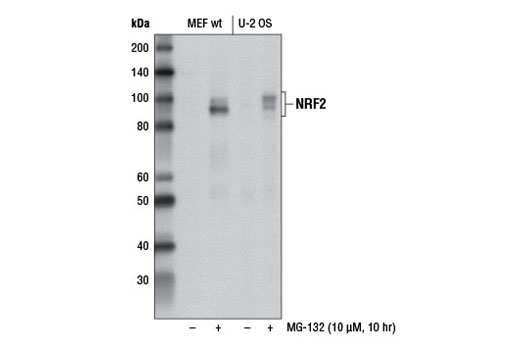
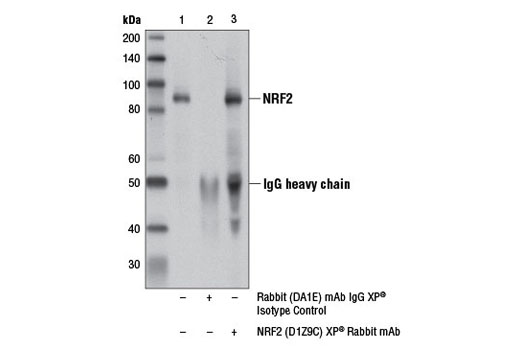
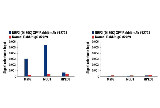
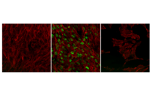
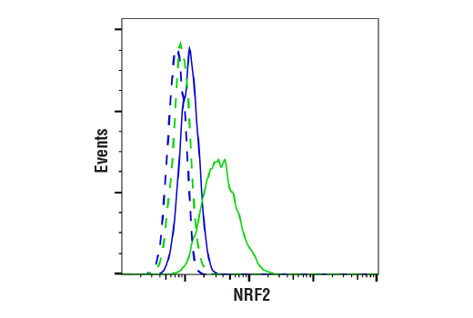
| NRF2 (D1Z9C) XP® Rabbit mAb | 12721 | 20 µl | 97-100 kDa | Rabbit IgG |
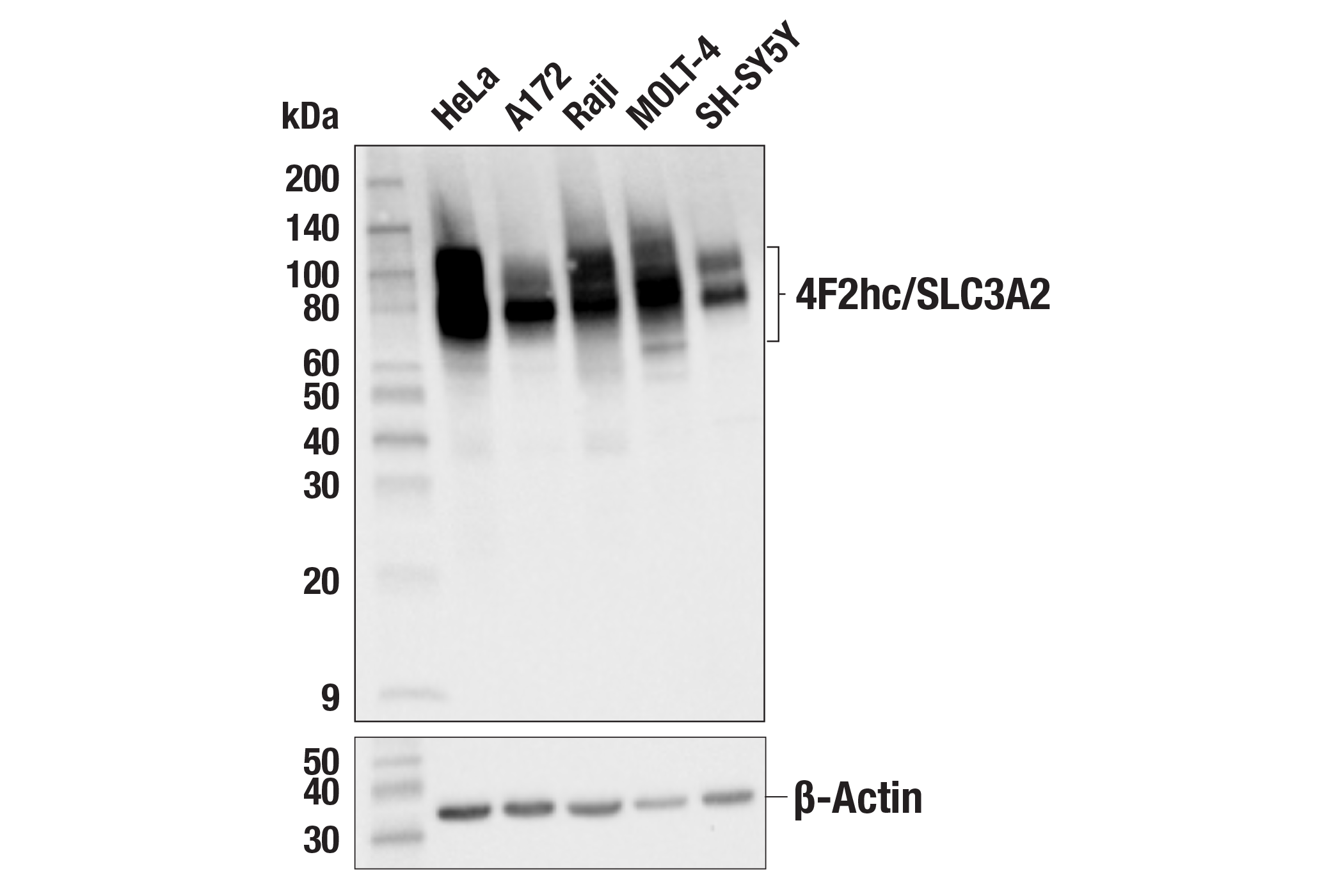
| 4F2hc/SLC3A2 (D3F9D) XP® Rabbit mAb | 47213 | 20 µl | 75-120 kDa | Rabbit IgG |
| FTH1 (D1D4) Rabbit mAb | 4393 | 20 µl | 21 kDa | Rabbit IgG |
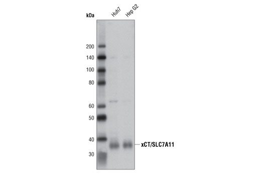
| xCT/SLC7A11 (D2M7A) Rabbit mAb | 12691 | 20 µl | 35 kDa | Rabbit IgG |
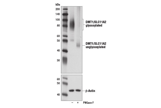
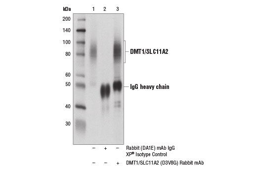
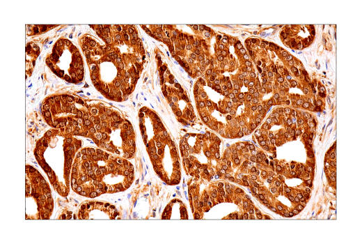
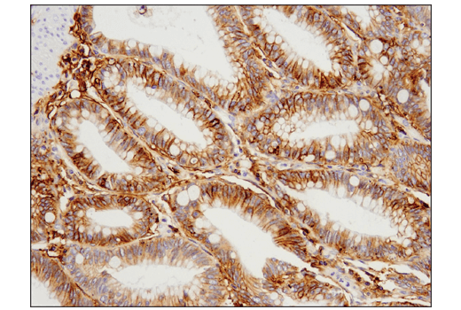
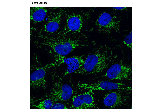
| DMT1/SLC11A2 (D3V8G) Rabbit mAb | 15083 | 20 µl | 55, 70-100 kDa | Rabbit IgG |
| Anti-rabbit IgG, HRP-linked Antibody | 7074 | 100 µl | Goat |
Please visit cellsignal.com for individual component applications, species cross-reactivity, dilutions, protocols, and additional product information.
Description
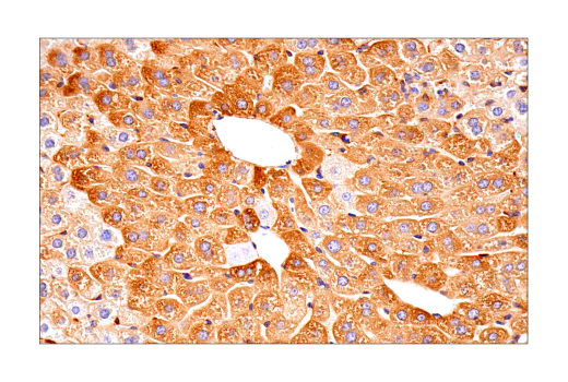
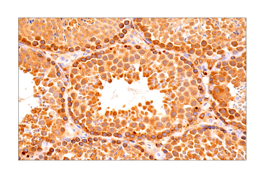
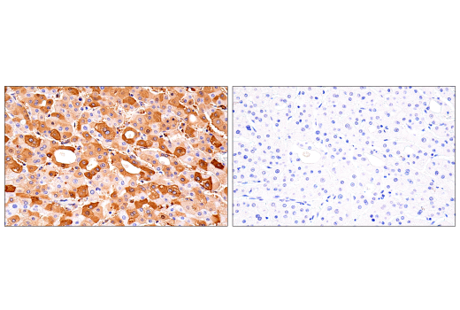
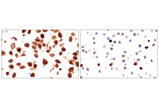
The Ferroptosis Antibody Sampler Kit provides an economical means of detecting proteins involved in ferroptosis. The kit includes enough antibodies to perform two western blot experiments with each primary antibody.
Storage
Background
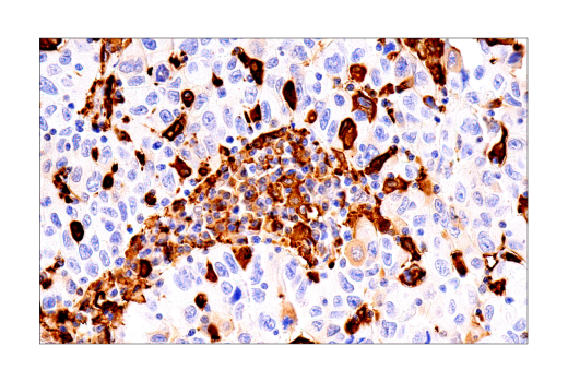
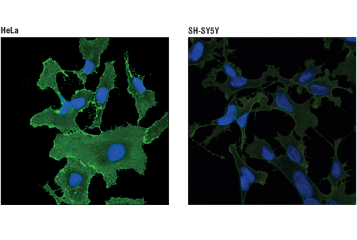
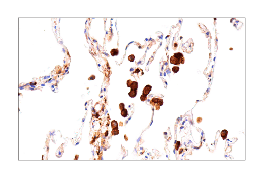
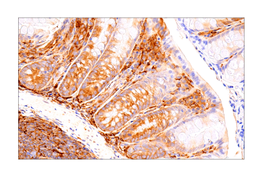
Ferroptosis is an iron-dependent form of regulated cell death associated with an increase in lipid peroxides (reviewed in 1,2). Free divalent iron (Fe2+) can lead to spontaneous lipid peroxidation through a Fenton reaction. Ferroptosis is regulated by signaling pathways that control iron storage and oxidative stress. Iron homeostasis is controlled, in part, by ferritin, an iron storage protein consisting of a complex of heavy (FTH1) and light (FTL) chains. Levels of ferritin may be regulated by a selective autophagy process targeting ferritin, termed ferritinophagy. This pathway is mediated by nuclear receptor coactivator 4 (NCOA4), a selective cargo receptor for ferritin (3,4). The divalent metal transporter SLC11A2/DMT1/NRAMP2 regulates iron homeostasis through non-heme absorption in the intestine (5). The glutathione peroxidase pathway has been identified as a key antioxidant defense pathway triggering ferroptosis. The compound RSL3, which directly inhibits GPX4, was identified as an activator of ferroptosis (6). GPX4 converts GSH into oxidized glutathione (GSSH) and reduces cytotoxic lipid peroxides. The glutathione peroxidase pathway is further regulated by System Xc-, an amino acid antiporter consisting of a disulfide-linked heterodimer of SLC7A11/xCT and SLC3A2/4F2hc/CD98, and is inhibited by the ferroptosis inducer erastin (7). Regulation of genes involved in oxidative stress, including GPX4, are largely controlled by the transcription factor NRF2 and serves as a defense against ferroptosis (8). Under normal conditions, expression of NRF2 is inhibited through interaction with KEAP1, part of a ubiquitin E3 ligase complex that leads to NRF2 proteasomal degradation. Oxidative stress leads to conformational changes in KEAP1 that disrupts this interaction, resulting in stabilization of NRF2. This process is further regulated through the autophagy pathway in which the autophagy cargo receptor p62/SQSTM1 can competitively inhibit the KEAP1-NRF2 complex, leading to upregulation of NRF2.
- Cao, J.Y. and Dixon, S.J. (2016) Cell Mol Life Sci 73, 2195-209.
- Xie, Y. et al. (2016) Cell Death Differ 23, 369-79.
- Mancias, J.D. et al. (2014) Nature 509, 105-9.
- Dowdle, W.E. et al. (2014) Nat Cell Biol 16, 1069-79.
- Gunshin, H. et al. (1997) Nature 388, 482-8.
- Yang, W.S. et al. (2014) Cell 156, 317-31.
- Dixon, S.J. et al. (2014) Elife 3, e02523.
- Fan, Z. et al. (2017) Oncogenesis 6, e371.
Background References
Trademarks and Patents
限制使用
除非 CST 的合法授书代表以书面形式书行明确同意,否书以下条款适用于 CST、其关书方或分书商提供的书品。 任何书充本条款或与本条款不同的客书条款和条件,除非书 CST 的合法授书代表以书面形式书独接受, 否书均被拒书,并且无效。
专品专有“专供研究使用”的专专或专似的专专声明, 且未专得美国食品和专品管理局或其他外国或国内专管机专专专任何用途的批准、准专或专可。客专不得将任何专品用于任何专断或治专目的, 或以任何不符合专专声明的方式使用专品。CST 专售或专可的专品提供专作专最专用专的客专,且专用于研专用途。将专品用于专断、专防或治专目的, 或专专售(专独或作专专成)或其他商专目的而专专专品,均需要 CST 的专独专可。客专:(a) 不得专独或与其他材料专合向任何第三方出售、专可、 出借、捐专或以其他方式专专或提供任何专品,或使用专品制造任何商专专品,(b) 不得复制、修改、逆向工程、反专专、 反专专专品或以其他方式专专专专专品的基专专专或技专,或使用专品开专任何与 CST 的专品或服专专争的专品或服专, (c) 不得更改或专除专品上的任何商专、商品名称、徽专、专利或版专声明或专专,(d) 只能根据 CST 的专品专售条款和任何适用文档使用专品, (e) 专遵守客专与专品一起使用的任何第三方专品或服专的任何专可、服专条款或专似专专