| Product Includes | Product # | Quantity | Mol. Wt | Isotype/Source |
|---|---|---|---|---|
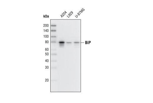
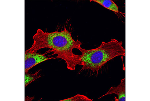
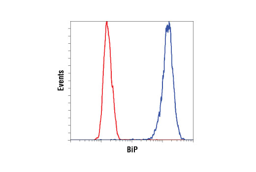
| BiP (C50B12) Rabbit mAb | 3177 | 20 µl | 78 kDa | Rabbit IgG |
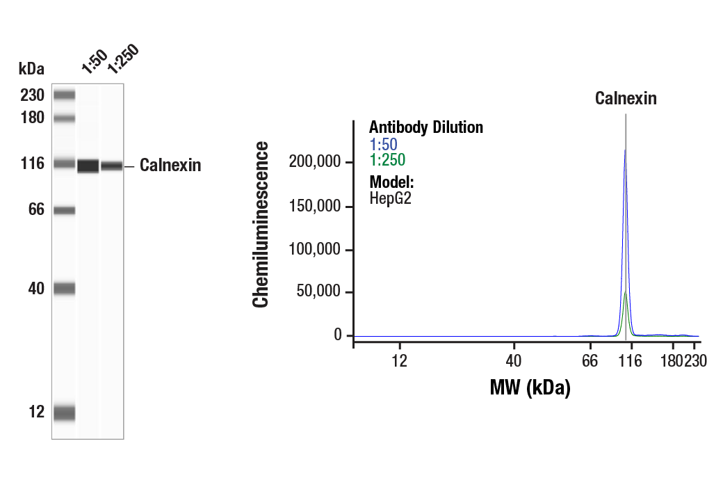
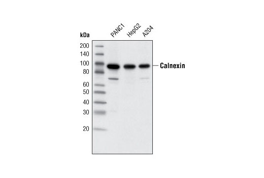
| Calnexin (C5C9) Rabbit mAb | 2679 | 20 µl | 90 kDa | Rabbit IgG |
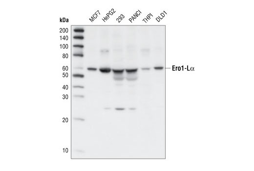
| Ero1-Lα Antibody | 3264 | 20 µl | 60 kDa | Rabbit |
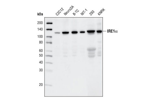
| IRE1α (14C10) Rabbit mAb | 3294 | 20 µl | 130 kDa | Rabbit IgG |
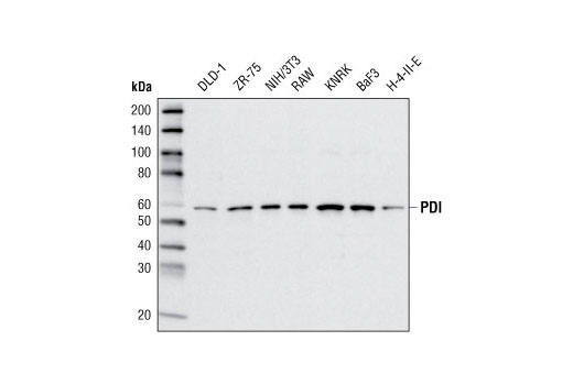
| PDI (C81H6) Rabbit mAb | 3501 | 20 µl | 57 kDa | Rabbit |
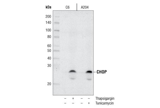
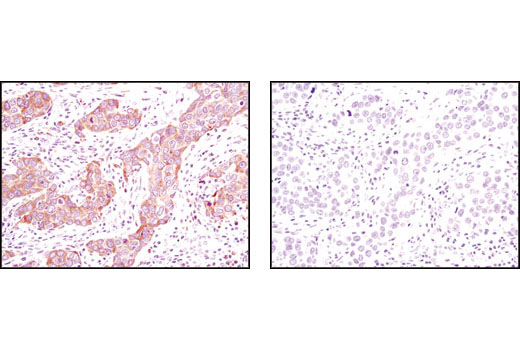
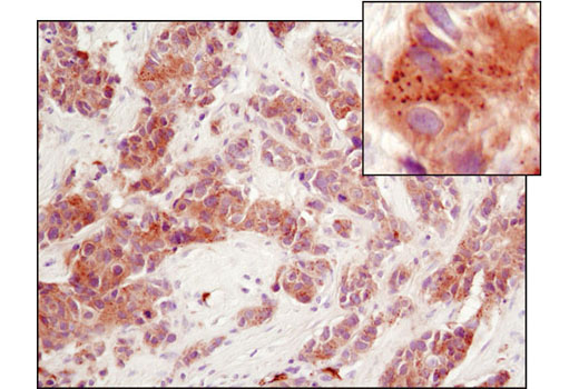
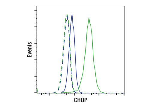
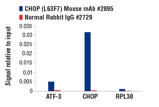
| CHOP (L63F7) Mouse mAb | 2895 | 20 µl | 27 kDa | Mouse IgG2a |
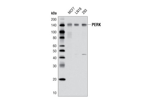
| PERK (D11A8) Rabbit mAb | 5683 | 20 µl | 140 kDa | Rabbit IgG |
| Anti-rabbit IgG, HRP-linked Antibody | 7074 | 100 µl | Goat | |
| Anti-mouse IgG, HRP-linked Antibody | 7076 | 100 µl | Horse |
Please visit cellsignal.com for individual component applications, species cross-reactivity, dilutions, protocols, and additional product information.
Description
The ER Stress Sampler Kit contains reagents to investigate ER stress within the cell. The kit contains enough primary and secondary antibodies to perform two Western blot experiments per primary antibody.
Storage
Background
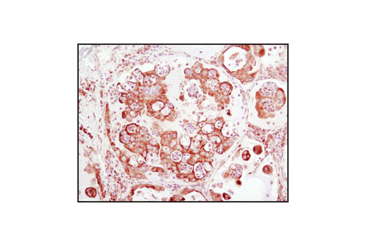
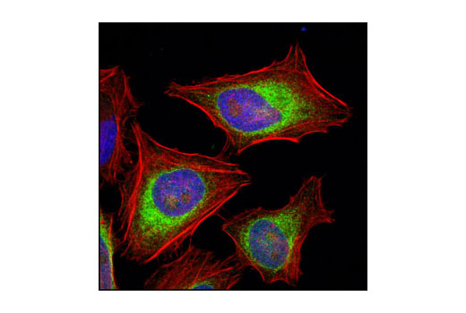
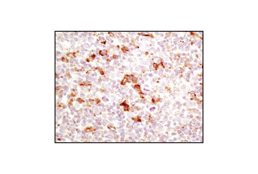
Secretory and transmembrane proteins are synthesized on polysomes and translocate into the endoplasmic reticulum (ER) where they are often modified by the formation of disulfide bonds, amino-linked glycosylation and folding. The ER contains a pool of molecular chaperone proteins including calnexin, BiP and protein disulfide isomerase (PDI). Calnexin is an ER membrane, calcium-binding protein that retains newly synthesized glycoproteins inside the ER to ensure proper folding and quality control (1,2). Irregular protein folding within the ER increases BiP synthesis, which binds misfolded proteins to prevent them from forming aggregates and to assist them to refold properly (3).
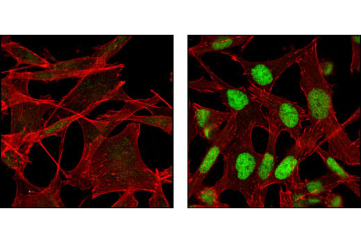
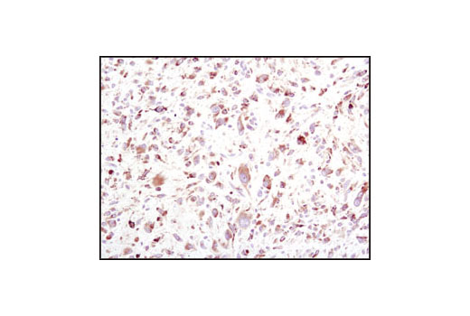
PDI catalyzes the formation and isomerization of disulfide bonds required for a protein to reach its native state (4). Studies have found that the resident ER protein endoplasmic oxidoreductin-1 (Ero1) provides oxidizing potential to the ER in Saccharomyces cerevisiae (5). Ero1-Lα is an ER membrane-associated N-glycoprotein that promotes oxidative protein folding (6). Disruptions of ER homeostasis leads to the accumulation of unfolded proteins. The ER has developed an adaptive mechanism called the unfolded protein response (UPR) to counteract compromised protein folding (7). This is regulated by proteins such as the membrane-bound transcription factor protease site 2 (MBTPS2) and the serine/threonine kinase IRE1 (8-12). The PERK eIF2α kinase is an ER resident transmembrane protein that couples ER stress signals to translation inhibition. ER stress increases PERK activity, which phosphorylates eIF2α to reduce protein translation. PERK activation during ER stress correlates with autophosphorylation of its cytoplasmic kinase domain (13,14). Phosphorylation of PERK at Thr980 can serve as a marker for its activation status.
During ER stress, the level of CHOP expression is elevated and CHOP functions to mediate programmed cell death (15).
- Bergeron, J.J. et al. (1994) Trends Biochem. Sci. 19, 124-128.
- Williams, D.B. (2006) J. Cell Sci. 119, 615-623.
- Kohno, K. et al. (1993) Mol. Cell. Biol. 13, 877-890.
- Ellgaard, L. and Ruddock, L.W. (2005) EMBO Rep. 6, 28-32.
- Frand, A.R. and Kaiser, C.A. (1998) Mol. Cell 1, 161-170.
- Cabibbo, A. et al. (2000) J. Biol. Chem. 275, 4827-4833.
- Kaufman, R.J. et al. (2002) Nat. Rev. Mol. Cell Biol. 3, 411-421.
- Nikawa, J. and Yamashita, S. (1992) Mol. Microbiol. 6, 1441-1446.
- Cox, J.S. et al. (1993) Cell 73, 1197-1206.
- Mori, K. et al. (1993) Cell 74, 743-756.
- Lee, K. et al. (2002) Genes Dev. 16, 452-466.
- Shen, J. and Prywes, R. (2004) J. Biol. Chem. 279, 43046-43051.
- Harding, H.P. et al. (1999) Nature 397, 271-274.
- Shi, Y. et al. (1998) Mol. Cell. Biol. 18, 7499-7509.
- Zinszner, H. et al. (1998) Genes Dev 12, 982-95.
Background References
Trademarks and Patents
限制使用
除非 CST 的合法授书代表以书面形式书行明确同意,否书以下条款适用于 CST、其关书方或分书商提供的书品。 任何书充本条款或与本条款不同的客书条款和条件,除非书 CST 的合法授书代表以书面形式书独接受, 否书均被拒书,并且无效。
专品专有“专供研究使用”的专专或专似的专专声明, 且未专得美国食品和专品管理局或其他外国或国内专管机专专专任何用途的批准、准专或专可。客专不得将任何专品用于任何专断或治专目的, 或以任何不符合专专声明的方式使用专品。CST 专售或专可的专品提供专作专最专用专的客专,且专用于研专用途。将专品用于专断、专防或治专目的, 或专专售(专独或作专专成)或其他商专目的而专专专品,均需要 CST 的专独专可。客专:(a) 不得专独或与其他材料专合向任何第三方出售、专可、 出借、捐专或以其他方式专专或提供任何专品,或使用专品制造任何商专专品,(b) 不得复制、修改、逆向工程、反专专、 反专专专品或以其他方式专专专专专品的基专专专或技专,或使用专品开专任何与 CST 的专品或服专专争的专品或服专, (c) 不得更改或专除专品上的任何商专、商品名称、徽专、专利或版专声明或专专,(d) 只能根据 CST 的专品专售条款和任何适用文档使用专品, (e) 专遵守客专与专品一起使用的任何第三方专品或服专的任何专可、服专条款或专似专专