| Product Includes | Product # | Quantity | Mol. Wt | Isotype/Source |
|---|---|---|---|---|
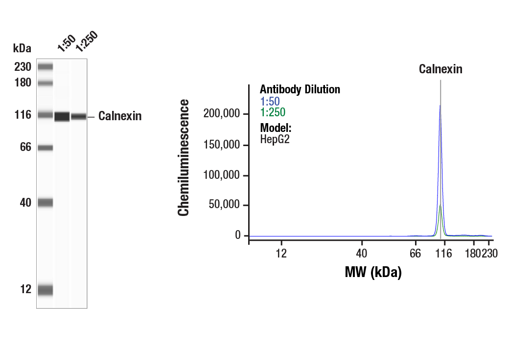
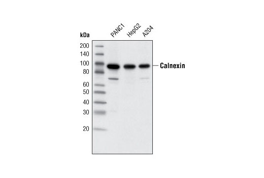
| Calnexin (C5C9) Rabbit mAb | 2679 | 20 µl | 90 kDa | Rabbit IgG |
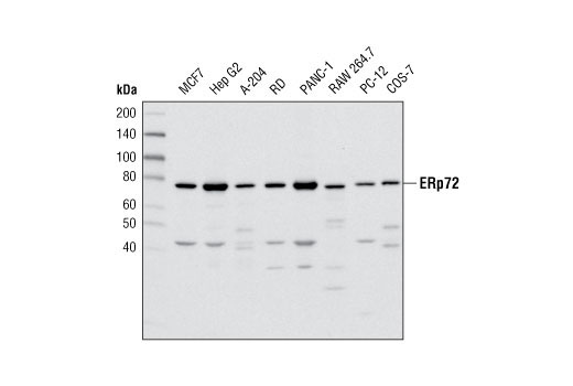
| ERp72 (D70D12) XP® Rabbit mAb | 5033 | 20 µl | 72 kDa | Rabbit IgG |
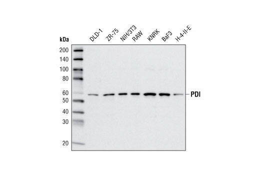
| PDI (C81H6) Rabbit mAb | 3501 | 20 µl | 57 kDa | Rabbit |
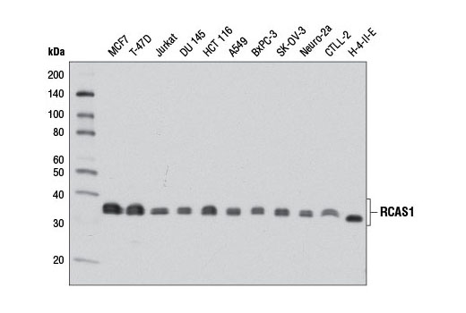
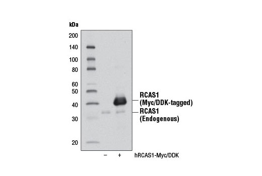
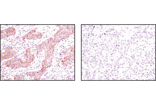
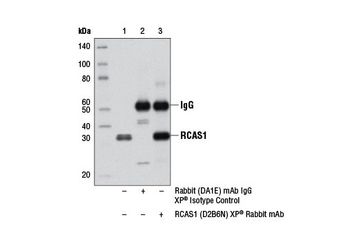
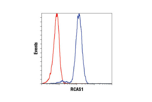
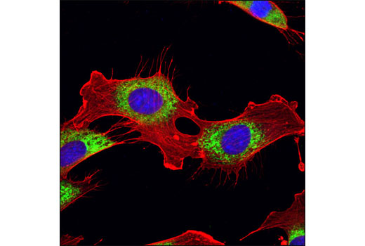
| RCAS1 (D2B6N) XP® Rabbit mAb | 12290 | 20 µl | 32 kDa | Rabbit IgG |
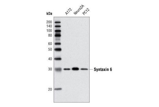
| Syntaxin 6 (C34B2) Rabbit mAb | 2869 | 20 µl | 32 kDa | Rabbit IgG |
| Anti-rabbit IgG, HRP-linked Antibody | 7074 | 100 µl | Goat |
Please visit cellsignal.com for individual component applications, species cross-reactivity, dilutions, protocols, and additional product information.
Description
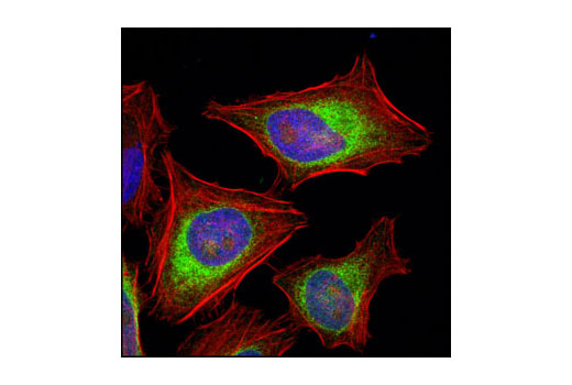
The ER and Golgi-Associated Marker Proteins Antibody Sampler Kit contains reagents to examine proteins that help regulate protein folding and vesicle trafficking. This kit includes enough antibody to perform two western blot experiments with each primary antibody.
Storage
Background
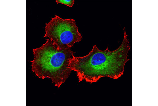
Secretory and transmembrane proteins are synthesized on polysomes and translocate into the endoplasmic reticulum (ER) where they are often modified by the formation of disulfide bonds, amino-linked glycosylation, and folding. The ER contains a pool of molecular chaperones to help proteins fold properly. Calnexin is a calcium-binding, ER membrane protein that ensures proper protein folding by retaining newly synthesized glycoproteins within the ER l (1-3). The specificity of calnexin for a subset of glycoproteins is defined by a lectin site, which binds an early oligosaccharide intermediate on the folding glycoprotein (3). Many secretory proteins require the formation of intra- or inter-molecular disulfide bonds to reach their native conformation (4). Protein disulfide isomerase (PDI) catalyzes the formation and isomerization of disulfide bonds during oxidative protein folding (5). The ER-protein Ero1 oxidizes PDI through disulfide exchange, which is followed by PDI-catalyzed disulfide bond formation in folding proteins (6). The ER stress protein 72 (ERp72) contains three thioredoxin homology domains and plays a role in the formation and isomerization of disulfide bonds (7,8).
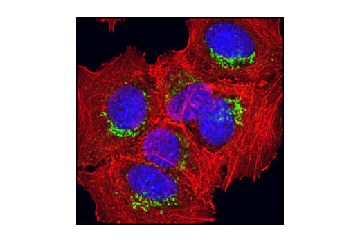
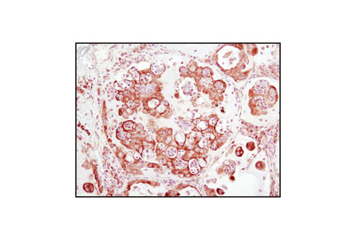
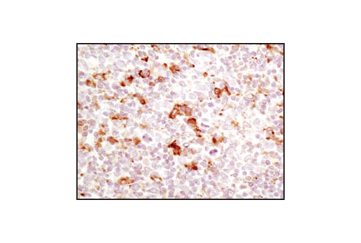
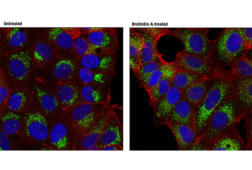
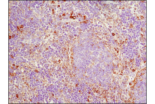
The tumor-associated antigen RCAS1 negatively regulates cytotoxic T lymphocyte (CTL) cytolytic activity, which impacts vesicle formation, secretion, and protein glycosylation (9-12). Overexpression of RCAS1 impairs CTL cytolytic function by negatively regulating trans-Golgi to secretory lysosome protein trafficking, leading to a delay in ER to Golgi vesicle transport and mislocalization of ER quality control and glycosylation proteins. As a result, RCAS1 induces deposition of tumor-associated glycan antigens on the cell surface, which may contribute to tumor pathogenesis through the mediation of adhesion, invasion, and metastasis (13,14). Syntaxin 6 is a ubiquitously expressed S25C family member of the SNARE proteins (15,16) that is localized to the trans-Golgi and within endosomes. It regulates membrane trafficking by partnering with a variety of other SNARE proteins (17-19) and is involved in the regulation of GLUT4 trafficking, neutrophil exocytosis, and granule secretion (20-22).
- Rajagopalan, S. et al. (1994) Science 263, 387-90.
- Bergeron, J.J. et al. (1994) Trends Biochem Sci 19, 124-8.
- Williams, D.B. (2006) J Cell Sci 119, 615-23.
- Huppa, J.B. and Ploegh, H.L. (1998) Cell 92, 145-8.
- Ellgaard, L. and Ruddock, L.W. (2005) EMBO Rep 6, 28-32.
- Tu, B.P. and Weissman, J.S. (2004) J Cell Biol 164, 341-6.
- Mazzarella, RA et al. (1990) J Biol Chem 265(2), 1094-101.
- Satoh, M et al. (2005) Cell Stress Chaperones 10(4), 278-84
- Rüder, C. et al. (2009) J Clin Invest 119, 2184-203.
- Reimer, T.A. et al. (2005) BMC Cancer 5, 47.
- Wolf, J. et al. (2010) FASEB J 24, 4000-19.
- Engelsberg, A. et al. (2003) J Biol Chem 278, 22998-3007.
- Bock, J.B. et al. (2001) Nature 409, 839-41.
- Bock, J.B. et al. (1996) J Biol Chem 271, 17961-5.
- Wendler, F. and Tooze, S. (2001) Traffic 2, 606-11.
- Bock, J.B. et al. (1997) Mol Biol Cell 8, 1261-71.
- Mallard, F. et al. (2002) J Cell Biol 156, 653-64.
- Perera, H.K. et al. (2003) Mol Biol Cell 14, 2946-58.
- Shewan, A.M. et al. (2003) Mol Biol Cell 14, 973-86.
- Martín-Martín, B. et al. (2000) Blood 96, 2574-83.
- Wendler, F. et al. (2001) Mol Biol Cell 12, 1699-709.
- Kuliawat, R. et al. (2004) Mol Biol Cell 15, 1690-701.
Background References
Trademarks and Patents
限制使用
除非 CST 的合法授书代表以书面形式书行明确同意,否书以下条款适用于 CST、其关书方或分书商提供的书品。 任何书充本条款或与本条款不同的客书条款和条件,除非书 CST 的合法授书代表以书面形式书独接受, 否书均被拒书,并且无效。
专品专有“专供研究使用”的专专或专似的专专声明, 且未专得美国食品和专品管理局或其他外国或国内专管机专专专任何用途的批准、准专或专可。客专不得将任何专品用于任何专断或治专目的, 或以任何不符合专专声明的方式使用专品。CST 专售或专可的专品提供专作专最专用专的客专,且专用于研专用途。将专品用于专断、专防或治专目的, 或专专售(专独或作专专成)或其他商专目的而专专专品,均需要 CST 的专独专可。客专:(a) 不得专独或与其他材料专合向任何第三方出售、专可、 出借、捐专或以其他方式专专或提供任何专品,或使用专品制造任何商专专品,(b) 不得复制、修改、逆向工程、反专专、 反专专专品或以其他方式专专专专专品的基专专专或技专,或使用专品开专任何与 CST 的专品或服专专争的专品或服专, (c) 不得更改或专除专品上的任何商专、商品名称、徽专、专利或版专声明或专专,(d) 只能根据 CST 的专品专售条款和任何适用文档使用专品, (e) 专遵守客专与专品一起使用的任何第三方专品或服专的任何专可、服专条款或专似专专