WB, IP, IHC-Bond, IHC-P, IF-IC
H M R Mk
Endogenous
125
Rabbit IgG
#P29317
1969
Product Information
Product Usage Information
| Application | Dilution |
|---|---|
| Western Blotting | 1:1000 |
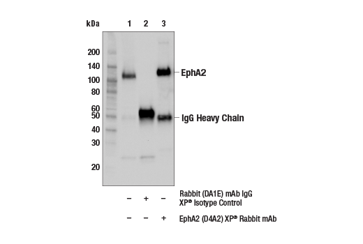
| Immunoprecipitation | 1:100 |
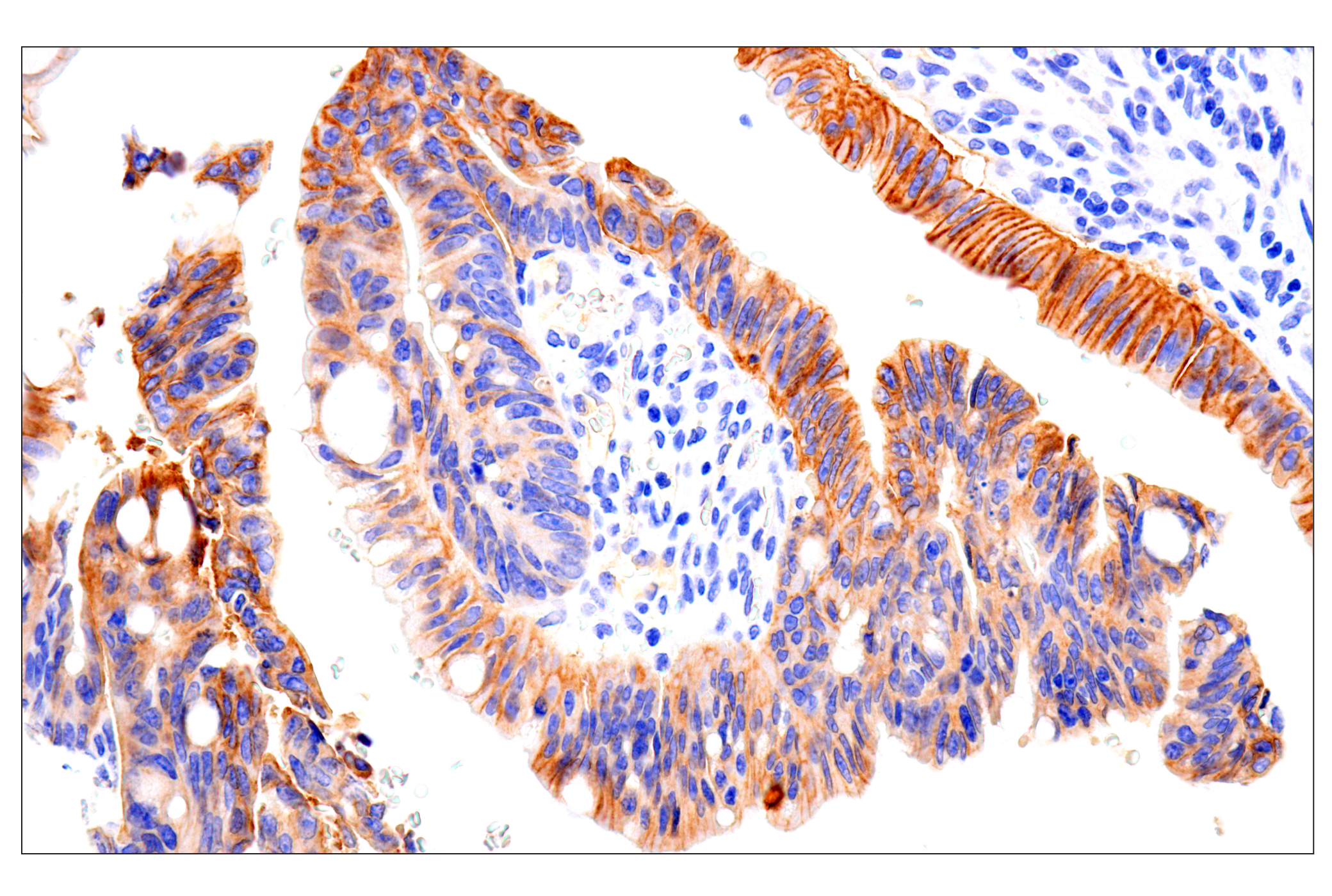
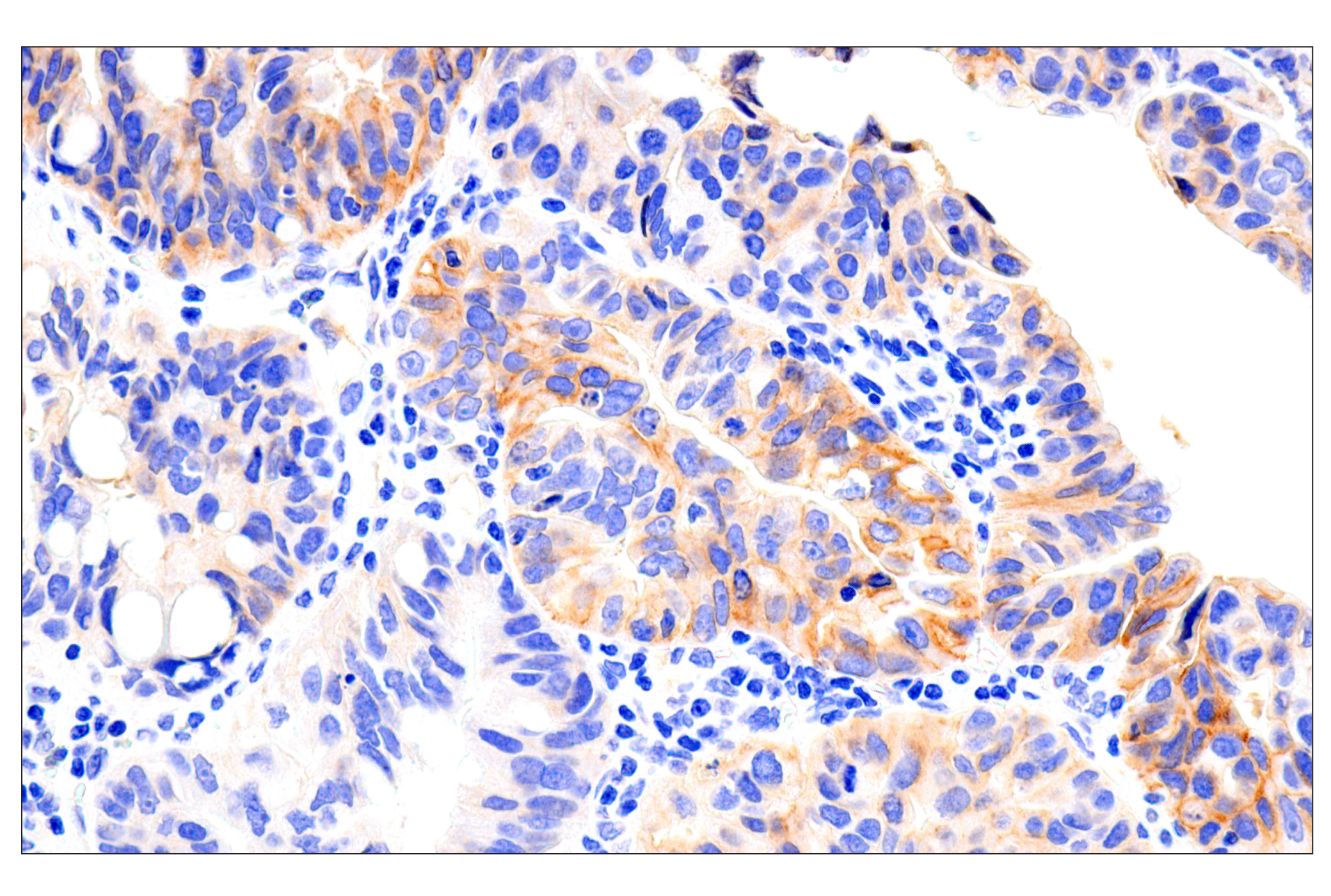
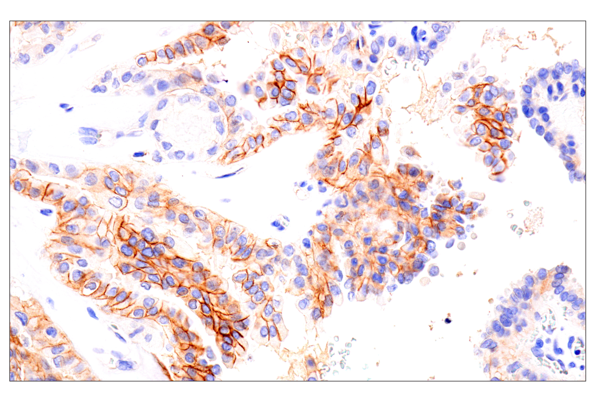
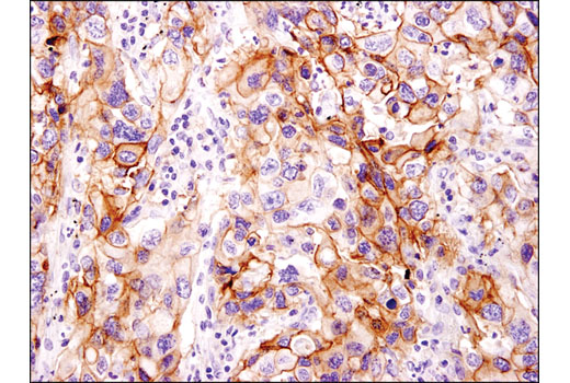
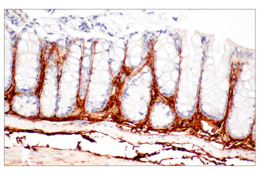
| IHC Leica Bond | 1:50 - 1:200 |
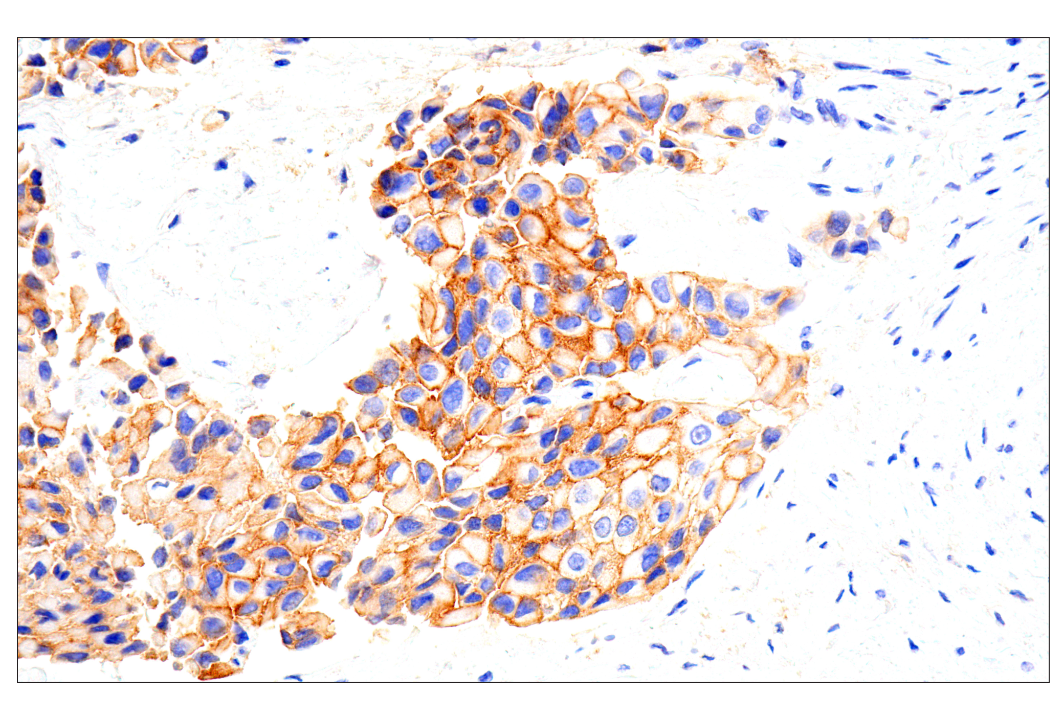
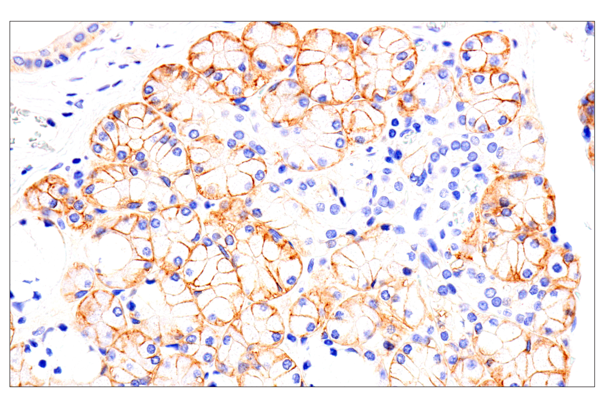
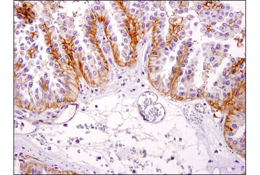
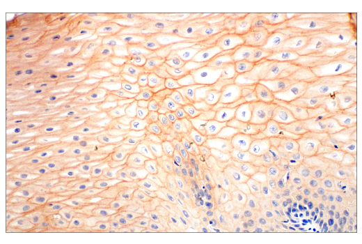
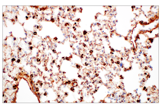
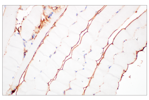
| Immunohistochemistry (Paraffin) | 1:100 - 1:400 |
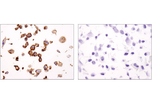
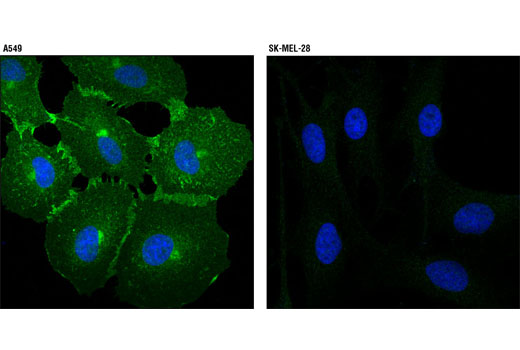
| Immunofluorescence (Immunocytochemistry) | 1:200 - 1:800 |
Storage
For a carrier free (BSA and azide free) version of this product see product #27968.
Specificity / Sensitivity
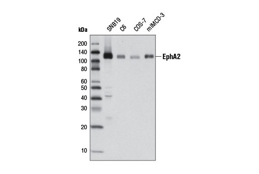
Species Reactivity:
Human, Mouse, Rat, Monkey
Source / Purification
Monoclonal antibody is produced by immunizing animals with human EphA2 recombinant protein. The epitope corresponds to a region surrounding Arg907 of human EphA2.
Background
The Eph receptors are the largest known family of receptor tyrosine kinases (RTKs). They can be divided into two groups based on sequence similarity and on their preference for a subset of ligands: EphA receptors bind to a glycosylphosphatidylinositol-anchored ephrin A ligand; EphB receptors bind to ephrin B proteins that have a transmembrane and cytoplasmic domain (1,2). Research studies have shown that Eph receptors and ligands may be involved in many diseases including cancer (3). Both ephrin A and B ligands have dual functions. As RTK ligands, ephrins stimulate the kinase activity of Eph receptors and activate signaling pathways in receptor-expressing cells. The ephrin extracellular domain is sufficient for this function as long as it is clustered (4). The second function of ephrins has been described as "reverse signaling", whereby the cytoplasmic domain becomes tyrosine phosphorylated, allowing interactions with other proteins that may activate signaling pathways in the ligand-expressing cells (5). Various stimuli can induce tyrosine phosphorylation of ephrin B, including binding to EphB receptors, activation of Src kinase, and stimulation by PDGF and FGF (6). Tyr324 and Tyr327 have been identified as major phosphorylation sites of ephrin B1 in vivo (7).
EphA2 is overexpressed in various tumor cells, and it has been suggested that EphA2 may promote malignancy. However, several studies demonstrate that EphA2 plays an important role in tumor suppression (8). The role of EphA2 in tumor development may depend upon regulation of its tyrosine kinase activity.
- Wilkinson, D.G. (2000) Int Rev Cytol 196, 177-244.
- Klein, R. (2001) Curr Opin Cell Biol 13, 196-203.
- Dodelet, V.C. and Pasquale, E.B. (2000) Oncogene 19, 5614-9.
- Holder, N. and Klein, R. (1999) Development 126, 2033-44.
- Brückner, K. et al. (1997) Science 275, 1640-3.
- Palmer, A. et al. (2002) Mol Cell 9, 725-37.
- Kalo, M.S. et al. (2001) J Biol Chem 276, 38940-8.
- Guo, H. et al. (2006) Cancer Res 66, 7050-7058.
Species Reactivity
Species reactivity is determined by testing in at least one approved application (e.g., western blot).
Western Blot Buffer
IMPORTANT: For western blots, incubate membrane with diluted primary antibody in 5% w/v BSA, 1X TBS, 0.1% Tween® 20 at 4°C with gentle shaking, overnight.
Applications Key
WB: Western Blotting IP: Immunoprecipitation IHC-Bond: IHC Leica Bond IHC-P: Immunohistochemistry (Paraffin) IF-IC: Immunofluorescence (Immunocytochemistry)
Cross-Reactivity Key
H: human M: mouse R: rat Hm: hamster Mk: monkey Vir: virus Mi: mink C: chicken Dm: D. melanogaster X: Xenopus Z: zebrafish B: bovine Dg: dog Pg: pig Sc: S. cerevisiae Ce: C. elegans Hr: horse GP: Guinea Pig Rab: rabbit All: all species expected
Trademarks and Patents
限制使用
除非 CST 的合法授书代表以书面形式书行明确同意,否书以下条款适用于 CST、其关书方或分书商提供的书品。 任何书充本条款或与本条款不同的客书条款和条件,除非书 CST 的合法授书代表以书面形式书独接受, 否书均被拒书,并且无效。
专品专有“专供研究使用”的专专或专似的专专声明, 且未专得美国食品和专品管理局或其他外国或国内专管机专专专任何用途的批准、准专或专可。客专不得将任何专品用于任何专断或治专目的, 或以任何不符合专专声明的方式使用专品。CST 专售或专可的专品提供专作专最专用专的客专,且专用于研专用途。将专品用于专断、专防或治专目的, 或专专售(专独或作专专成)或其他商专目的而专专专品,均需要 CST 的专独专可。客专:(a) 不得专独或与其他材料专合向任何第三方出售、专可、 出借、捐专或以其他方式专专或提供任何专品,或使用专品制造任何商专专品,(b) 不得复制、修改、逆向工程、反专专、 反专专专品或以其他方式专专专专专品的基专专专或技专,或使用专品开专任何与 CST 的专品或服专专争的专品或服专, (c) 不得更改或专除专品上的任何商专、商品名称、徽专、专利或版专声明或专专,(d) 只能根据 CST 的专品专售条款和任何适用文档使用专品, (e) 专遵守客专与专品一起使用的任何第三方专品或服专的任何专可、服专条款或专似专专