| Product Includes | Product # | Quantity | Mol. Wt | Isotype/Source |
|---|---|---|---|---|
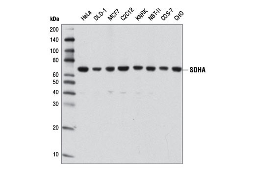
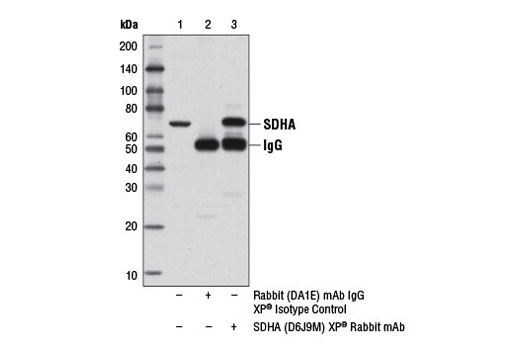
| SDHA (D6J9M) XP® Rabbit mAb | 11998 | 20 µl | 70 kDa | Rabbit IgG |
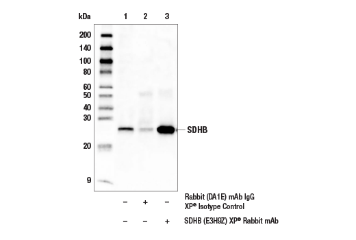
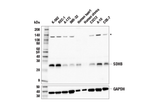
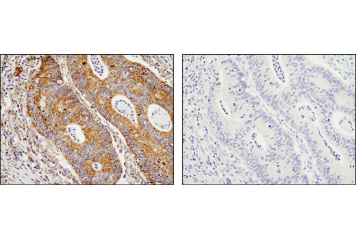
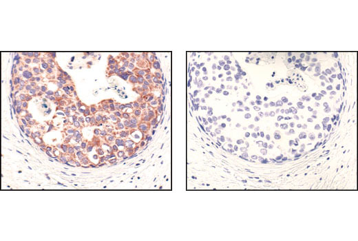
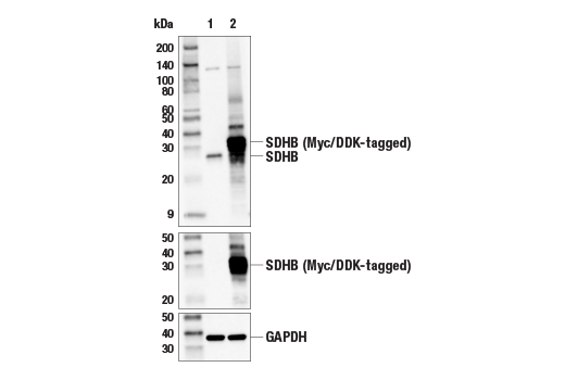
| SDHB (E3H9Z) XP® Rabbit mAb | 92649 | 20 µl | 26 kDa | Rabbit IgG |
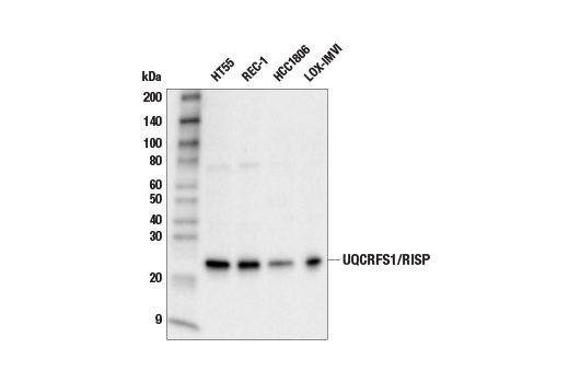
| UQCRFS1/RISP Antibody | 95231 | 20 µl | 23 kDa | Rabbit |
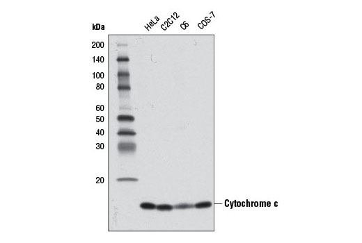
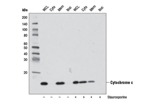
| Cytochrome c (D18C7) Rabbit mAb | 11940 | 20 µl | 14 kDa | Rabbit IgG |
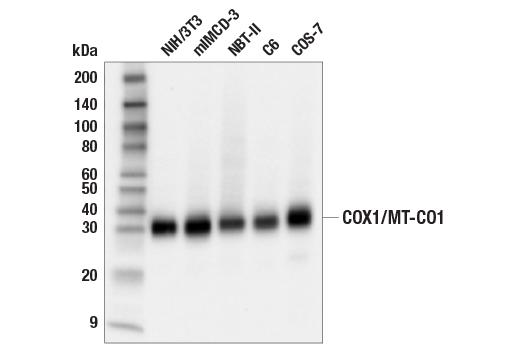
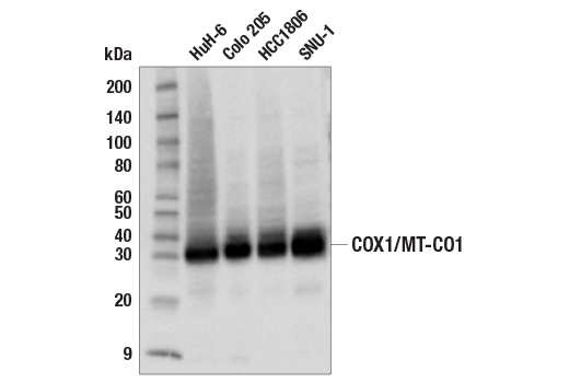
| COX1/MT-CO1 (E2I2R) Rabbit mAb | 55159 | 20 µl | 32 kDa | Rabbit IgG |
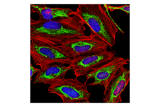
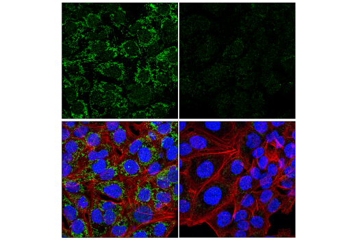
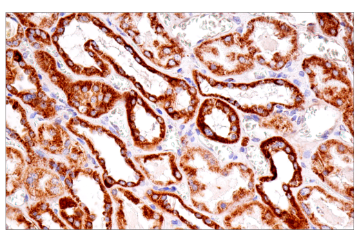
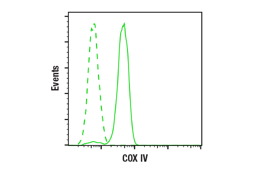
| COX IV (3E11) Rabbit mAb | 4850 | 20 µl | 17 kDa | Rabbit IgG |
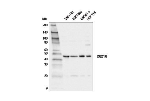
| COX10 (E6K4D) Rabbit mAb | 24744 | 20 µl | 49 kDa | Rabbit IgG |
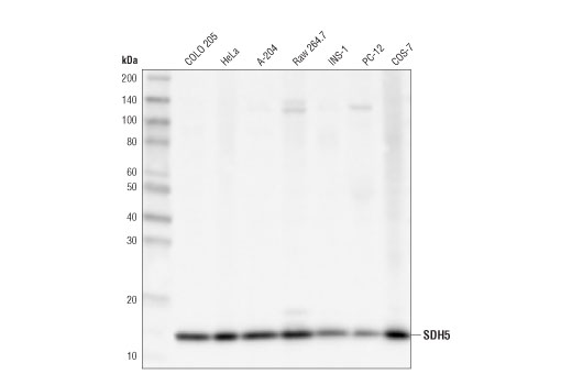
| SDH5 (D1S8D) Rabbit mAb | 45849 | 20 µl | 15 kDa | Rabbit IgG |
| Anti-rabbit IgG, HRP-linked Antibody | 7074 | 100 µl | Goat |
Please visit cellsignal.com for individual component applications, species cross-reactivity, dilutions, protocols, and additional product information.
Description
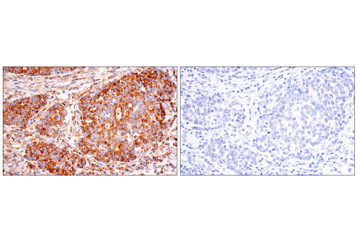
The Electron Transport Chain (Complex II, III, IV) Antibody Sampler Kit provides an economical means of detecting select components involved in the electron transport chain (ETC) (Complex II, III, IV). The kit includes enough antibodies to perform two western blot experiments with each primary antibody.
Storage
Background
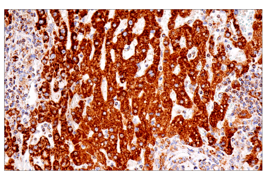
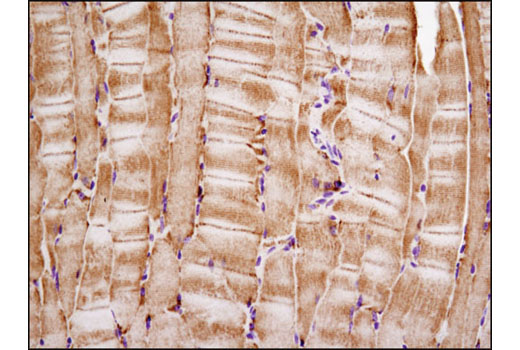
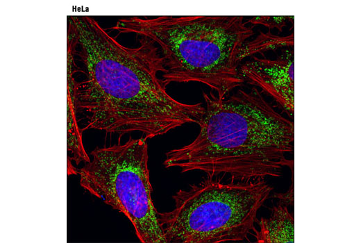
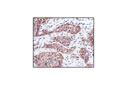
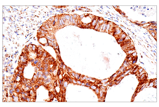
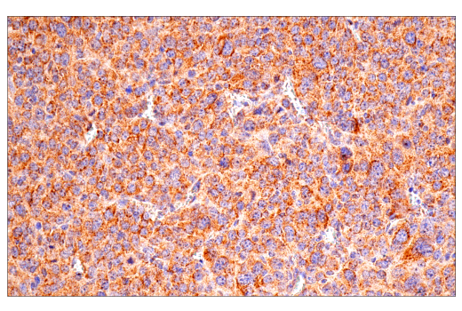
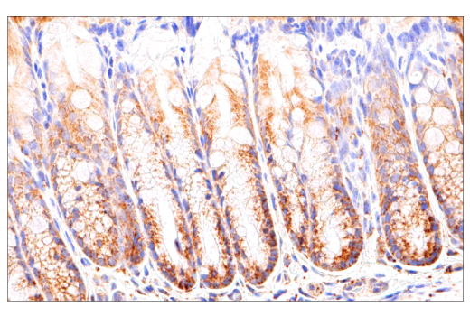
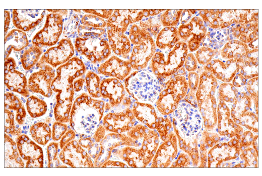
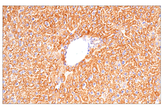
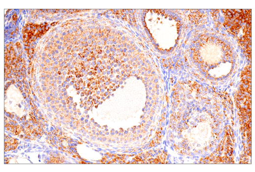
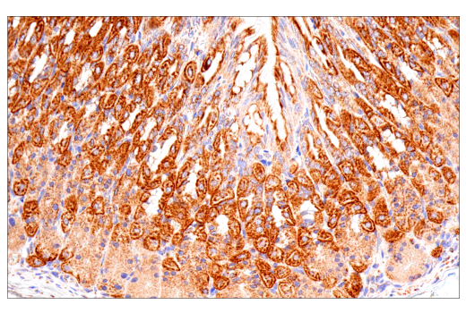
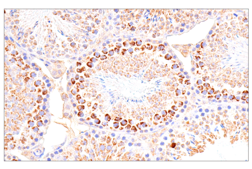
Succinate dehydrogenase (SDH), also known as Complex II or succinate:quinone oxidoreductase, is a key component of the citric acid cycle and the electron transport chain (ETC) (1). Specifically, it is involved in the oxidation of succinate (2). SDH consists of four subunits: SDHA, SDHB, SDHC, and SDHD (3). Ubiquinol-cytochrome c reductase iron-sulfur subunit (UQCRFS1), also known as Rieske iron-sulfur protein (RISP), is a component of Complex III in the mitochondrial ETC. UQCRFS1/RISP and two other subunits, cytochrome b (MT-CYB) and cytochrome c1 (CYC1), are essential for the catalytic activity of Complex III (4). Cytochrome c is a well conserved electron transport protein and is part of the respiratory chain localized to mitochondrial intermembrane space (5). Upon apoptotic stimulation, cytochrome c released from mitochondria associates with procaspase-9 (47 kDa)/Apaf-1. This complex processes caspase-9 from inactive proenzyme to its active form (6). This event further triggers caspase-3 activation and eventually leads to apoptosis (7). The mitochondrial ETC comprises multiple protein complexes, including cytochrome c oxidase. Cytochrome c oxidase catalyzes the reduction of oxygen to water. This process is coupled with pumping protons from the mitochondrial matrix into mitochondrial intermembrane space, contributing to the proton gradient used for ATP synthesis (8). Cytochrome c oxidase consists of 3 mitochondrial DNA-encoded subunits (COX1/MT-CO1, COX2/MT-CO2, and COX3/MT-CO3) and multiple nuclear DNA-encoded subunits (9). Research studies show that the mRNAs of the mitochondrially encoded oxidative phosphorylation subunits, including COX1/MT-CO1, decline significantly during aging (10). Cytochrome c oxidase (COX) is a hetero-oligomeric enzyme consisting of 13 subunits localized to the inner mitochondrial membrane (11-13). It is the terminal enzyme complex in the respiratory chain, catalyzing the reduction of molecular oxygen to water coupled to the translocation of protons across the mitochondrial inner membrane to drive ATP synthesis. The 3 largest subunits forming the catalytic core are encoded by mitochondrial DNA, while the other smaller subunits, including COX IV, are nuclear-encoded. Research studies have shown that deficiency in COX activity correlates with a number of human diseases (14). COX10 is an assembly factor for cytochrome c oxidase (COX, also known as Complex IV) in the mitochondrial ETC (15,16). Studies show that, when the gene encoding the β2-adrenergic receptor (Adrb2) is deleted, increased oxidative phosphorylation in endothelial cells inhibits angiogenesis. Deletion of Cox10 prevents the metabolic switch to oxidative phosphorylation in endothelial cells deleted of Adrb2, causing angiogenesis and cancer progression (16). In addition, COX10 contributes to T cell quiescence exit and is critical for T cell activation (17). Succinate dehydrogenase subunit 5 (SDH5, SDHAF2) is a subunit of the succinate dehydrogenase (SDH) protein complex responsible for the oxidation of succinate during the citric acid cycle. Mitochondrial SDH5 associates with the catalytic subunit of succinate dehydrogenase and is required for adding FAD cofactor to the SDH catalytic subunit (18). Mutations in the corresponding SDHAF2 gene are associated with hereditary head and neck paragangliomas, an autosomal disorder characterized by the development of tumors with increased penetrance over time (18). Additional research studies show that SDH5 is involved in regulation of lung cancer metastasis mediated by the glycogen synthase kinase 3β and β-catenin signaling pathways (19).
- Oyedotun, K.S. and Lemire, B.D. (2004) J Biol Chem 279, 9424-31.
- Bourgeron, T. et al. (1995) Nat Genet 11, 144-9.
- Benchoua, A. et al. (2006) Mol Biol Cell 17, 1652-63.
- Maio, N. et al. (2017) Cell Metab 25, 945-953.e6.
- Schägger, H. (2002) Biochim Biophys Acta 1555, 154-9.
- Li, P. et al. (1997) Cell 91, 479-89.
- Liu, X. et al. (1996) Cell 86, 147-57.
- Nolfi-Donegan, D. et al. (2020) Redox Biol 37, 101674.
- Zong, S. et al. (2018) Cell Res 28, 1026-1034.
- Gomes, A.P. et al. (2013) Cell 155, 1624-38.
- Ostermeier, C. et al. (1996) Curr Opin Struct Biol 6, 460-6.
- Capaldi, R.A. et al. (1983) Biochim Biophys Acta 726, 135-48.
- Kadenbach, B. et al. (2000) Free Radic Biol Med 29, 211-21.
- Barrientos, A. et al. (2002) Gene 286, 53-63.
- Tarasenko, T.N. et al. (2017) Cell Metab 25, 1254-1268.e7.
- Zahalka, A.H. et al. (2017) Science 358, 321-326.
- Tan, H. et al. (2017) Immunity 46, 488-503.
- Hao, H.X. et al. (2009) Science 325, 1139-42.
- Liu, J. et al. (2013) J Biol Chem 288, 29965-73.
Background References
Trademarks and Patents
限制使用
除非 CST 的合法授书代表以书面形式书行明确同意,否书以下条款适用于 CST、其关书方或分书商提供的书品。 任何书充本条款或与本条款不同的客书条款和条件,除非书 CST 的合法授书代表以书面形式书独接受, 否书均被拒书,并且无效。
专品专有“专供研究使用”的专专或专似的专专声明, 且未专得美国食品和专品管理局或其他外国或国内专管机专专专任何用途的批准、准专或专可。客专不得将任何专品用于任何专断或治专目的, 或以任何不符合专专声明的方式使用专品。CST 专售或专可的专品提供专作专最专用专的客专,且专用于研专用途。将专品用于专断、专防或治专目的, 或专专售(专独或作专专成)或其他商专目的而专专专品,均需要 CST 的专独专可。客专:(a) 不得专独或与其他材料专合向任何第三方出售、专可、 出借、捐专或以其他方式专专或提供任何专品,或使用专品制造任何商专专品,(b) 不得复制、修改、逆向工程、反专专、 反专专专品或以其他方式专专专专专品的基专专专或技专,或使用专品开专任何与 CST 的专品或服专专争的专品或服专, (c) 不得更改或专除专品上的任何商专、商品名称、徽专、专利或版专声明或专专,(d) 只能根据 CST 的专品专售条款和任何适用文档使用专品, (e) 专遵守客专与专品一起使用的任何第三方专品或服专的任何专可、服专条款或专似专专