Revision 3

#33437Store at -20C

1 个试剂盒

(9 x 20 microliters)

Cell Signaling Technology

Orders: 877-616-CELL (2355) [email protected]

Support: 877-678-TECH (8324)

Web: [email protected] cellsignal.com

3 Trask LaneDanversMassachusetts01923USA
For Research Use Only. Not for Use in Diagnostic Procedures.
Product Includes Product # Quantity Mol. Wt Isotype/Source
MMP-1 (E9S9N) Rabbit mAb 54376 20 µl 55 kDa Rabbit IgG
MMP-2 (D4M2N) Rabbit mAb 40994 20 µl 64,72 kDa Rabbit IgG
MMP-9 (D6O3H) XP® Rabbit mAb 13667 20 µl 84, 92 kDa Rabbit IgG
MMP-13 (E4W3T) Rabbit mAb 69926 20 µl 60 kDa Rabbit IgG
Fibronectin/FN1 (E5H6X) Rabbit mAb 26836 20 µl 300 kDa Rabbit IgG
MMP-3 (D7F5B) Rabbit mAb 14351 20 µl 60 kDa Rabbit IgG
LOX (D8F2K) Rabbit mAb 58135 20 µl 54, 56 kDa Rabbit IgG
COL11A1 (E6O7R) Rabbit mAb 70458 20 µl 250 kDa Rabbit IgG
COL1A1 (E6A8E) Rabbit mAb 39952 20 µl 220 kDa Rabbit IgG
Anti-rabbit IgG, HRP-linked Antibody 7074 100 µl Goat 

Please visit cellsignal.com for individual component applications, species cross-reactivity, dilutions, protocols, and additional product information.

Description

The ECM Profiling Antibody Sampler Kit provides an economical means of detecting endogenous levels of the specific ECM components using the corresponding antibodies. The kit includes enough antibodies to perform two western blot experiments with each primary antibody.

Storage

Supplied in 10 mM sodium HEPES (pH 7.5), 150 mM NaCl, 100 µg/ml BSA, 50% glycerol and less than 0.02% sodium azide. Store at –20°C. Do not aliquot the antibody.

Background

The extracellular matrix (ECM) is a cell surface associated three-dimensional macromolecular network composed of collagens, proteoglycans/glycosaminoglycans, elastin, fibronectin, laminins, and several other glycoproteins (1). The network provides a dynamic microenvironment surrounding the cell enabling it to carry on its function. Among the ECM proteins, fibronectin functions as a mediator to bridge distinct ECM components such as collagens, growth factors, as well as cell surface integrins to regulate ECM structural change and initiate signaling pathways (2). During normal development and pathological conditions, the ECM network is highly dynamic and continuously undergoes remodeling marked by the change of the ECM structural components, such as COL1A1, COL11A1, fibronectin, and versican. The matrix-degrading enzyme MMPs such as MMP1, MMP2, and MMP9 are highly involved in this process (4). Additional players in this process are the LOX family members of lysyl oxidase. They catalyze the first step of the covalent cross-linking of ECM proteins, collagens, and elastin, which contributes to ECM stiffness and mechanical properties (5).

  1. Theocharis, A.D. et al. (2019) FEBS J 286, 2830-69.
  2. Zollinger, A.J. and Smith, M.L. (2017) Matrix Biol 60-1, 27-37.
  3. Paolillo, M. and Schinelli, S. (2019) Int J Mol Sci 20, pii: E4947. doi: 10.3390/ijms20194947.
  4. Bonnans, C. et al. (2014) Nat Rev Mol Cell Biol 15, 786-801.
  5. Amendola, P.G. et al. (2019) Cancers (Basel) 11, pii: E729. doi: 10.3390/cancers11050729.

Background References

    Trademarks and Patents

    Cell Signaling Technology is a trademark of Cell Signaling Technology, Inc.
    XP is a registered trademark of Cell Signaling Technology, Inc.
    All other trademarks are the property of their respective owners. Visit cellsignal.com/trademarks for more information.

    限制使用

    除非 CST 的合法授书代表以书面形式书行明确同意,否书以下条款适用于 CST、其关书方或分书商提供的书品。 任何书充本条款或与本条款不同的客书条款和条件,除非书 CST 的合法授书代表以书面形式书独接受, 否书均被拒书,并且无效。

    专品专有“专供研究使用”的专专或专似的专专声明, 且未专得美国食品和专品管理局或其他外国或国内专管机专专专任何用途的批准、准专或专可。客专不得将任何专品用于任何专断或治专目的, 或以任何不符合专专声明的方式使用专品。CST 专售或专可的专品提供专作专最专用专的客专,且专用于研专用途。将专品用于专断、专防或治专目的, 或专专售(专独或作专专成)或其他商专目的而专专专品,均需要 CST 的专独专可。客专:(a) 不得专独或与其他材料专合向任何第三方出售、专可、 出借、捐专或以其他方式专专或提供任何专品,或使用专品制造任何商专专品,(b) 不得复制、修改、逆向工程、反专专、 反专专专品或以其他方式专专专专专品的基专专专或技专,或使用专品开专任何与 CST 的专品或服专专争的专品或服专, (c) 不得更改或专除专品上的任何商专、商品名称、徽专、专利或版专声明或专专,(d) 只能根据 CST 的专品专售条款和任何适用文档使用专品, (e) 专遵守客专与专品一起使用的任何第三方专品或服专的任何专可、服专条款或专似专专
    Revision 3
    #33437

    ECM Profiling Antibody Sampler Kit

    ECM Profiling Antibody Sampler Kit: Image 1 Expand Image
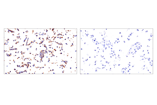
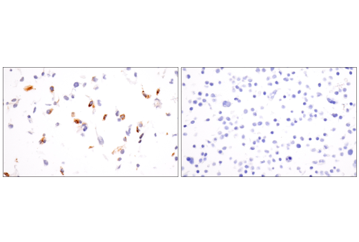
    使用 MMP-1 (E9S9N) Rabbit mAb 对石蜡包埋的 SCaBER 细胞沉淀物(左图,阳性)或 HT-29 细胞沉淀物(右图,阴性)进行免疫组织化学分析。
    ECM Profiling Antibody Sampler Kit: Image 2 Expand Image
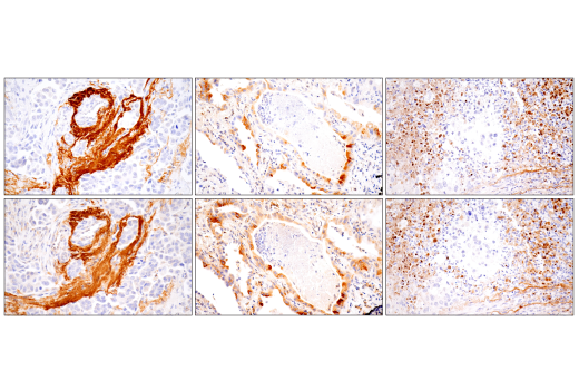
    使用 MMP-1 (E9S9N) Rabbit mAb(上图)或 MMP-1 Rabbit mAb(下图)对石蜡包埋的人导管乳腺癌组织(左图)、肺腺癌组织(中图)或鳞状细胞肺癌组织(右图)进行免疫组织化学分析。这两种抗体可检测人 MMP-1 上独特的非重叠表位。使用两种抗体获得的相似染色模式有助于确认染色特异性。
    ECM Profiling Antibody Sampler Kit: Image 3 Expand Image
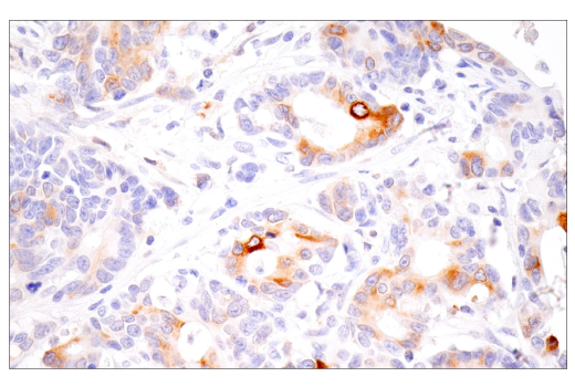
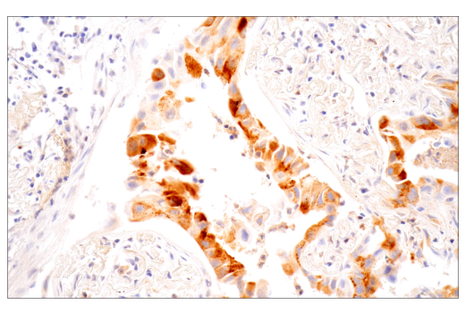
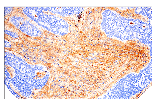
    使用 MMP-1 (E9S9N) Rabbit mAb 对石蜡包埋的人食管癌组织进行免疫组织化学分析。
    ECM Profiling Antibody Sampler Kit: Image 4 Expand Image
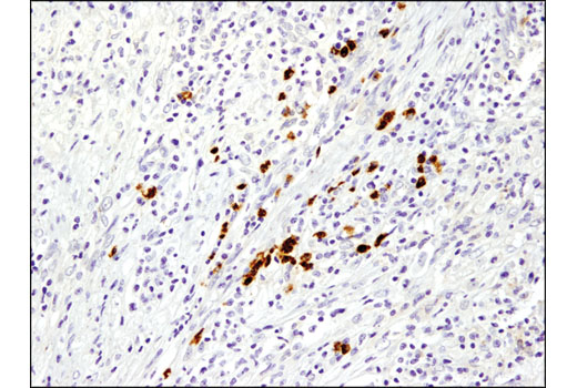
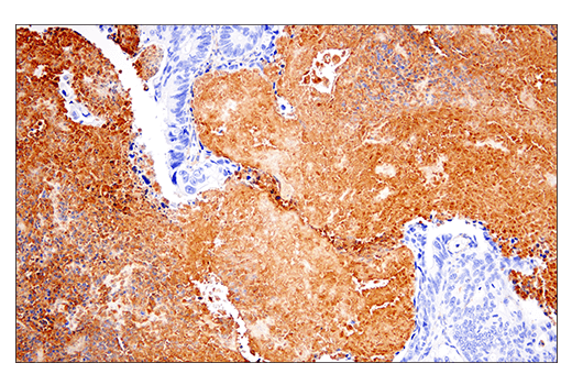
    使用 MMP-1 (E9S9N) Rabbit mAb 对石蜡包埋的人肺腺癌组织进行免疫组织化学分析。
    ECM Profiling Antibody Sampler Kit: Image 5 Expand Image
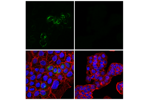
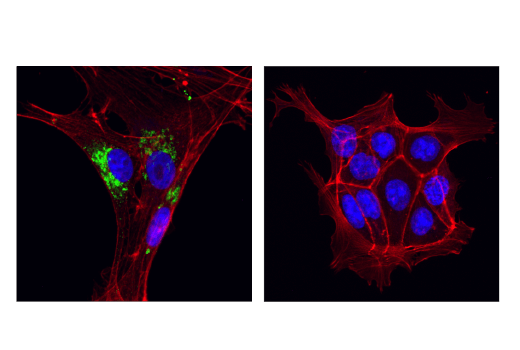
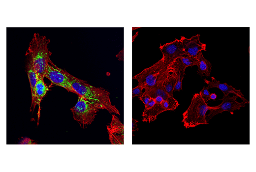
    使用 MMP-1 (E9S9N) Rabbit mAb(绿色)、DyLight 650 Phalloidin #12956(红色)和 DAPI #4083(蓝色)对 SCaBER 细胞(左图,阳性)或 HT-29 细胞(右图,阴性)进行共聚焦免疫荧光分析。
    ECM Profiling Antibody Sampler Kit: Image 6 Expand Image
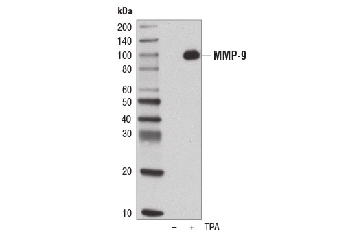
    使用 MMP-9 (D6O3H) XP® Rabbit mAb ,对未处理的 (-) 或已经过 TPA #4174 (200 nM,48 hr;+) 处理的 U-2 OS 细胞的浓缩、无血清培养基进行蛋白质印迹分析。
    ECM Profiling Antibody Sampler Kit: Image 7 Expand Image
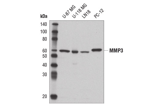
    使用 MMP-3 (D7F5B) 兔单抗对不同细胞系提取物进行蛋白质印迹分析。
    ECM Profiling Antibody Sampler Kit: Image 8 Expand Image
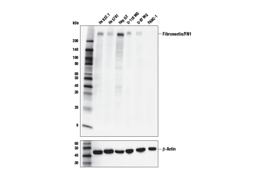
    使用 Fibronectin/FN1 (E5H6X) Rabbit mAb(上图)和 β-Actin (D6A8) Rabbit mAb #8457(下图)对不同细胞系的提取物进行蛋白印迹分析。PANC-1 细胞提取物中缺失纤连蛋白/FN1 表达与分子和蛋白质组表达谱数据一致,证实了该抗体的特异性。
    ECM Profiling Antibody Sampler Kit: Image 9 Expand Image
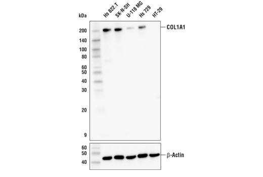
    使用 COL1A1 (E6A8E) 兔单克隆抗体(上图)和 β-Actin (D6A8) Rabbit mAb #8457(下图),对不同细胞系的提取物进行蛋白印迹分析。
    ECM Profiling Antibody Sampler Kit: Image 10 Expand Image
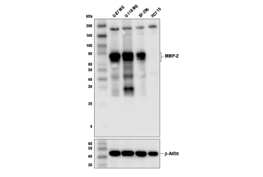
    使用 MMP-2 (D4M2N) Rabbit mAb(上图)和 β-Actin (D6A8) Rabbit mAb #8457(下图),对不同细胞系的提取物进行蛋白质印迹分析。
    ECM Profiling Antibody Sampler Kit: Image 11 Expand Image
    使用 MMP-1 (E9S9N) 兔单克隆抗体(上图)和 β-肌动蛋白 (D6A8) 兔单克隆抗体 #8457(下图)对不同细胞系的提取物进行蛋白印迹分析。
    ECM Profiling Antibody Sampler Kit: Image 12 Expand Image
    使用 LOX (D8F2K) Rabbit mAb(上图)和 β-Actin (D6A8) Rabbit mAb #8457(下图)对不同细胞系的提取物进行蛋白质印迹分析。
    ECM Profiling Antibody Sampler Kit: Image 13 Expand Image
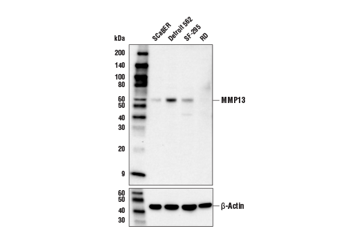
    使用 MMP-13 (E4W3T) Rabbit mAb(上图)和 β-Actin (D6A8) Rabbit mAb #8457(下图)对不同细胞系的提取物进行蛋白印迹分析。
    ECM Profiling Antibody Sampler Kit: Image 14 Expand Image
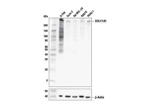
    使用 COL11A1 (E6O7R) Rabbit mAb(上图)或

    β-Actin (D6A8) Rabbit mAb #8457(下图)对不同细胞系的提取物进行蛋白印迹分析。如所预期的一样,与所示的其他细胞系相比,A-204 细胞具有更高水平的 COL11A1 表达。

    ECM Profiling Antibody Sampler Kit: Image 15 Expand Image
    一抗与靶标蛋白结合之后,与偶联 HRP 的二抗形成复合体。添加 LumiGLO®,在酶催化分解期间发光。
    ECM Profiling Antibody Sampler Kit: Image 16 Expand Image
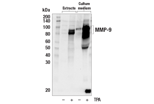
    使用 MMP-9 (D6O3H) XP® Rabbit mAb 对未经处理 (-) 或已经过 TPA #4174(200 nM,48 小时;+)处理的 U-2 OS 细胞的提取物和浓缩培养基进行蛋白质印迹法分析。正如预期的那样,MMP-9 是由 TPA 处理诱导的。
    ECM Profiling Antibody Sampler Kit: Image 17 Expand Image
    对 Hep G2 细胞提取物纤连蛋白/FN1 蛋白进行免疫沉淀。泳道 1 为 10% 输入,泳道 2 为 Rabbit (DA1E) mAb IgG XP® Isotype Control #3900,泳道 3 为 Fibronectin/FN1 (E5H6X) Rabbit mAb。使用 Fibronectin/FN1 (E5H6X) Rabbit mAb 进行蛋白印迹分析。Anti-rabbit IgG, HRP-linked Antibody #7074 用作二抗。
    ECM Profiling Antibody Sampler Kit: Image 18 Expand Image
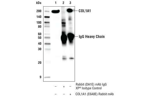
    对 SK-N-SH 细胞提取物 COL1A1 蛋白进行免疫沉淀。泳道 1 为 10% 输入,泳道 2 为 Rabbit (DA1E) mAb IgG XP® Isotype Control #3900,泳道 3 为 COL1A1 (E6A8E) 兔单克隆抗体。使用 COL1A1 (E6A8E) 兔单克隆抗体进行蛋白印迹分析。Anti-rabbit IgG, HRP-linked Antibody #7074 用作二抗。

    .€‚

    ECM Profiling Antibody Sampler Kit: Image 19 Expand Image
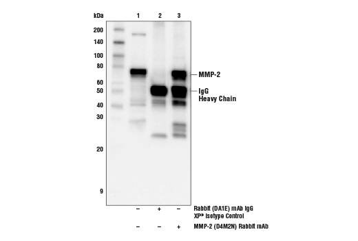
    对 U-118MG 细胞提取物的 MMP-2 蛋白质进行免疫沉淀。泳道 1 是 10% 输入,泳道 2 是 Rabbit (DA1E) mAb IgG XP® Isotype Control #3900,而泳道 3 是 MMP-2 (D4M2N) Rabbit mAb。使用 MMP-2 (D4M2N) Rabbit mAb 作为一抗,进行蛋白质印迹分析。Anti-rabbit IgG, HRP-linked Antibody #7074 用作二抗。
    ECM Profiling Antibody Sampler Kit: Image 20 Expand Image
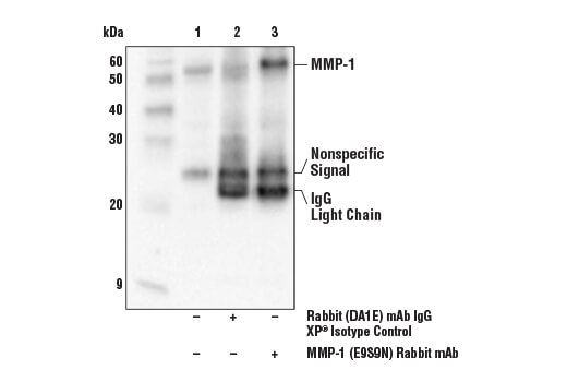
    对 SCaBER 细胞提取物 MMP-1 蛋白进行免疫沉淀。泳道 1 为 10%输入,泳道 2 为兔 (DA1E) 单克隆抗体 IgG XP® 同型对照 #3900,泳道 3 为 MMP-1 (E9S9N) 兔单克隆抗体。使用 MMP-1 (E9S9N) 兔单克隆抗体进行蛋白印迹分析。抗兔 IgG (特定轻链), HRP-偶联抗体用作二抗。
    ECM Profiling Antibody Sampler Kit: Image 21 Expand Image
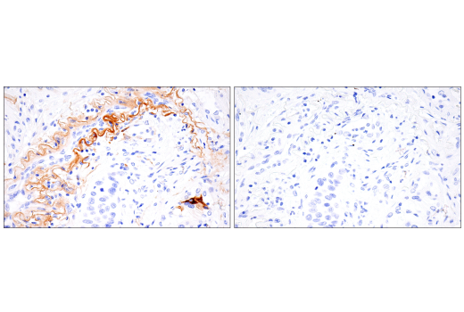
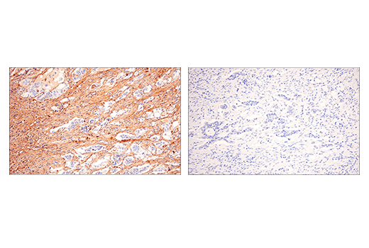
    以浓度匹配的 Rabbit (DA1E) mAb IgG XP® Isotype Control #3900(右图)作为对照,使用 MMP-1 (E9S9N) Rabbit mAb(左图)对石蜡包埋的人鳞状细胞肺癌组织进行免疫组织化学分析。
    ECM Profiling Antibody Sampler Kit: Image 22 Expand Image
    使用 MMP-9 (D6O3H) XP® Rabbit mAb 对石蜡包埋的人乳腺癌进行免疫组织化学分析。
    ECM Profiling Antibody Sampler Kit: Image 23 Expand Image
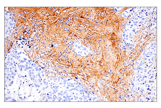
    在 Leica® BOND Rx 上使用 Fibronectin/FN1 (E5H6X) Rabbit mAb 对石蜡包埋的人肺神经内分泌癌细胞进行免疫组织化学分析。
    ECM Profiling Antibody Sampler Kit: Image 24 Expand Image
    使用 COL1A1 (E6A8E) 兔单克隆抗体(绿色)对 U-118 MG 细胞(左图,阳性)和 HT-29 细胞(右图,阴性)进行共聚焦免疫荧光分析。肌动蛋白微丝由 DyLight 554 Phalloidin #13054(红色)标记。样本用 ProLong® Gold Antifade Reagent with DAPI #8961(蓝色)封片。
    ECM Profiling Antibody Sampler Kit: Image 25 Expand Image
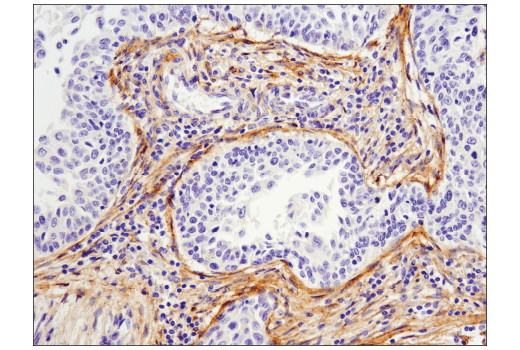
    使用 MMP-2 (D4M2N) Rabbit mAb 对石蜡包埋的人结膀胱癌细胞进行免疫组织化学分析。
    ECM Profiling Antibody Sampler Kit: Image 26 Expand Image
    使用 MMP-9 (D6O3H) XP® Rabbit mAb 对石蜡包埋的人肺癌进行免疫组织化学分析。
    ECM Profiling Antibody Sampler Kit: Image 27 Expand Image
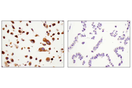
    使用 Fibronectin/FN1 (E5H6X) Rabbit mAb 对石蜡包埋的 Hep G2 细胞沉淀物(左图,阳性)或 PANC-1 细胞沉淀物(右图,阴性)进行免疫组织化学分析。
    ECM Profiling Antibody Sampler Kit: Image 28 Expand Image
    使用 MMP-2 (D4M2N) Rabbit mAb 对石蜡包埋的 U-87 MG 细胞沉淀物(左图,阳性)或 HCT-15 细胞沉淀物(右图,阴性)进行免疫组织化学分析。
    ECM Profiling Antibody Sampler Kit: Image 29 Expand Image
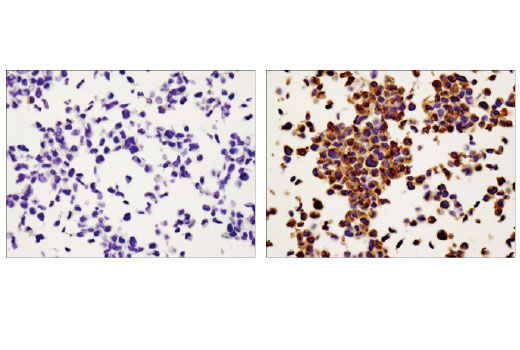
    使用 MMP-9 (D6O3H) XP® Rabbit mAb 对未处理的(左)或已经过 TPA #4174(右)处理的石蜡包埋的 U-2 OS 细胞沉淀物进行免疫组织化学分析。
    ECM Profiling Antibody Sampler Kit: Image 30 Expand Image
    使用 Fibronectin/FN1 (E5H6X) Rabbit mAb 对石蜡包埋的人结肠腺癌细胞进行免疫组织化学分析。
    ECM Profiling Antibody Sampler Kit: Image 31 Expand Image
    使用 MMP-2 (D4M2N) Rabbit mAb 对石蜡包埋的人宫颈癌细胞进行免疫组织化学分析。
    ECM Profiling Antibody Sampler Kit: Image 32 Expand Image
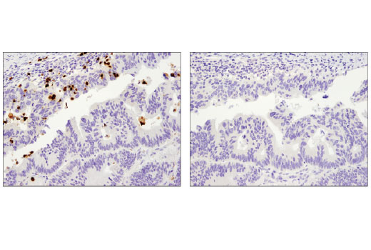
    在对照肽(左)或抗原特异性肽(右)存在的情况下,使用 MMP-9 (D6O3H) XP® Rabbit mAb 对石蜡包埋的人结肠癌进行免疫组织化学分析。
    ECM Profiling Antibody Sampler Kit: Image 33 Expand Image
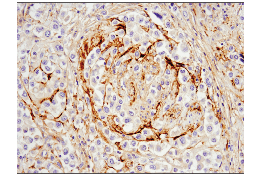
    使用 Fibronectin/FN1 (E5H6X) Rabbit mAb 对石蜡包埋的人乳腺导管癌细胞进行免疫组织化学分析。
    ECM Profiling Antibody Sampler Kit: Image 34 Expand Image
    使用 MMP-2 (D4M2N) Rabbit mAb 对石蜡包埋的人尿路上皮癌细胞进行免疫组织化学分析。
    ECM Profiling Antibody Sampler Kit: Image 35 Expand Image
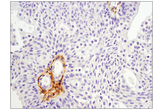
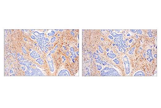
    使用 Fibronectin/FN1 (E5H6X) Rabbit mAb(左图)或 Fibronectin Rabbit mAb(右图)对石蜡包埋的人气管腺样囊性癌细胞进行免疫组织化学分析。这两种抗体可检测人纤连蛋白/FN1 蛋白上的独立、独特表位。使用两种抗体获得的相似染色模式有助于确认染色特异性。
    ECM Profiling Antibody Sampler Kit: Image 36 Expand Image
    使用 MMP-2 (D4M2N) Rabbit mAb(绿色)对 U-87MG细胞(左图,阳性)和 HCT-15 细胞(右图,阴性)进行共聚焦免疫荧光分析。肌动蛋白微丝由 DyLight 554 Phalloidin #13054(红色)标记。蓝色伪彩 = DRAQ5® #4084(DNA 荧光染料)。
    ECM Profiling Antibody Sampler Kit: Image 37 Expand Image
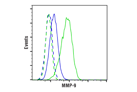
    使用 MMP-9 (D6O3H) XP® Rabbit mAb(实线)或浓度匹配的 Rabbit (DA1E) mAb IgG XP® Isotype Control #3900(虚线)对未经处理(蓝色)或经过 TPA (12-O-Tetradecanoylphorbol-13-Acetate) #4174(200 nM,24 小时;蓝色)或处理的 U-2 OS 细胞进行流式细胞分析。Anti-rabbit IgG (H+L), F(ab')2 Fragment (Alexa Fluor® 488 Conjugate) #4412 用作二抗。
    ECM Profiling Antibody Sampler Kit: Image 38 Expand Image
    以浓度匹配的 Rabbit (DA1E) mAb IgG XP® Isotype Control #3900(右图)作为对照,使用 Fibronectin/FN1 (E5H6X) Rabbit mAb(左图)对石蜡包埋的人结肠腺癌细胞进行免疫组织化学分析。
    ECM Profiling Antibody Sampler Kit: Image 39 Expand Image
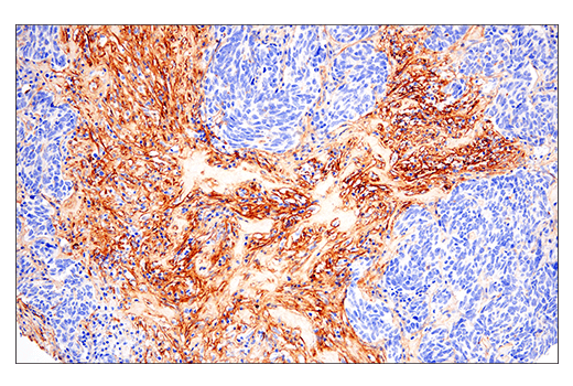
    使用 Fibronectin/FN1 (E5H6X) Rabbit mAb 对石蜡包埋的人肺腺癌细胞进行免疫组织化学分析。
    ECM Profiling Antibody Sampler Kit: Image 40 Expand Image
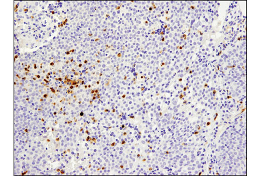
    使用 Fibronectin/FN1 (E5H6X) Rabbit mAb 对石蜡包埋的人 B 细胞非霍奇金氏淋巴瘤细胞进行免疫组织化学分析。
    ECM Profiling Antibody Sampler Kit: Image 41 Expand Image
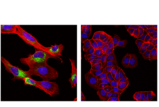
    使用 Fibronectin/FN1 (E5H6X) Rabbit mAb(绿色)对 U-118 MG 细胞(左图,阳性)和 PANC-1 细胞(右图,阴性)进行共聚焦免疫荧光分析。肌动蛋白微丝由 DyLight 650 Phalloidin #12956(红色)标记。样本用 ProLong® Gold Antifade Reagent with DAPI #8961(蓝色)封片。