| Product Includes | Product # | Quantity | Mol. Wt | Isotype/Source |
|---|---|---|---|---|
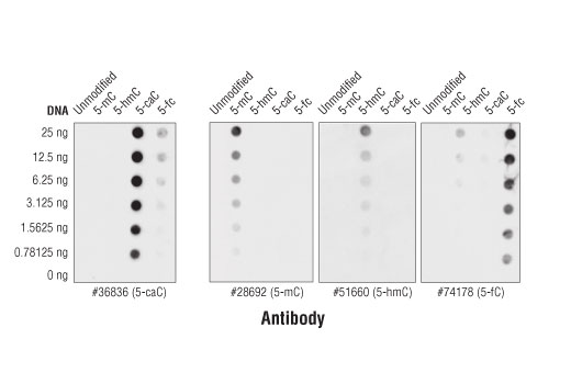
| 5-Methylcytosine (5-mC) (D3S2Z) Rabbit mAb | 28692 | 20 µl | Rabbit IgG | |
| 5-Hydroxymethylcytosine (5-hmC) (HMC31) Mouse mAb | 51660 | 20 µl | Mouse IgG1 | |
| 5-Carboxylcytosine (5-caC) (D7S8U) Rabbit mAb | 36836 | 20 µl | Rabbit IgG | |
| 5-Formylcytosine (5-fC) (D5D4K) Rabbit mAb | 74178 | 20 µl | Rabbit IgG | |
| Anti-rabbit IgG, HRP-linked Antibody | 7074 | 100 µl | Goat | |
| Anti-mouse IgG, HRP-linked Antibody | 7076 | 100 µl | Horse |
Please visit cellsignal.com for individual component applications, species cross-reactivity, dilutions, protocols, and additional product information.
Description
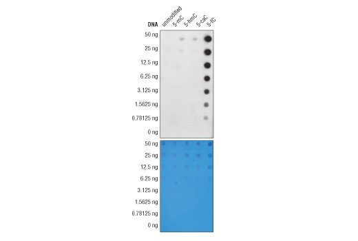
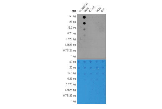
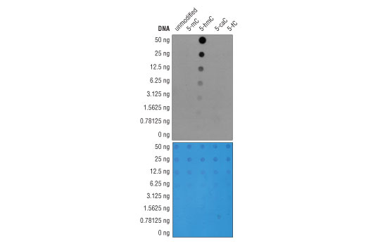
The DNA Cytosine Modification Antibody Sampler Kit provides an economical means of detecting the levels of cytosine modifications in DNA by dot blot using antibodies against 5-methylcytosine, 5-hydroxymethylcytosine, 5-formylcytosine, and 5-carboxylcytosine.
Storage
Background
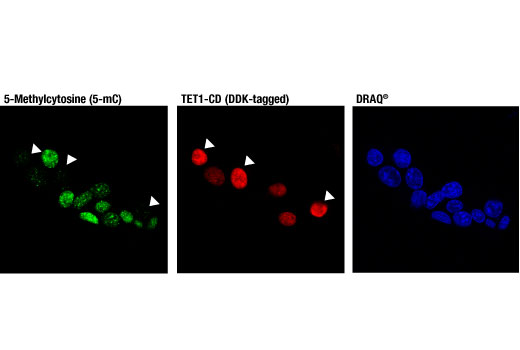
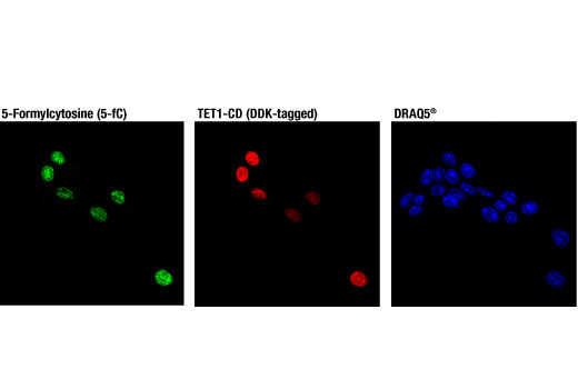
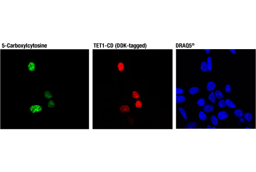
Methylation of DNA at cytosine residues is a heritable, epigenetic modification that is critical for proper regulation of gene expression, genomic imprinting, and mammalian development (1,2). 5-methylcytosine is a repressive epigenetic mark established de novo by two enzymes, DNMT3a and DNMT3b, and is maintained by DNMT1 (3, 4). 5-methylcytosine was originally thought to be passively depleted during DNA replication. However, subsequent studies have shown that Ten-Eleven Translocation (TET) proteins TET1, TET2, and TET3 can catalyze the oxidation of methylated cytosine to 5-hydroxymethylcytosine (5-hmC) (5). Additionally, TET proteins can further oxidize 5-hmC to form 5-formylcytosine (5-fC) and 5-carboxylcytosine (5-caC), both of which are excised by thymine-DNA glycosylase (TDG), effectively linking cytosine oxidation to the base excision repair pathway and supporting active cytosine demethylation (6,7).
TET protein-mediated cytosine hydroxymethylation was initially demonstrated in mouse brain and embryonic stem cells (5, 8). Since then this modification has been discovered in many tissues, with the highest levels found in the brain (9). While 5-fC and 5-caC appear to be short-lived intermediate species, there is mounting evidence showing that 5-hmC is a distinct epigenetic mark with various unique functions (10,11). The modified base itself is stable in vivo and interacts with various readers, including MeCP2 (11,12). The global level of 5-hmC increases during brain development and 5-hmC is enriched at promoter regions and poised enhancers. Furthermore, there is an inverse correlation between levels of 5-hmC and histone H3K9 and H3K27 trimethylation, suggesting a role for 5-hmC in gene activation (12). Lower amounts of 5-hmC have been reported in various cancers, including myeloid leukemia and melanoma (13,14).
- Hermann, A. et al. (2004) Cell Mol Life Sci 61, 2571-87.
- Turek-Plewa, J. and Jagodziński, P.P. (2005) Cell Mol Biol Lett 10, 631-47.
- Okano, M. et al. (1999) Cell 99, 247-57.
- Li, E. et al. (1992) Cell 69, 915-26.
- Tahiliani, M. et al. (2009) Science 324, 930-5.
- He, Y.F. et al. (2011) Science 333, 1303-7.
- Ito, S. et al. (2011) Science 333, 1300-3.
- Kriaucionis, S. and Heintz, N. (2009) Science 324, 929-30.
- Globisch, D. et al. (2010) PLoS One 5, e15367.
- Gao, Y. et al. (2013) Cell Stem Cell 12, 453-69.
- Mellén, M. et al. (2012) Cell 151, 1417-30.
- Wen, L. et al. (2014) Genome Biol 15, R49.
- Delhommeau, F. et al. (2009) N Engl J Med 360, 2289-301.
- Lian, C.G. et al. (2012) Cell 150, 1135-46.
Background References
Trademarks and Patents
限制使用
除非 CST 的合法授书代表以书面形式书行明确同意,否书以下条款适用于 CST、其关书方或分书商提供的书品。 任何书充本条款或与本条款不同的客书条款和条件,除非书 CST 的合法授书代表以书面形式书独接受, 否书均被拒书,并且无效。
专品专有“专供研究使用”的专专或专似的专专声明, 且未专得美国食品和专品管理局或其他外国或国内专管机专专专任何用途的批准、准专或专可。客专不得将任何专品用于任何专断或治专目的, 或以任何不符合专专声明的方式使用专品。CST 专售或专可的专品提供专作专最专用专的客专,且专用于研专用途。将专品用于专断、专防或治专目的, 或专专售(专独或作专专成)或其他商专目的而专专专品,均需要 CST 的专独专可。客专:(a) 不得专独或与其他材料专合向任何第三方出售、专可、 出借、捐专或以其他方式专专或提供任何专品,或使用专品制造任何商专专品,(b) 不得复制、修改、逆向工程、反专专、 反专专专品或以其他方式专专专专专品的基专专专或技专,或使用专品开专任何与 CST 的专品或服专专争的专品或服专, (c) 不得更改或专除专品上的任何商专、商品名称、徽专、专利或版专声明或专专,(d) 只能根据 CST 的专品专售条款和任何适用文档使用专品, (e) 专遵守客专与专品一起使用的任何第三方专品或服专的任何专可、服专条款或专似专专