Revision 4

#85786Store at -20C

Cell Signaling Technology

Orders: 877-616-CELL (2355) [email protected]

Support: 877-678-TECH (8324)

Web: [email protected] cellsignal.com

3 Trask LaneDanversMassachusetts01923USA
For Research Use Only. Not for Use in Diagnostic Procedures.
Applications:

WB, IHC-P

REACTIVITY:

H

SENSITIVITY:

Endogenous

MW (kDa):

40, 100

Source/Isotype:

Rabbit IgG

UniProt ID:

#P07585

Entrez-Gene Id:

1634

Product Information

Product Usage Information

Application Dilution
Western Blotting 1:1000
Immunohistochemistry (Paraffin) 1:100 - 1:400

Storage

Supplied in 10 mM sodium HEPES (pH 7.5), 150 mM NaCl, 100 µg/mL BSA, 50% glycerol, and less than 0.02% sodium azide. Store at –20°C. Do not aliquot the antibody.

For a carrier free (BSA and azide free) version of this product see product #55868.

Specificity / Sensitivity

Decorin (E2N2C) XP® Rabbit mAb recognizes endogenous levels of total decorin protein. This protein is heavily glycosylated, therefore, multiple bands or a smear may result from western blot analysis using this antibody.

Species Reactivity:

Human

Source / Purification

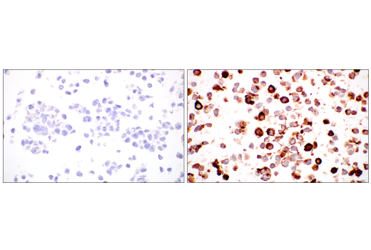
Monoclonal antibody is produced by immunizing animals with a synthetic peptide corresponding to residues surrounding Pro216 of human decorin protein.

Background

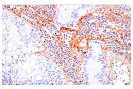
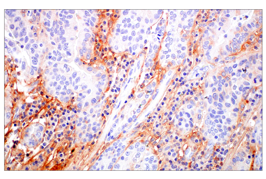
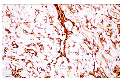
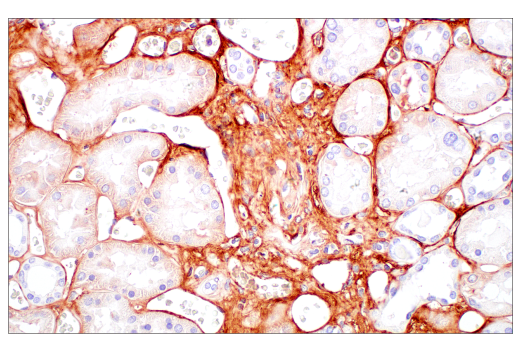
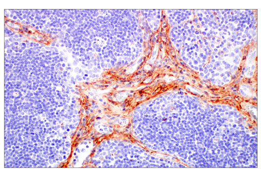
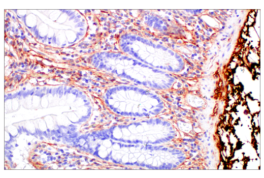
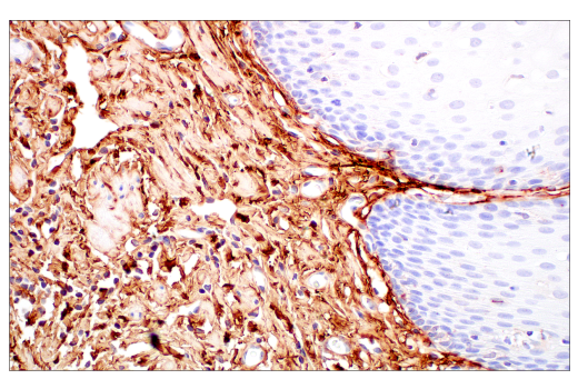
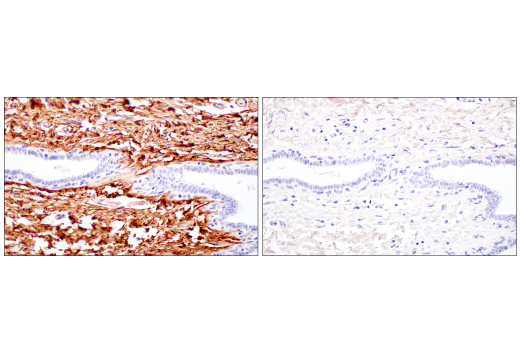
Decorin, named for its role in “decorating” type I collagen fibrils, is a collagen-binding proteoglycan that belongs to the small leucine-rich proteoglycan (SLRP) family (1). Through its binding to type I collagens and other important matrix proteins (e.g., fibronectin, thrombospondin, TGF-β), decorin plays a critically important role in assembly, maintenance, and physiological functions of the extracellular matrix. Notably, multiple research studies have reported anti-tumor functions for decorin and the related protein biglycan (2-4), implicating SLRP proteins as functionally important components of the tumor microenvironment (5). The anti-tumor effects of decorin have been ascribed to its interactions with cell surface receptors and associated signaling pathways (6), including those mediating inflammation, autophagy, and cell proliferation (7-9).

  1. Neill, T. et al. (2012) Am J Pathol 181, 380-7.
  2. Neill, T. et al. (2015) Mol Cell Oncol 2, e975645.
  3. Horváth, Z. et al. (2014) Matrix Biol 35, 194-205.
  4. Reszegi, A. et al. (2020) Biomolecules 10, 1199. doi: 10.3390/biom10081199.
  5. Baghy, K. et al. (2020) Adv Exp Med Biol 1272, 17-38.
  6. Xie, C. et al. (2022) Am J Physiol Cell Physiol 322, C554-C566.
  7. Schaefer, L. et al. (2017) FEBS J 284, 10-26.
  8. Merline, R. et al. (2011) Sci Signal 4, ra75.
  9. Gubbiotti, M.A. et al. (2015) Matrix Biol 48, 14-25.

Species Reactivity

Species reactivity is determined by testing in at least one approved application (e.g., western blot).

Western Blot Buffer

IMPORTANT: For western blots, incubate membrane with diluted primary antibody in 5% w/v nonfat dry milk, 1X TBS, 0.1% Tween® 20 at 4°C with gentle shaking, overnight.

Applications Key

WB: Western Blotting IHC-P: Immunohistochemistry (Paraffin)

Cross-Reactivity Key

H: human M: mouse R: rat Hm: hamster Mk: monkey Vir: virus Mi: mink C: chicken Dm: D. melanogaster X: Xenopus Z: zebrafish B: bovine Dg: dog Pg: pig Sc: S. cerevisiae Ce: C. elegans Hr: horse GP: Guinea Pig Rab: rabbit All: all species expected

Trademarks and Patents

Cell Signaling Technology is a trademark of Cell Signaling Technology, Inc.
SignalStain is a registered trademark of Cell Signaling Technology, Inc.
XP is a registered trademark of Cell Signaling Technology, Inc.
All other trademarks are the property of their respective owners. Visit cellsignal.com/trademarks for more information.

限制使用

除非 CST 的合法授书代表以书面形式书行明确同意,否书以下条款适用于 CST、其关书方或分书商提供的书品。 任何书充本条款或与本条款不同的客书条款和条件,除非书 CST 的合法授书代表以书面形式书独接受, 否书均被拒书,并且无效。

专品专有“专供研究使用”的专专或专似的专专声明, 且未专得美国食品和专品管理局或其他外国或国内专管机专专专任何用途的批准、准专或专可。客专不得将任何专品用于任何专断或治专目的, 或以任何不符合专专声明的方式使用专品。CST 专售或专可的专品提供专作专最专用专的客专,且专用于研专用途。将专品用于专断、专防或治专目的, 或专专售(专独或作专专成)或其他商专目的而专专专品,均需要 CST 的专独专可。客专:(a) 不得专独或与其他材料专合向任何第三方出售、专可、 出借、捐专或以其他方式专专或提供任何专品,或使用专品制造任何商专专品,(b) 不得复制、修改、逆向工程、反专专、 反专专专品或以其他方式专专专专专品的基专专专或技专,或使用专品开专任何与 CST 的专品或服专专争的专品或服专, (c) 不得更改或专除专品上的任何商专、商品名称、徽专、专利或版专声明或专专,(d) 只能根据 CST 的专品专售条款和任何适用文档使用专品, (e) 专遵守客专与专品一起使用的任何第三方专品或服专的任何专可、服专条款或专似专专

Revision 4
#85786

Decorin (E2N2C) XP® Rabbit mAb

Western Blotting Image 1: Decorin (E2N2C) XP® Rabbit mAb Expand Image
使用 Decorin (E2N2C) XP® Rabbit mAb(上图)或 GAPDH (D16H11) XP® Rabbit mAb #5174(下图)对人子宫组织、结肠组织和肝组织的提取物进行蛋白质印迹分析。核心蛋白聚糖蛋白在人肝组织中低表达与预测的表达模式一致。
Western Blotting Image 2: Decorin (E2N2C) XP® Rabbit mAb Expand Image
使用 Decorin (E2N2C) XP® Rabbit mAb(上图)、 Myc-Tag (71D10) Rabbit mAb #2278(中图)或 GAPDH (D16H11) XP® Rabbit mAb #5174(下图),对空载转染(泳道 1)或经编码 Myc/DDK 标记的核心蛋白聚糖质粒瞬时转染(泳道 2)的 293T 细胞的提取物进行蛋白质印迹分析。
Immunohistochemistry Image 1: Decorin (E2N2C) XP® Rabbit mAb Expand Image
使用 Decorin (E2N2C) XP® Rabbit mAb 对石蜡包埋的人前列腺癌组织进行免疫组织化学分析。
Immunohistochemistry Image 2: Decorin (E2N2C) XP® Rabbit mAb Expand Image
使用 Decorin (E2N2C) XP® Rabbit mAb 对石蜡包埋的人宫颈鳞状细胞癌组织进行免疫组织化学分析。
Immunohistochemistry Image 3: Decorin (E2N2C) XP® Rabbit mAb Expand Image
使用 Decorin (E2N2C) XP® Rabbit mAb 对石蜡包埋的人结肠腺癌组织进行免疫组织化学分析。
Immunohistochemistry Image 4: Decorin (E2N2C) XP® Rabbit mAb Expand Image
使用 Decorin (E2N2C) XP® Rabbit mAb 对石蜡包埋的人食管腺癌组织进行免疫组织化学分析。
Immunohistochemistry Image 5: Decorin (E2N2C) XP® Rabbit mAb Expand Image
使用 Decorin (E2N2C) XP® Rabbit mAb 对石蜡包埋的人乳头状甲状腺癌组织进行免疫组织化学分析。
Immunohistochemistry Image 6: Decorin (E2N2C) XP® Rabbit mAb Expand Image
使用 Decorin (E2N2C) XP® Rabbit mAb 对石蜡包埋的人正常心脏组织进行免疫组织化学分析。
Immunohistochemistry Image 7: Decorin (E2N2C) XP® Rabbit mAb Expand Image
使用 Decorin (E2N2C) XP® Rabbit mAb 对石蜡包埋的人正常前列腺组织进行免疫组织化学分析。
Immunohistochemistry Image 8: Decorin (E2N2C) XP® Rabbit mAb Expand Image
使用 Decorin (E2N2C) XP® Rabbit mAb 对石蜡包埋的人正常肾脏组织进行免疫组织化学分析。
Immunohistochemistry Image 9: Decorin (E2N2C) XP® Rabbit mAb Expand Image
使用 Decorin (E2N2C) XP® Rabbit mAb 对石蜡包埋的人正常胸腺组织进行免疫组织化学分析。
Immunohistochemistry Image 10: Decorin (E2N2C) XP® Rabbit mAb Expand Image
使用 Decorin (E2N2C) XP® Rabbit mAb 对石蜡包埋的人正常小肠组织进行免疫组织化学分析。
Immunohistochemistry Image 11: Decorin (E2N2C) XP® Rabbit mAb Expand Image
使用 Decorin (E2N2C) XP® Rabbit mAb 对石蜡包埋的人正常食道组织进行免疫组织化学分析。
Immunohistochemistry Image 12: Decorin (E2N2C) XP® Rabbit mAb Expand Image
与浓度匹配的 Rabbit (DA1E) mAb IgG XP® Isotype Control #3900(右图)比较,使用 Decorin (E2N2C) XP® Rabbit mAb(左图)对石蜡包埋的人正常乳腺组织进行免疫组织化学分析。
Immunohistochemistry Image 13: Decorin (E2N2C) XP® Rabbit mAb Expand Image
使用 Decorin (E2N2C) XP® Rabbit mAb 对未转染(左图)或核心蛋白聚糖转染的(右图)的石蜡包埋的 293T 细胞沉淀物进行免疫组织化学分析。