| Product Includes | Product # | Quantity | Mol. Wt | Isotype/Source |
|---|---|---|---|---|
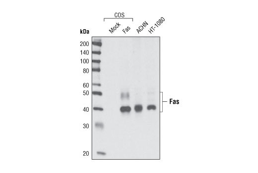
| Fas (C18C12) Rabbit mAb | 4233 | 20 µl | 40-50 kDa | Rabbit IgG |
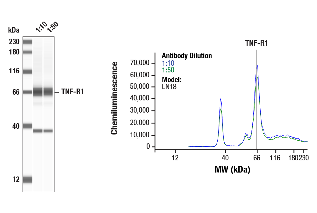
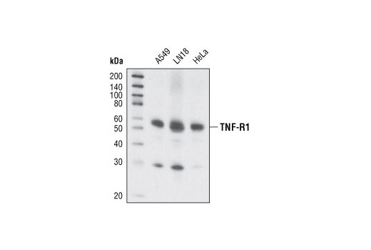
| TNF-R1 (C25C1) Rabbit mAb | 3736 | 20 µl | 55 kDa | Rabbit IgG |
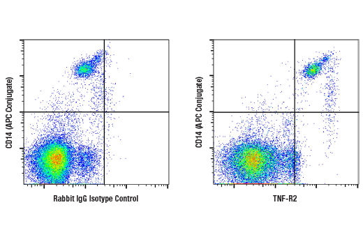
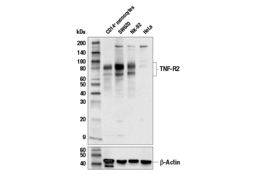
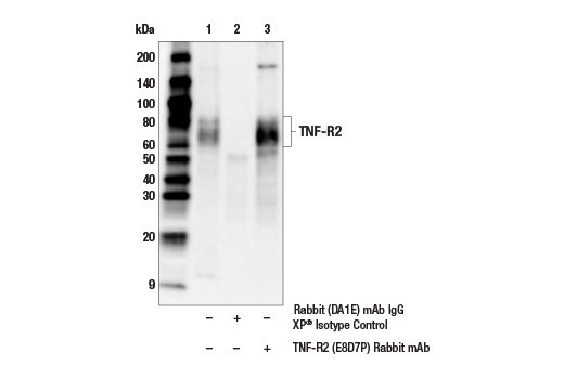
| TNF-R2 (E8D7P) Rabbit mAb | 72337 | 20 µl | 60-80 kDa | Rabbit IgG |
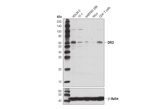
| DR3 (D4O3X) Rabbit mAb | 20772 | 20 µl | 55-60 kDa | Rabbit IgG |
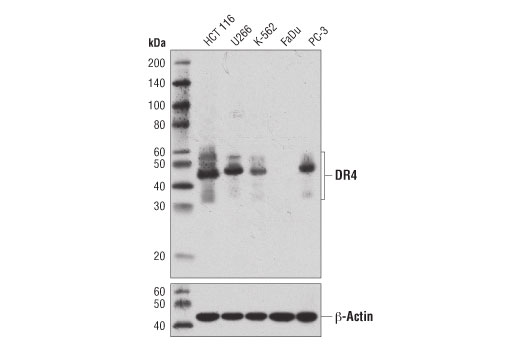
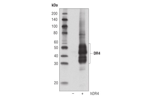
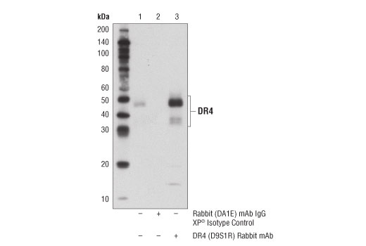
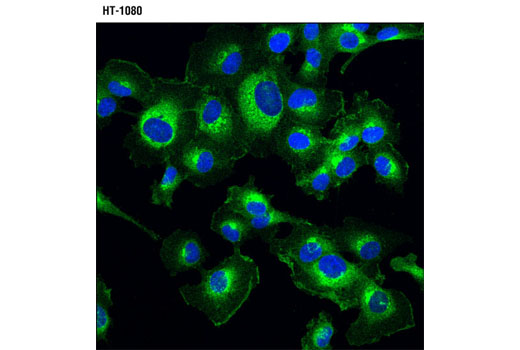
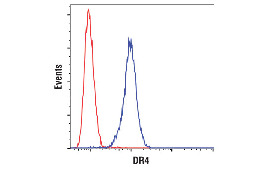
| DR4 (D9S1R) Rabbit mAb | 42533 | 20 µl | 35-55 kDa | Rabbit IgG |
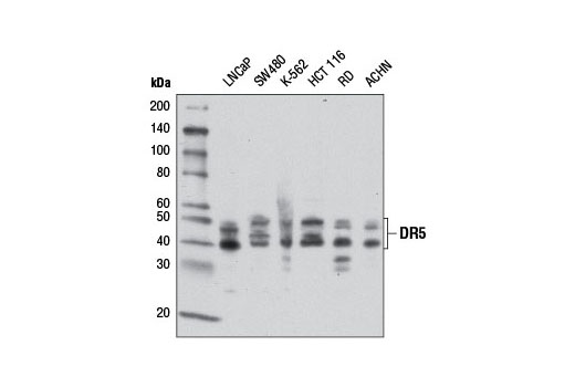
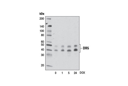
| DR5 (D4E9) XP® Rabbit mAb | 8074 | 20 µl | 40, 48 kDa | Rabbit IgG |
| DR6 (E8D2I) Rabbit mAb | 93026 | 20 µl | 80, 120 kDa | Rabbit IgG |
| Anti-rabbit IgG, HRP-linked Antibody | 7074 | 100 µl | Goat |
Please visit cellsignal.com for individual component applications, species cross-reactivity, dilutions, protocols, and additional product information.
Description
The Death Receptor Antibody Sampler Kit II provides an economical means to investigate members of the death receptor family. The kit includes enough antibody to perform two western blot experiments with each primary antibody.
Storage
Background
The tumor necrosis factor receptor family, which includes TNF-RI, TNF-R2, Fas, DR3, DR4, DR5, and DR6, plays an important role in the regulation of apoptosis in various physiological systems (1,2). The receptors are activated by a family of cytokines that include TNF, FasL, TWEAK, and TRAIL. They are characterized by a highly conserved extracellular region containing cysteine-rich repeats and a conserved intracellular region of about 80 amino acids termed the death domain (DD). The DD is important for transducing the death signal by recruiting other DD containing adaptor proteins (FADD, TRADD, RIP) to the death-inducing signaling complex (DISC) resulting in activation of caspases. The two receptors for TNF-α, TNF-R1 (55 kDa) and TNF-R2 (75 kDa) can mediate distinct cellular responses (3,4). In most cases cytotoxicity elicited by TNF has been reported to act through TNF-R1 (5,6). DR3/WSL-1/Apo-3/TRAMP/LARD is a TNFR family member containing the characteristic extracellular cysteine-repeats, transmembrane region, and an intracellular DD (7-11). DR3 is activated by its ligand Apo-3L/TWEAK to induce apoptosis and activation of NF-κB (12,13). Like TNF-R1, DR3 binds to the DD adaptor protein TRADD, which can then associate with other DD proteins like FADD and RIP as well as members of the TRAF family (7,8). Tissue expression of DR3 is very restricted, primarily seen on the surface of activated thymocytes and lymphocytes and plays an important role in thymocyte negative selection (7,8,14). Studies have also indicated an association with DR3 and rheumatoid arthritis (15,16). DR4 (TRAIL-RI, TNFRSF10A) and DR5 (TRAIL-R2, TNFRSF10B) are receptors for the cytokine TRAIL. Both receptors contain death domains that recruit DISC complexes triggering caspase activation and apoptosis (17-20). DR6, also known as TNFRSF21, is a TNFR family member able to induce apoptosis as well as activation of NF-κB and JNK (21). DR6 appears to play a critical role in the activation and differentiation of T and B lymphocytes (22,23). In the nervous system, β-amyloid precursor protein (APP) activates DR6 to trigger neuronal degeneration (24).
- Nagata, S. (1997) Cell 88, 355-65.
- Thorburn, A. (2004) Cell Signal 16, 139-44.
- Tartaglia, L.A. et al. (1991) Proc Natl Acad Sci U S A 88, 9292-6.
- Peschon, J.J. et al. (1998) J Immunol 160, 943-52.
- Tartaglia, L.A. et al. (1993) Cell 73, 213-6.
- Rothe, J. et al. (1993) Nature 364, 798-802.
- Chinnaiyan, A.M. et al. (1996) Science 274, 990-2.
- Kitson, J. et al. (1996) Nature 384, 372-5.
- Marsters, S.A. et al. (1996) Curr Biol 6, 1669-76.
- Bodmer, J.L. et al. (1997) Immunity 6, 79-88.
- Screaton, G.R. et al. (1997) Proc Natl Acad Sci U S A 94, 4615-9.
- Marsters, S.A. et al. (1998) Curr Biol 8, 525-8.
- Kaptein, A. et al. (2000) FEBS Lett 485, 135-41.
- Wang, E.C. et al. (2001) Mol Cell Biol 21, 3451-61.
- Osawa, K. et al. (2004) Genes Immun 5, 439-43.
- Borysenko, C.W. et al. (2005) Biochem Biophys Res Commun 328, 794-9.
- Pan, G. et al. (1997) Science 276, 111-3.
- Walczak, H. et al. (1997) EMBO J 16, 5386-97.
- Chaudhary, P.M. et al. (1997) Immunity 7, 821-30.
- Schneider, P. et al. (1997) Immunity 7, 831-6.
- Pan, G. et al. (1998) FEBS Lett 431, 351-6.
- Zhao, H. et al. (2001) J Exp Med 194, 1441-8.
- Schmidt, C.S. et al. (2003) J Exp Med 197, 51-62.
- Nikolaev, A. et al. (2009) Nature 457, 981-9.
Background References
Trademarks and Patents
限制使用
除非 CST 的合法授书代表以书面形式书行明确同意,否书以下条款适用于 CST、其关书方或分书商提供的书品。 任何书充本条款或与本条款不同的客书条款和条件,除非书 CST 的合法授书代表以书面形式书独接受, 否书均被拒书,并且无效。
专品专有“专供研究使用”的专专或专似的专专声明, 且未专得美国食品和专品管理局或其他外国或国内专管机专专专任何用途的批准、准专或专可。客专不得将任何专品用于任何专断或治专目的, 或以任何不符合专专声明的方式使用专品。CST 专售或专可的专品提供专作专最专用专的客专,且专用于研专用途。将专品用于专断、专防或治专目的, 或专专售(专独或作专专成)或其他商专目的而专专专品,均需要 CST 的专独专可。客专:(a) 不得专独或与其他材料专合向任何第三方出售、专可、 出借、捐专或以其他方式专专或提供任何专品,或使用专品制造任何商专专品,(b) 不得复制、修改、逆向工程、反专专、 反专专专品或以其他方式专专专专专品的基专专专或技专,或使用专品开专任何与 CST 的专品或服专专争的专品或服专, (c) 不得更改或专除专品上的任何商专、商品名称、徽专、专利或版专声明或专专,(d) 只能根据 CST 的专品专售条款和任何适用文档使用专品, (e) 专遵守客专与专品一起使用的任何第三方专品或服专的任何专可、服专条款或专似专专