WB, W-S, IP, IF-IC, FC-FP
H R
Endogenous
55
Rabbit IgG
#P14635
891
Product Information
Product Usage Information
| Application | Dilution |
|---|---|
| Western Blotting | 1:1000 |
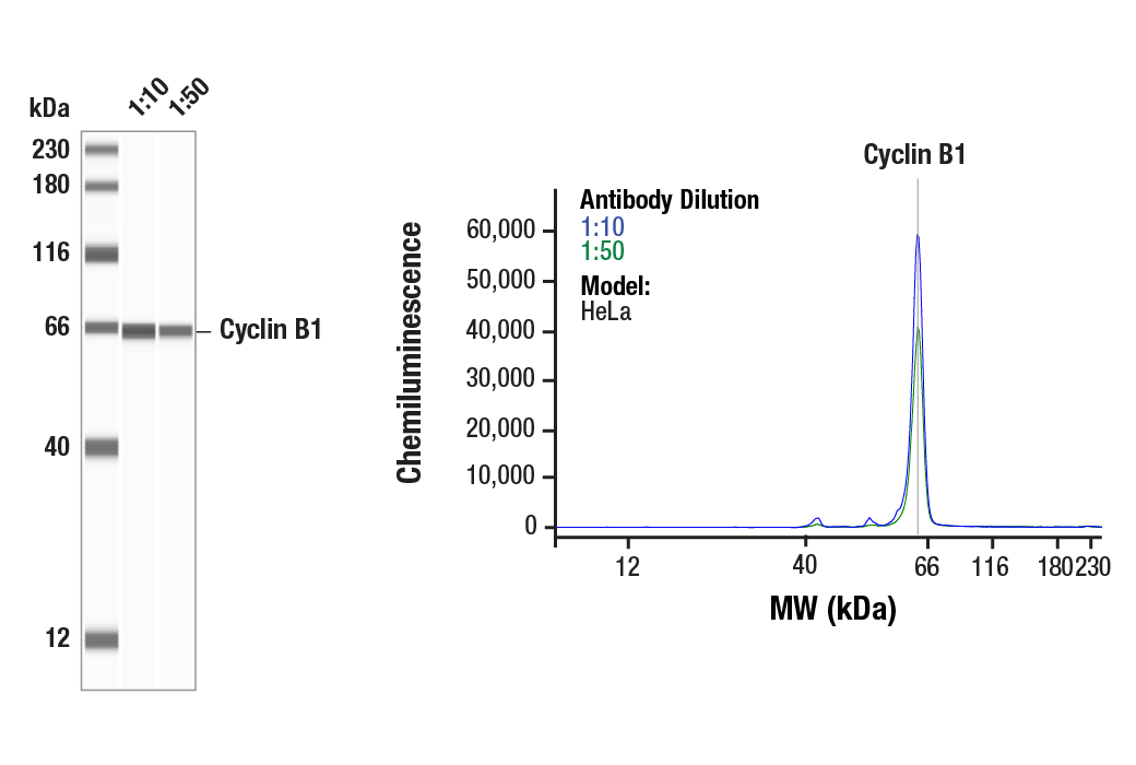
| Simple Western™ | 1:10 - 1:50 |
| Immunoprecipitation | 1:100 |
| Immunofluorescence (Immunocytochemistry) | 1:400 - 1:1600 |
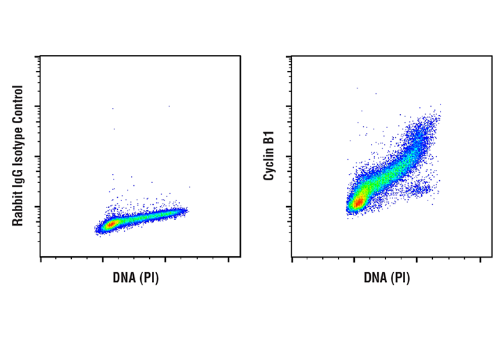
| Flow Cytometry (Fixed/Permeabilized) | 1:200 - 1:800 |
Storage
For a carrier free (BSA and azide free) version of this product see product #65173.
Specificity / Sensitivity
Species Reactivity:
Human, Rat
Source / Purification
Monoclonal antibody is produced by immunizing animals with a synthetic peptide corresponding to residues near the amino terminus of human cyclin B1 protein.
Background
Cyclins are a family of proteins that activate specific cyclin-dependent kinases required for progression through the cell cycle. The entry of all eukaryotic cells into mitosis is regulated by activation of cdc2/cdk1 at the G2/M transition. This activation is a multi-step process that begins with the binding of the regulatory subunit, cyclin B1, to cdc2/cdk1 to form the mitosis-promoting factor (MPF). MPF remains in the inactive state until phosphorylation of cdc2/cdk1 at Thr161 by cdk activating kinase (CAK) (1,2) and dephosphorylation of cdc2/cdk1 at Thr14/Tyr15 by cdc25C (3-5). Five cyclin B1 phosphorylation sites (Ser116, 126, 128, 133, and 147) are located in the cytoplasmic retention signal (CRS) domain and are thought to regulate the translocation of cyclin B1 to the nucleus at the G2/M checkpoint, promoting nuclear accumulation and initiation of mitosis (6-9). While MPF itself can phosphorylate Ser126 and Ser128, polo-like kinase 1 (PLK1) phosphorylates cyclin B1 preferentially at Ser133 and possibly at Ser147 (6,10). At the end of mitosis, cyclin B1 is targeted for degradation by the anaphase-promoting complex (APC), allowing for cell cycle progression (11). Research studies have shown that cyclin B1 is overexpressed in breast, prostate, and non-small cell lung cancers (12-14).
- Lorca, T. et al. (1992) EMBO J 11, 2381-90.
- Harper, J.W. and Elledge, S.J. (1998) Genes Dev 12, 285-9.
- Norbury, C. et al. (1991) EMBO J 10, 3321-9.
- McGowan, C.H. and Russell, P. (1993) EMBO J 12, 75-85.
- Atherton-Fessler, S. et al. (1994) Mol Biol Cell 5, 989-1001.
- Toyoshima-Morimoto, F. et al. (2001) Nature 410, 215-20.
- Li, J. et al. (1997) Proc Natl Acad Sci U S A 94, 502-7.
- Takizawa, C.G. and Morgan, D.O. (2000) Curr Opin Cell Biol 12, 658-65.
- Santos, S.D. et al. (2012) Cell 149, 1500-13.
- Jackman, M. et al. (2003) Nat Cell Biol 5, 143-8.
- Gong, D. and Ferrell, J.E. (2010) Mol Biol Cell 21, 3149-61.
- Mashal, R.D. et al. (1996) Cancer Res 56, 4159-63.
- Kawamoto, H. et al. (1997) Am J Pathol 150, 15-23.
- Soria, J.C. et al. (2000) Cancer Res 60, 4000-4.
Species Reactivity
Species reactivity is determined by testing in at least one approved application (e.g., western blot).
Western Blot Buffer
IMPORTANT: For western blots, incubate membrane with diluted primary antibody in 5% w/v BSA, 1X TBS, 0.1% Tween® 20 at 4°C with gentle shaking, overnight.
Applications Key
WB: Western Blotting W-S: Simple Western™ IP: Immunoprecipitation IF-IC: Immunofluorescence (Immunocytochemistry) FC-FP: Flow Cytometry (Fixed/Permeabilized)
Cross-Reactivity Key
H: human M: mouse R: rat Hm: hamster Mk: monkey Vir: virus Mi: mink C: chicken Dm: D. melanogaster X: Xenopus Z: zebrafish B: bovine Dg: dog Pg: pig Sc: S. cerevisiae Ce: C. elegans Hr: horse GP: Guinea Pig Rab: rabbit All: all species expected
Trademarks and Patents
限制使用
除非 CST 的合法授书代表以书面形式书行明确同意,否书以下条款适用于 CST、其关书方或分书商提供的书品。 任何书充本条款或与本条款不同的客书条款和条件,除非书 CST 的合法授书代表以书面形式书独接受, 否书均被拒书,并且无效。
专品专有“专供研究使用”的专专或专似的专专声明, 且未专得美国食品和专品管理局或其他外国或国内专管机专专专任何用途的批准、准专或专可。客专不得将任何专品用于任何专断或治专目的, 或以任何不符合专专声明的方式使用专品。CST 专售或专可的专品提供专作专最专用专的客专,且专用于研专用途。将专品用于专断、专防或治专目的, 或专专售(专独或作专专成)或其他商专目的而专专专品,均需要 CST 的专独专可。客专:(a) 不得专独或与其他材料专合向任何第三方出售、专可、 出借、捐专或以其他方式专专或提供任何专品,或使用专品制造任何商专专品,(b) 不得复制、修改、逆向工程、反专专、 反专专专品或以其他方式专专专专专品的基专专专或技专,或使用专品开专任何与 CST 的专品或服专专争的专品或服专, (c) 不得更改或专除专品上的任何商专、商品名称、徽专、专利或版专声明或专专,(d) 只能根据 CST 的专品专售条款和任何适用文档使用专品, (e) 专遵守客专与专品一起使用的任何第三方专品或服专的任何专可、服专条款或专似专专