Revision 2
Cell Signaling Technology

Orders: 877-616-CELL (2355) [email protected]

Support: 877-678-TECH (8324)

Web: [email protected] cellsignal.com

3 Trask LaneDanversMassachusetts01923USA
For Research Use Only. Not for Use in Diagnostic Procedures.
:

:

:

:

UniProt ID:

#O14757

Entrez-Gene Id:

1111

Product Includes Quantity Reactivity MW(kDa) Isotype
Chk1 (2G1D5) Mouse mAb 2360 100 µl H M R Mk 56 Mouse IgG1
Phospho-Chk1 (Ser317) (D12H3) XP® Rabbit mAb 12302 100 µl H M Mk 56 Rabbit IgG

Please visit cellsignal.com for individual component applications, species cross-reactivity, dilutions, protocols, and additional product information.

Description

PhosphoPlus® Duets from Cell Signaling Technology (CST) provide a means to assess protein activation status. Each Duet contains an activation-state and total protein antibody to your target of interest. These antibodies have been selected from CST's product offering based upon superior performance in specified applications. 

Storage

Supplied in 10 mM sodium HEPES (pH 7.5), 150 mM NaCl, 100 µg/ml BSA, 50% glycerol and less than 0.02% sodium azide. Store at –20°C. Do not aliquot the antibodies.

Background

Chk1 kinase acts downstream of ATM/ATR kinase and plays an important role in DNA damage checkpoint control, embryonic development, and tumor suppression (1). Activation of Chk1 involves phosphorylation at Ser317 and Ser345 by ATM/ATR, followed by autophosphorylation of Ser296. Activation occurs in response to blocked DNA replication and certain forms of genotoxic stress (2). While phosphorylation at Ser345 serves to localize Chk1 to the nucleus following checkpoint activation (3), phosphorylation at Ser317 along with site-specific phosphorylation of PTEN allows for re-entry into the cell cycle following stalled DNA replication (4). Chk1 exerts its checkpoint mechanism on the cell cycle, in part, by regulating the cdc25 family of phosphatases. Chk1 phosphorylation of cdc25A targets it for proteolysis and inhibits its activity through 14-3-3 binding (5). Activated Chk1 can inactivate cdc25C via phosphorylation at Ser216, blocking the activation of cdc2 and transition into mitosis (6). Centrosomal Chk1 has been shown to phosphorylate cdc25B and inhibit its activation of CDK1-cyclin B1, thereby abrogating mitotic spindle formation and chromatin condensation (7). Furthermore, Chk1 plays a role in spindle checkpoint function through regulation of aurora B and BubR1 (8). Research studies have implicated Chk1 as a drug target for cancer therapy as its inhibition leads to cell death in many cancer cell lines (9).

  1. Liu, Q. et al. (2000) Genes Dev 14, 1448-59.
  2. Zhao, H. and Piwnica-Worms, H. (2001) Mol Cell Biol 21, 4129-39.
  3. Jiang, K. et al. (2003) J Biol Chem 278, 25207-17.
  4. Martin, S.A. and Ouchi, T. (2008) Mol Cancer Ther 7, 2509-16.
  5. Chen, M.S. et al. (2003) Mol Cell Biol 23, 7488-97.
  6. Zeng, Y. et al. (1998) Nature 395, 507-10.
  7. Löffler, H. et al. (2006) Cell Cycle 5, 2543-7.
  8. Zachos, G. et al. (2007) Dev Cell 12, 247-60.
  9. Garber, K. (2005) J Natl Cancer Inst 97, 1026-8.

Background References

    Trademarks and Patents

    Cell Signaling Technology is a trademark of Cell Signaling Technology, Inc.
    PhosphoPlus is a registered trademark of Cell Signaling Technology, Inc.
    XP is a registered trademark of Cell Signaling Technology, Inc.
    All other trademarks are the property of their respective owners. Visit cellsignal.com/trademarks for more information.

    限制使用

    除非 CST 的合法授书代表以书面形式书行明确同意,否书以下条款适用于 CST、其关书方或分书商提供的书品。 任何书充本条款或与本条款不同的客书条款和条件,除非书 CST 的合法授书代表以书面形式书独接受, 否书均被拒书,并且无效。

    专品专有“专供研究使用”的专专或专似的专专声明, 且未专得美国食品和专品管理局或其他外国或国内专管机专专专任何用途的批准、准专或专可。客专不得将任何专品用于任何专断或治专目的, 或以任何不符合专专声明的方式使用专品。CST 专售或专可的专品提供专作专最专用专的客专,且专用于研专用途。将专品用于专断、专防或治专目的, 或专专售(专独或作专专成)或其他商专目的而专专专品,均需要 CST 的专独专可。客专:(a) 不得专独或与其他材料专合向任何第三方出售、专可、 出借、捐专或以其他方式专专或提供任何专品,或使用专品制造任何商专专品,(b) 不得复制、修改、逆向工程、反专专、 反专专专品或以其他方式专专专专专品的基专专专或技专,或使用专品开专任何与 CST 的专品或服专专争的专品或服专, (c) 不得更改或专除专品上的任何商专、商品名称、徽专、专利或版专声明或专专,(d) 只能根据 CST 的专品专售条款和任何适用文档使用专品, (e) 专遵守客专与专品一起使用的任何第三方专品或服专的任何专可、服专条款或专似专专
    Revision 2
    #51664

    PhosphoPlus® Chk1 (Ser317) Antibody Duet

    PhosphoPlus® Chk1 (Ser317) Antibody Duet: Image 1 Expand Image
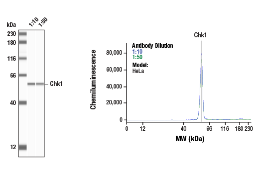
    使用 Chk1 (2G1D5) Mouse mAb #2360 对 HeLa 细胞的裂解物 (1.0 mg/mL) 进行 Simple Western™ 分析。虚拟泳道式图像(左图)显示一抗稀释比例为 1:10 和 1:50 时的单一靶标条带(如图所示)。对应的电泳图(右图)为一抗稀释比例在 1:10(蓝线)和 1:50(绿线)时沿毛细血管内分子量的化学发光结果。在还原条件下,使用 12-230 kDa 分离模块在 ProteinSimple(BioTechne 品牌)的 Jess™ Simple Western 仪器上进行该实验。
    PhosphoPlus® Chk1 (Ser317) Antibody Duet: Image 2 Expand Image
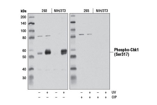
    使用 Phospho-Chk1 (Ser317) (D12H3) XP® Rabbit mAb,对未经处理 (-) 或经 UV 处理 (100 mJ, 1 小时恢复;+) 的293和 NIH/3T3 细胞提取物进行蛋白质印迹分析。在蛋白质印迹前,右侧的印迹经小牛肠磷酸酶处理。
    PhosphoPlus® Chk1 (Ser317) Antibody Duet: Image 3 Expand Image
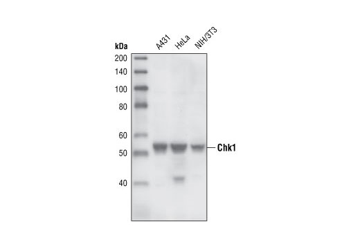
    使用 Chk1 (2G1D5) Mouse mAb 对来自不同细胞系的提取物进行蛋白质印迹分析。
    PhosphoPlus® Chk1 (Ser317) Antibody Duet: Image 4 Expand Image

    Immunohistochemical analysis of paraffin-embedded human oropharyngeal squamous cell carcinoma using Chk1 (2G1D5) Mouse mAb.

    PhosphoPlus® Chk1 (Ser317) Antibody Duet: Image 5 Expand Image
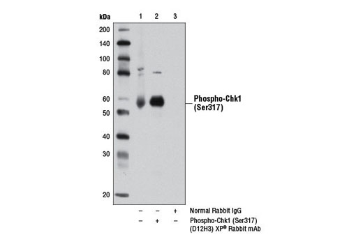
    使用 Phospho-Chk1 (Ser317) (D12H3) XP® Rabbit mAb (泳道2) 或 Rabbit (D1AG) mAb IgG XP® Isotype Control #3900 (泳道3) ,对经紫外线 (100 mJ, 1 小时恢复) 处理的293 细胞提取物中的 phospho-Chk1 (Ser317) 进行免疫沉淀。泳道 1 是 10% 输入对照。
    PhosphoPlus® Chk1 (Ser317) Antibody Duet: Image 6 Expand Image
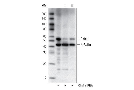
    使用 Chk1 (2G1D5) Mouse mAb #2360和 β-Actin (13E5) Rabbit mAb #4970 对来自被 100 nM SignalSilence® Control siRNA (Fluorescein Conjugate) #6201 (-) 或 SignalSilence® Chk1 siRNA I #6241 或 SignalSilence® Chk1 siRNA II (+) 转染的 Hela 细胞的提取物进行蛋白质印迹分析。Chk1 (2G1D5) Mouse mAb 可证实 Chk1 表达的沉默并且 β-Actin (13E5) Rabbit mAb 可用作 Chk1 siRNA 的载量和特异性的对照。
    PhosphoPlus® Chk1 (Ser317) Antibody Duet: Image 7 Expand Image

    Immunohistochemical analysis of paraffin-embedded human thymoma using Chk1 (2G1D5) Mouse mAb.

    PhosphoPlus® Chk1 (Ser317) Antibody Duet: Image 8 Expand Image
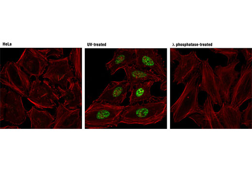
    使用 Phospho-Chk1 (Ser317) (D12H3) XP® Rabbit mAb (绿色) ,对未经处理 (左图) 、经紫外线处理的 (中图) 或经紫外线和λ磷酸酶同时处理 (右图) 的 Hela 细胞进行共聚焦免疫荧光分析。使用 DY-554 phalloidin(红色)标记肌动蛋白纤丝。
    PhosphoPlus® Chk1 (Ser317) Antibody Duet: Image 9 Expand Image
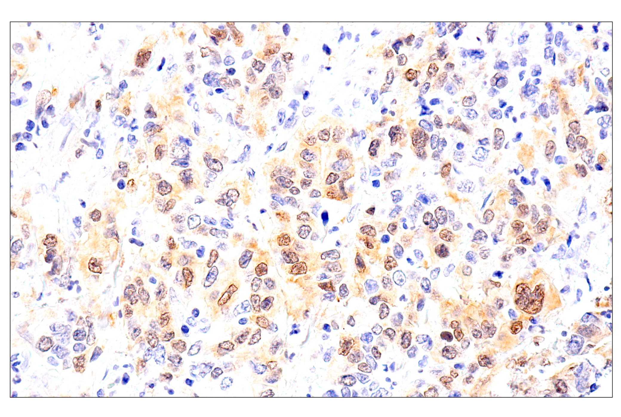
    Immunohistochemical analysis of paraffin-embedded human squamous cell lung carcinoma using Chk1 (2G1D5) Mouse mAb.
    PhosphoPlus® Chk1 (Ser317) Antibody Duet: Image 10 Expand Image

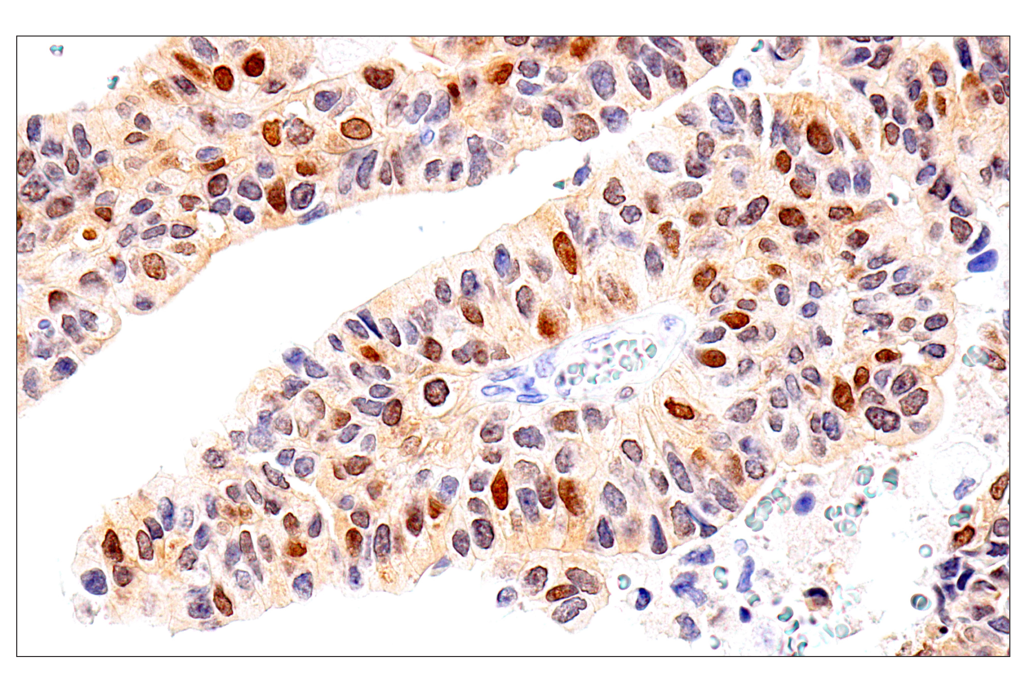
    Immunohistochemical analysis of paraffin-embedded human esophageal adenocarcinoma using Chk1 (2G1D5) Mouse mAb.

    PhosphoPlus® Chk1 (Ser317) Antibody Duet: Image 11 Expand Image

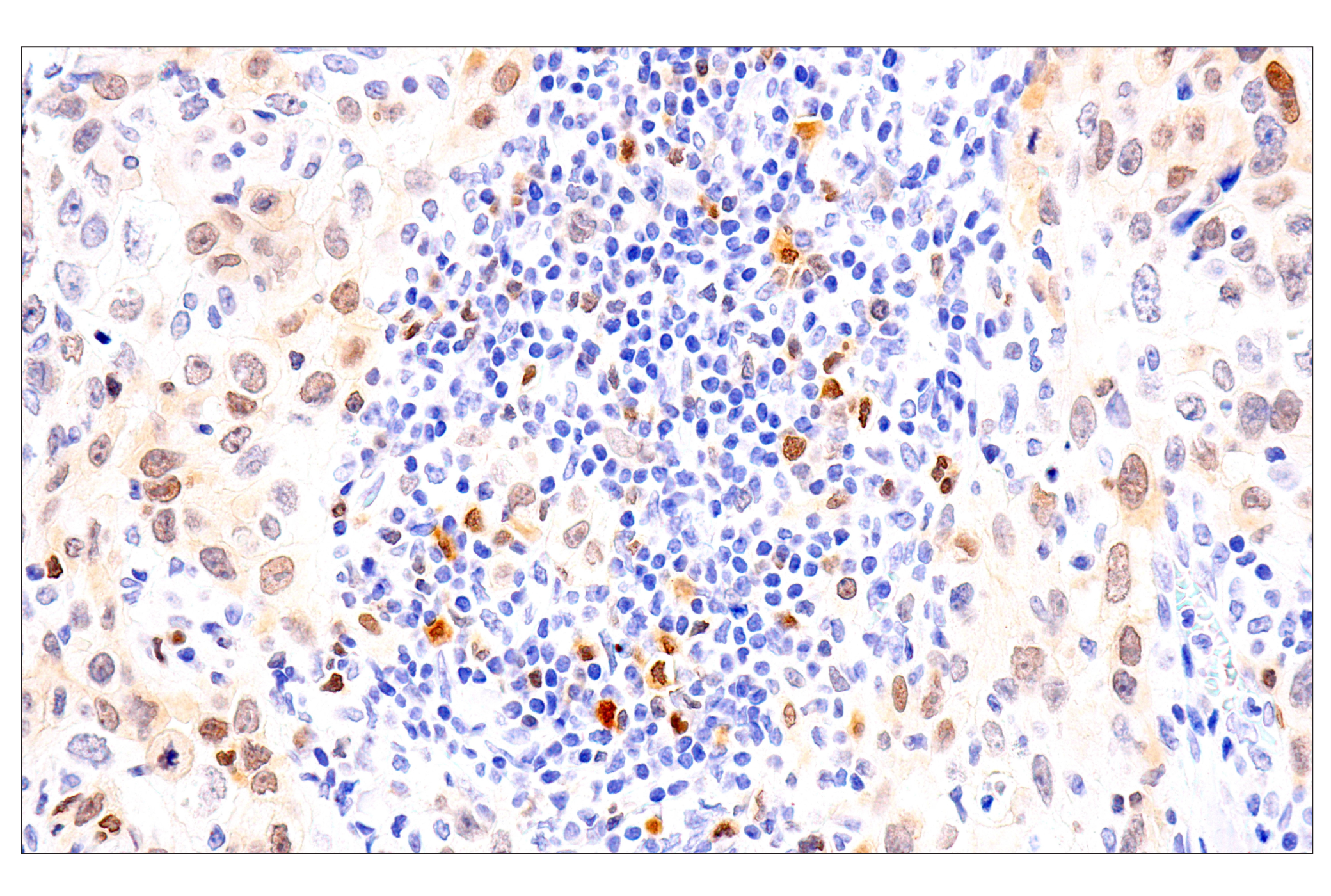
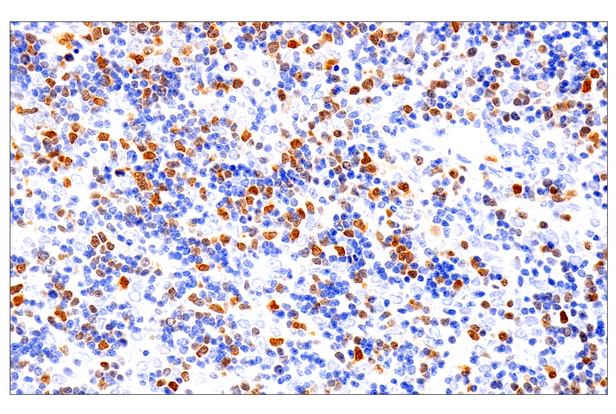
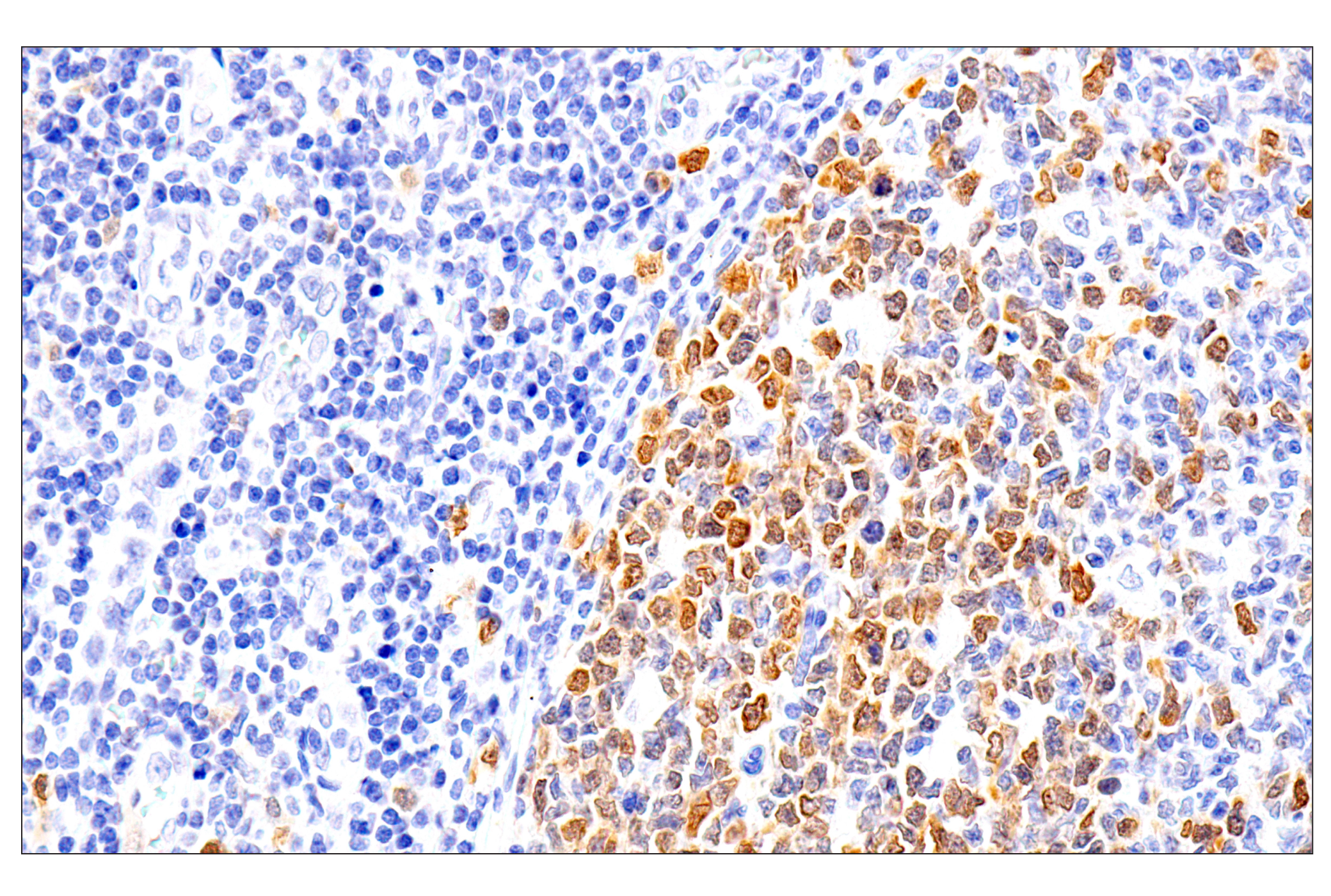
    Immunohistochemical analysis of paraffin-embedded human Non-Hodgkin lymphoma using Chk1 (2G1D5) Mouse mAb.

    PhosphoPlus® Chk1 (Ser317) Antibody Duet: Image 12 Expand Image

    Immunohistochemical analysis of paraffin-embedded human urothelial carcinoma using Chk1 (2G1D5) Mouse mAb.

    PhosphoPlus® Chk1 (Ser317) Antibody Duet: Image 13 Expand Image

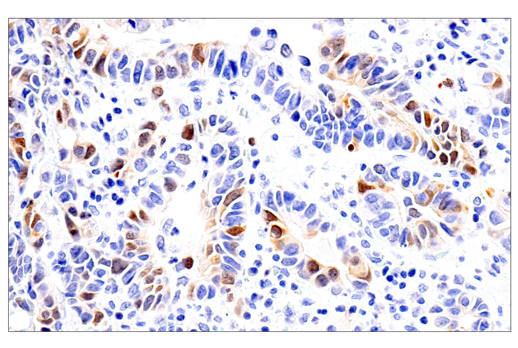
    Immunohistochemical analysis of paraffin-embedded human gastric adenocarcinoma using Chk1 (2G1D5) Mouse mAb.

    PhosphoPlus® Chk1 (Ser317) Antibody Duet: Image 14 Expand Image
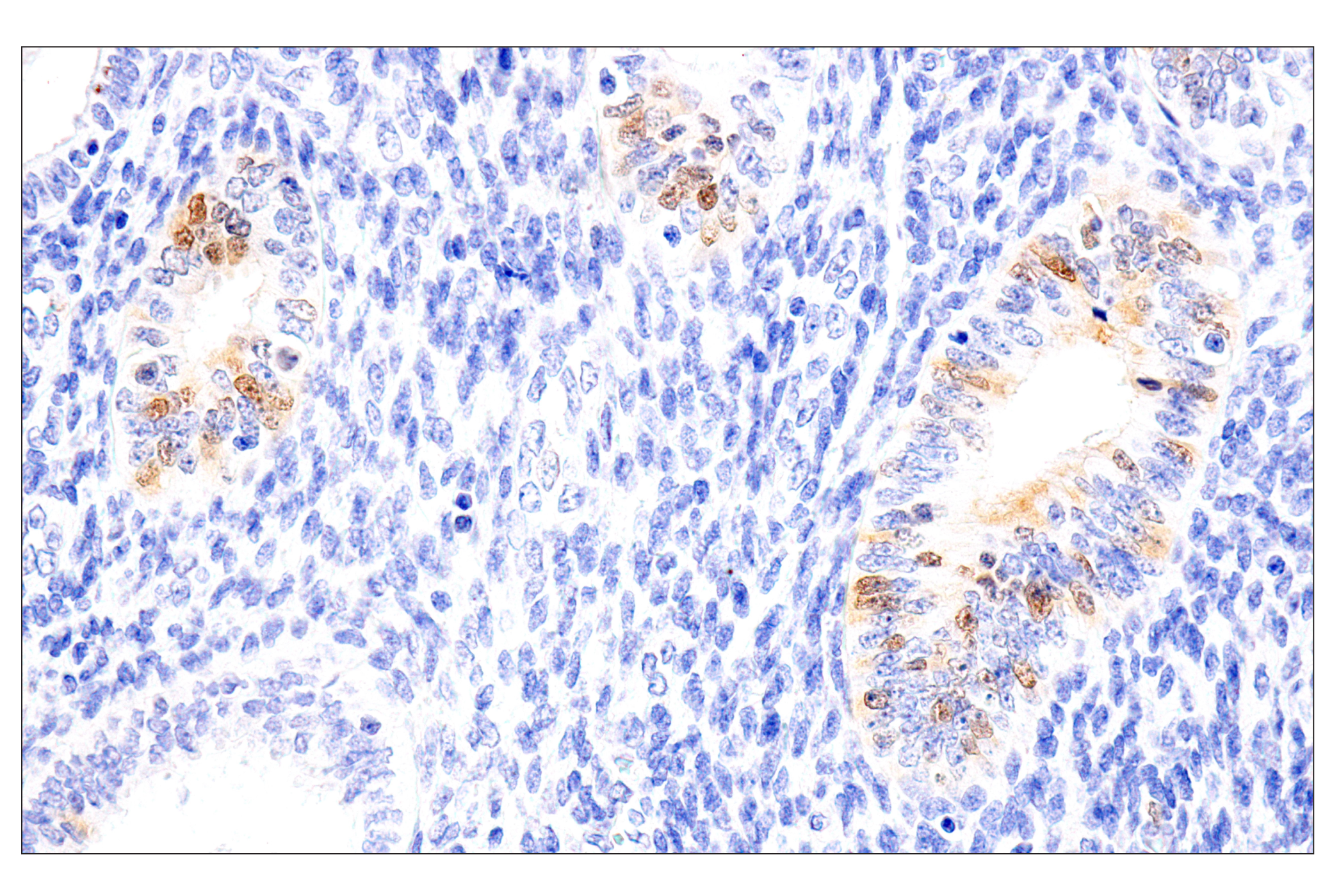
    Immunohistochemical analysis of paraffin-embedded human endometrial cancer using Chk1 (2G1D5) Mouse mAb.
    PhosphoPlus® Chk1 (Ser317) Antibody Duet: Image 15 Expand Image

    Immunohistochemical analysis of paraffin-embedded normal human uterus using Chk1 (2G1D5) Mouse mAb.

    PhosphoPlus® Chk1 (Ser317) Antibody Duet: Image 16 Expand Image

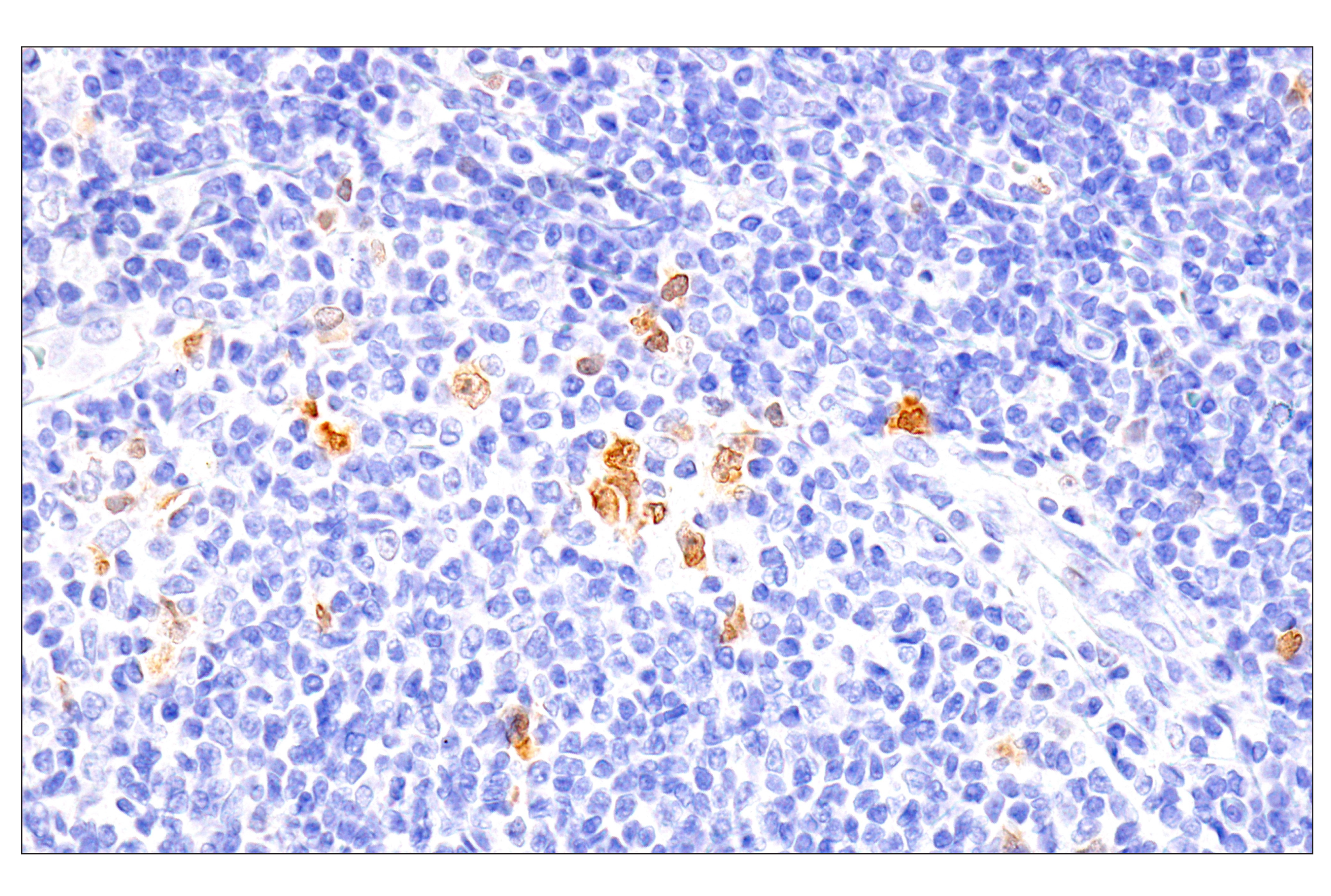
    Immunohistochemical analysis of paraffin-embedded human tonsil using Chk1 (2G1D5) Mouse mAb.

    PhosphoPlus® Chk1 (Ser317) Antibody Duet: Image 17 Expand Image

    Immunohistochemical analysis of paraffin-embedded normal human esophagus using Chk1 (2G1D5) Mouse mAb.

    PhosphoPlus® Chk1 (Ser317) Antibody Duet: Image 18 Expand Image

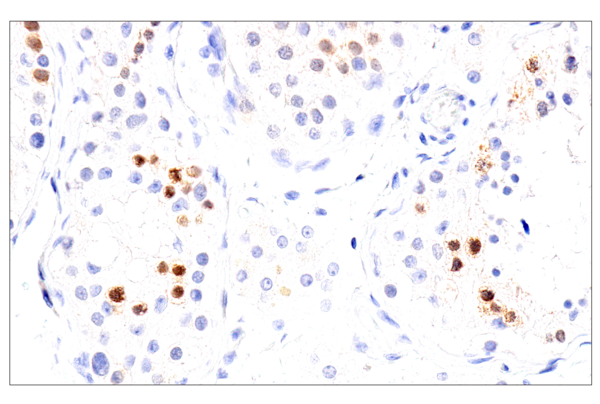
    Immunohistochemical analysis of paraffin-embedded normal human testis using Chk1 (2G1D5) Mouse mAb.

    PhosphoPlus® Chk1 (Ser317) Antibody Duet: Image 19 Expand Image
    Immunohistochemical analysis of paraffin-embedded normal human small intestine using Chk1 (2G1D5) Mouse mAb (left) compared to concentration-matched Mouse (G3A1) mAb IgG1 Isotype Control #5415 (right). 
    PhosphoPlus® Chk1 (Ser317) Antibody Duet: Image 20 Expand Image
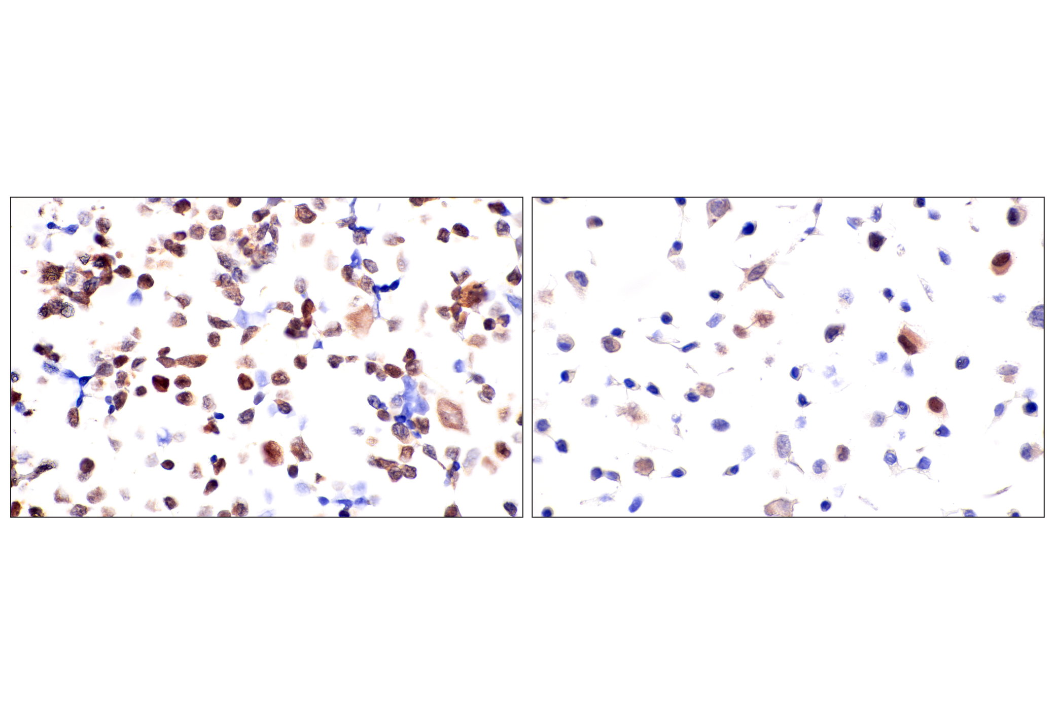
    Immunohistochemical analysis of paraffin-embedded 293T cell pellet (left, high-expressing) or SK-MEL-28 cell pellet (right, low-expressing) using Chk1 (2G1D5) Mouse mAb.