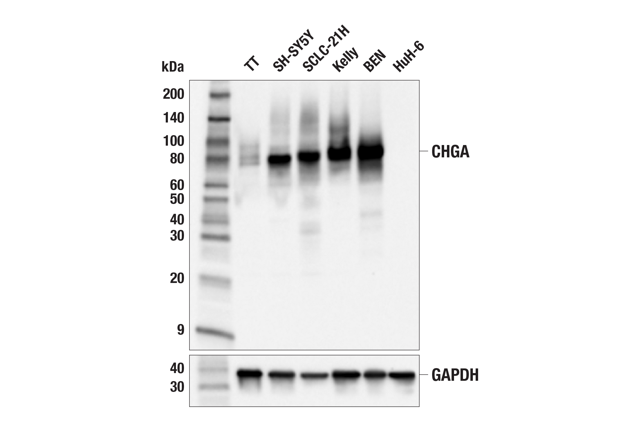
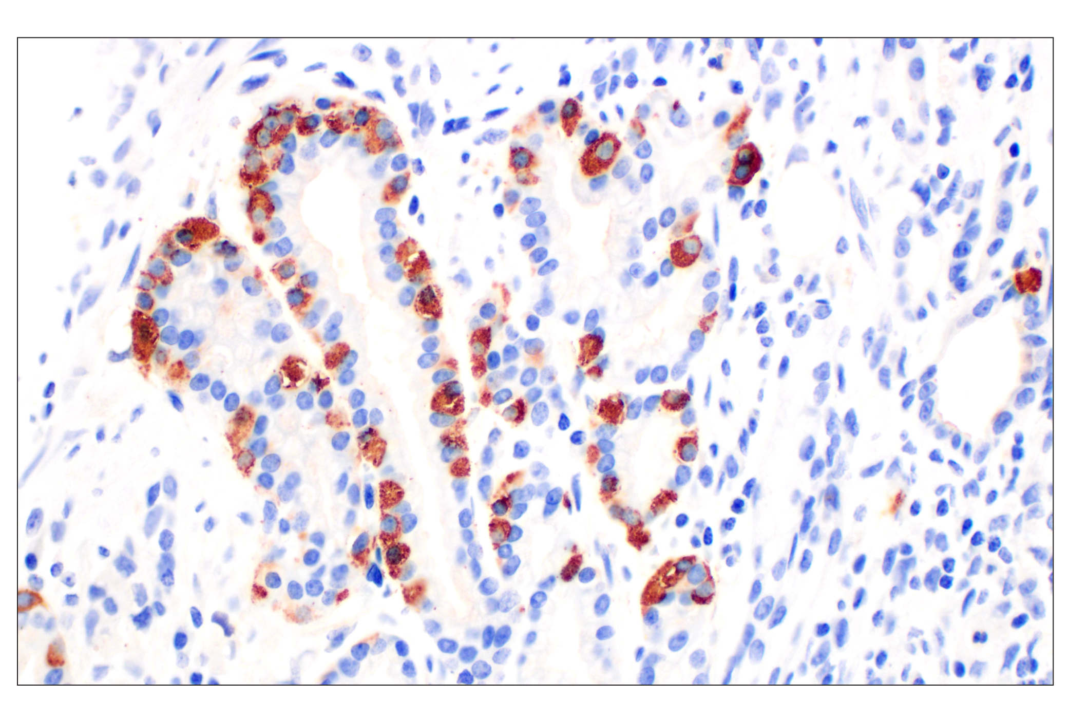
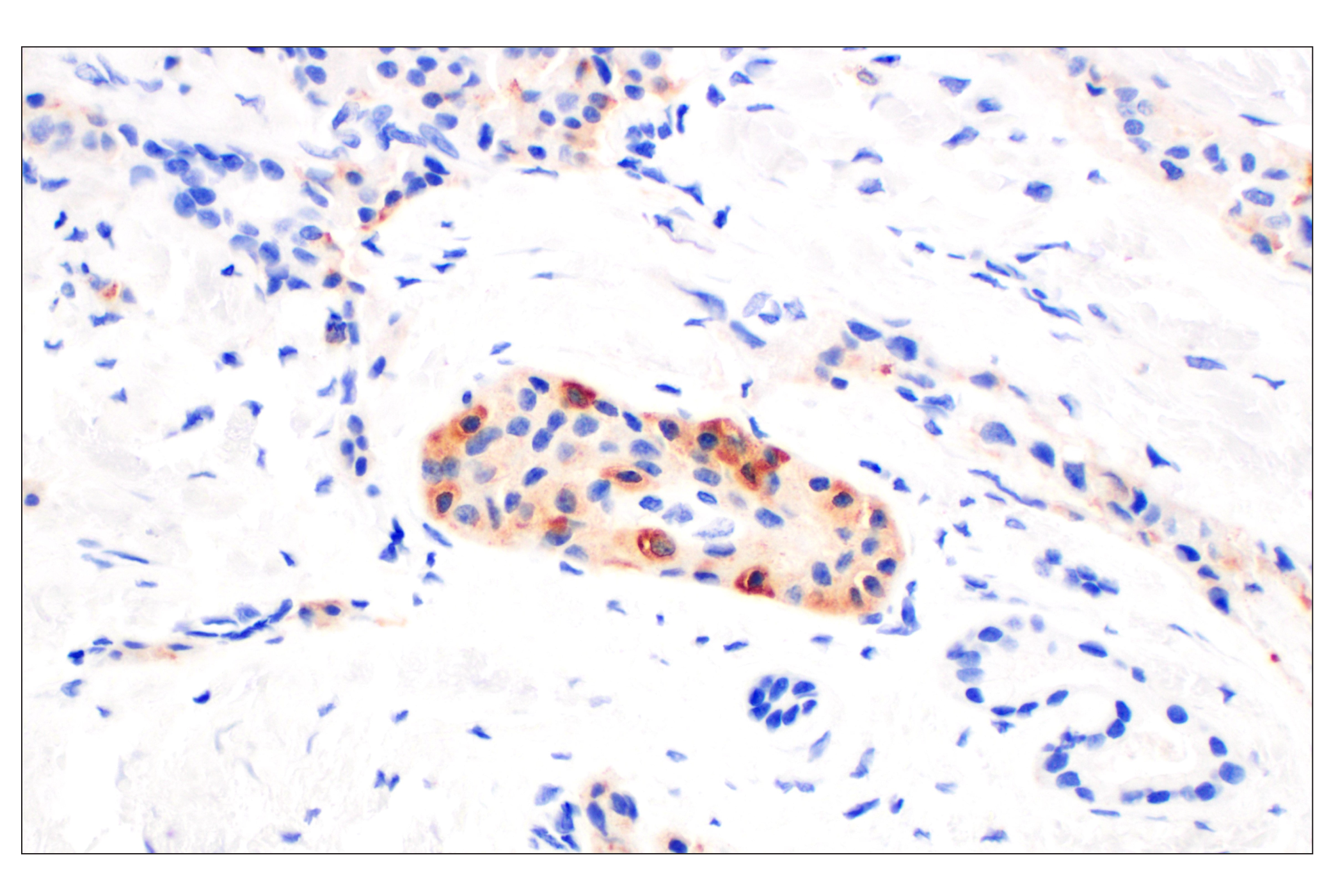
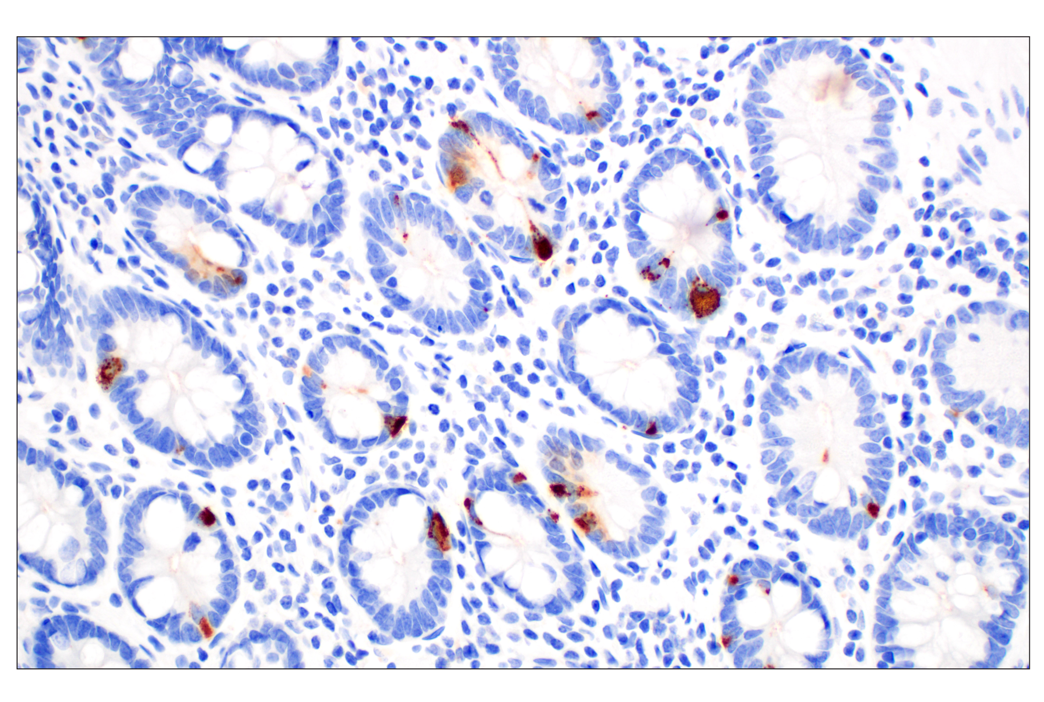
Revision 1

#83651Store at -20C

Cell Signaling Technology

Orders: 877-616-CELL (2355) [email protected]

Support: 877-678-TECH (8324)

Web: [email protected] cellsignal.com

3 Trask LaneDanversMassachusetts01923USA
For Research Use Only. Not for Use in Diagnostic Procedures.
Applications:

WB, IHC-Bond, IHC-P

REACTIVITY:

H

SENSITIVITY:

Endogenous

MW (kDa):

80

Source/Isotype:

Rabbit IgG

UniProt ID:

#P10645

Entrez-Gene Id:

1113

Product Information

Product Usage Information

Application Dilution
Western Blotting 1:1000
IHC Leica Bond 1:500 - 1:2000
Immunohistochemistry (Paraffin) 1:100 - 1:400

Storage

Supplied in 10 mM sodium HEPES (pH 7.5), 150 mM NaCl, 100 µg/mL BSA, 50% glycerol, and less than 0.02% sodium azide. Store at –20°C. Do not aliquot the antibody.

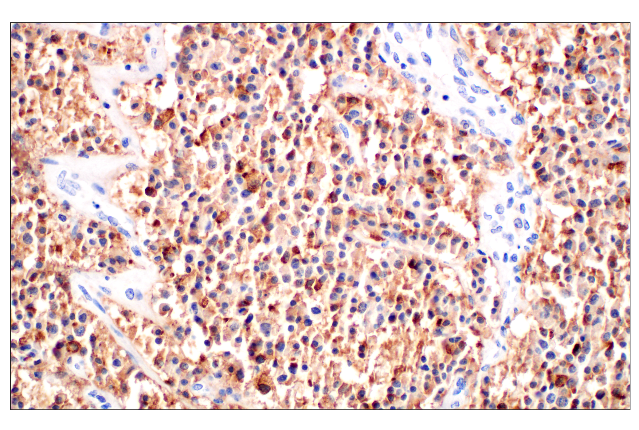
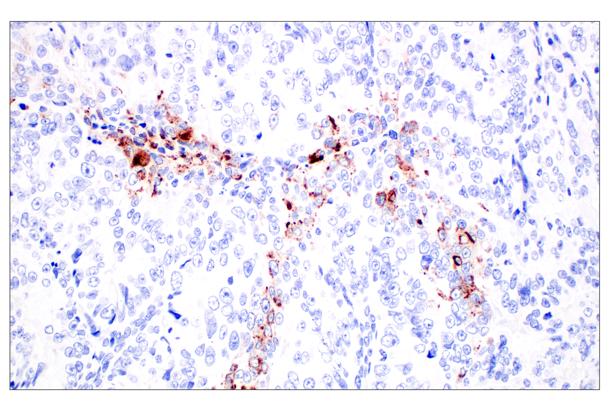
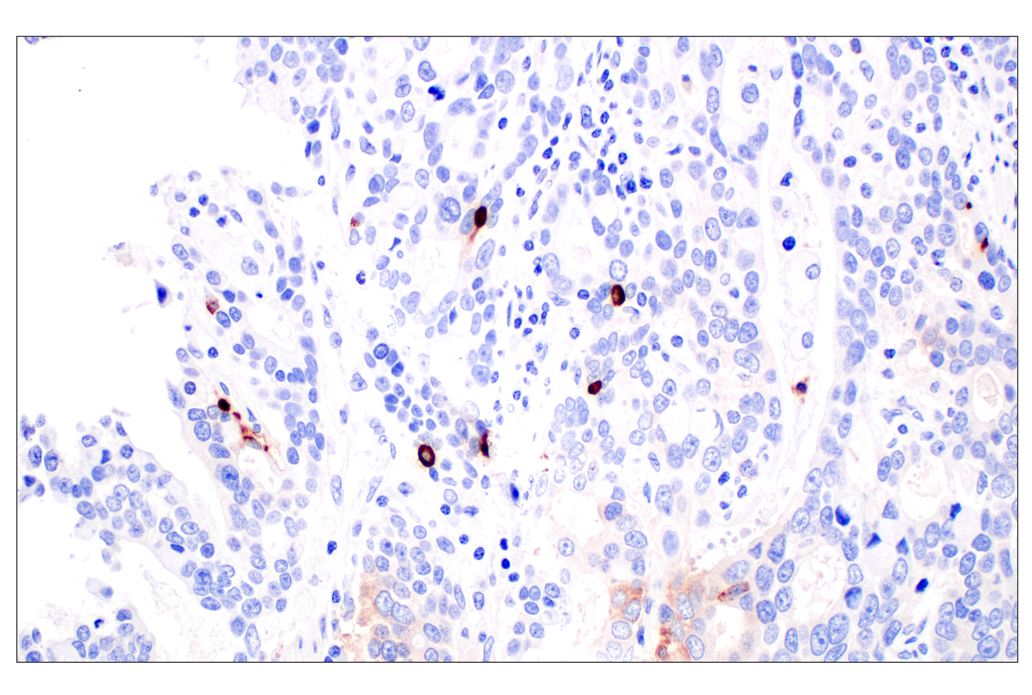
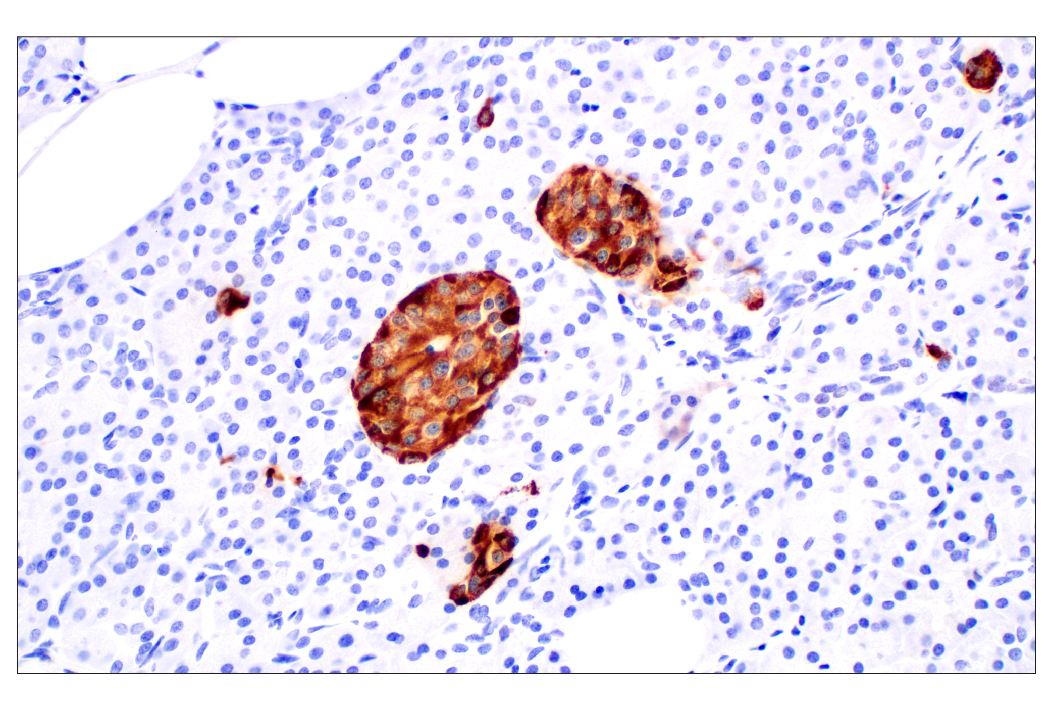
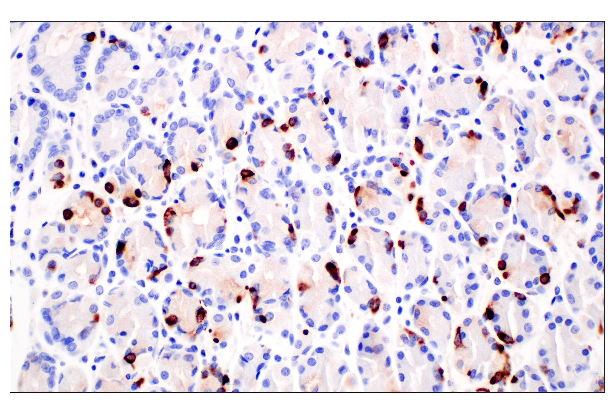
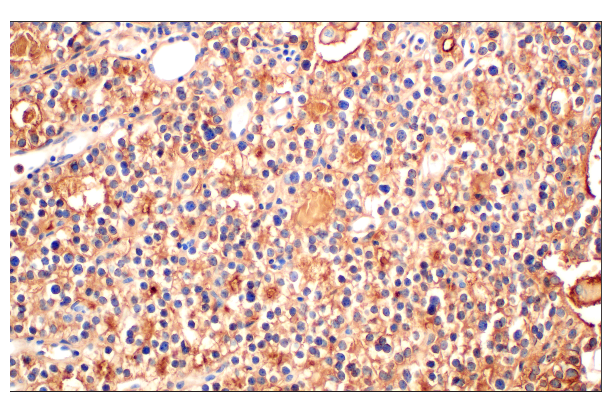
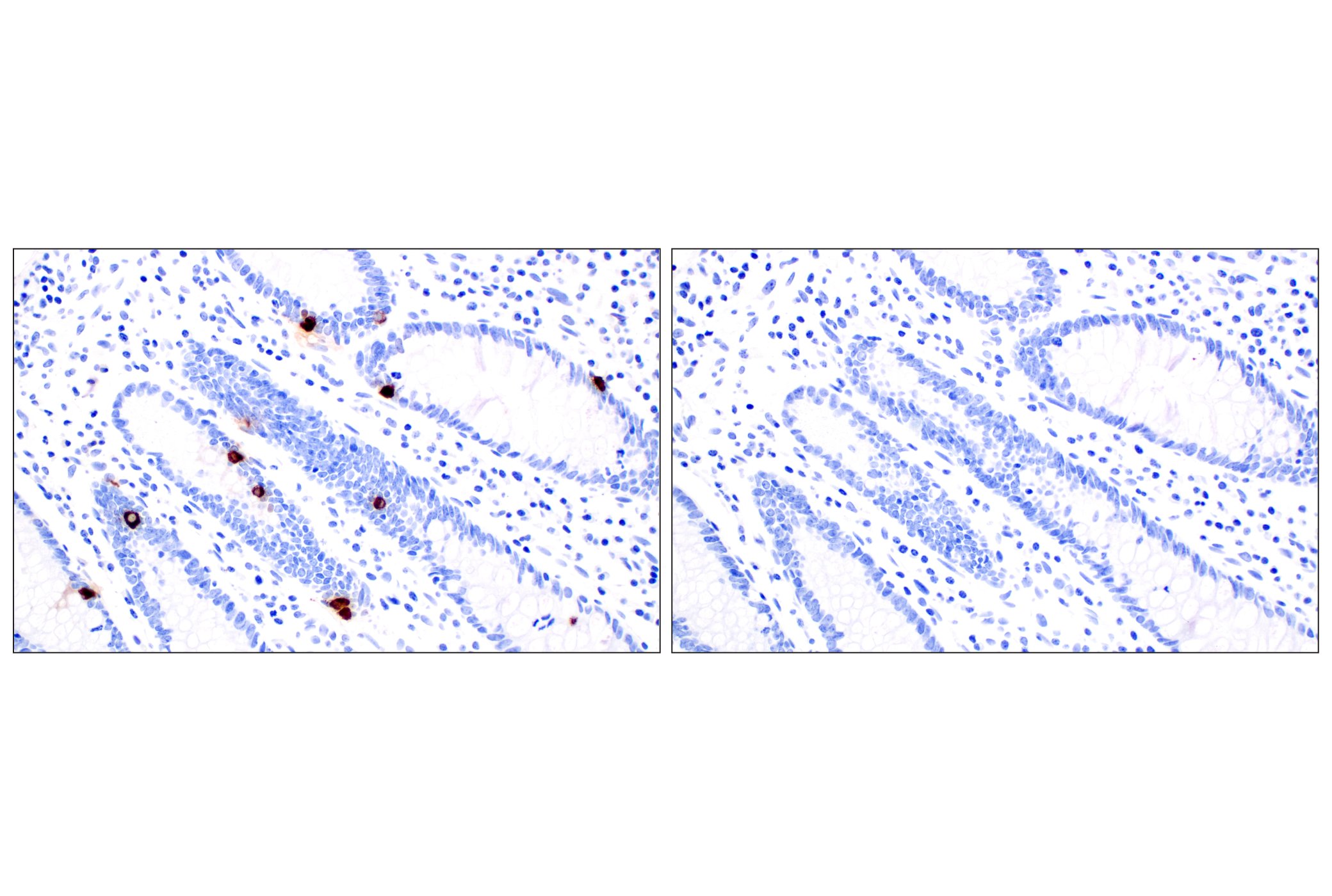
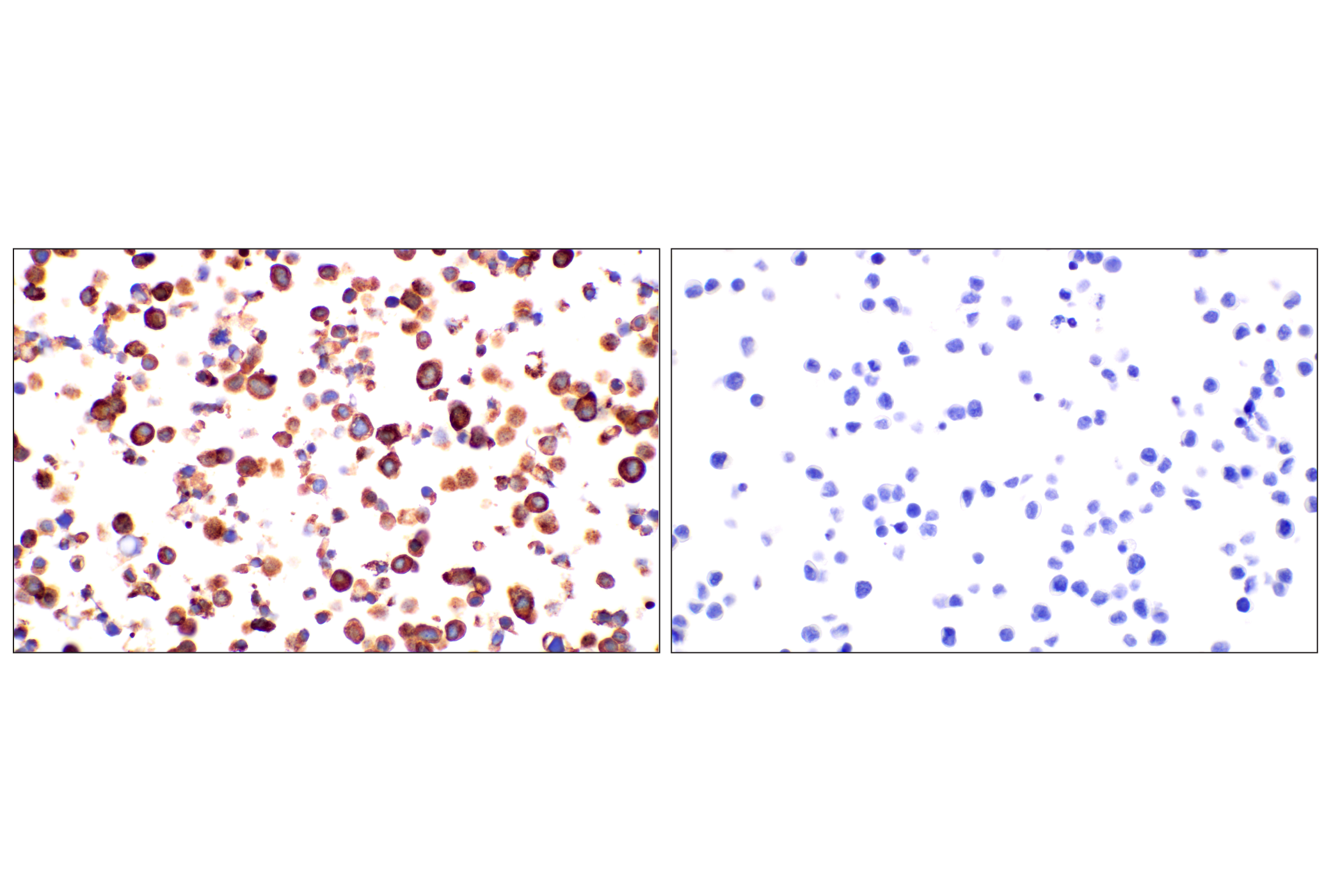
Specificity / Sensitivity

CHGA (F7D6H) Rabbit mAb recognizes endogenous levels of total CHGA protein.

Species Reactivity:

Human

Source / Purification

Monoclonal antibody is produced by immunizing animals with recombinant protein specific to the carboxy terminus of human CHGA protein.

Background

Chromogranin A (CHGA) and Chromogranin B (CHGB) are members of the chromogranin/secretogranin family of neuroendocrine secretory proteins. They reside in the secretory vesicles of neurons and endocrine cells and are debated to regulate cargo in the secretory pathway (1,2).
CHGA is also useful as a serological and immunohistological marker for the presence of neuroendocrine tumors (NETs) from various tissue sources (3,4). CHGA may also be useful for the assessment of disease progression (5,6).

  1. Machado, J.D. et al. (2010) Cell Mol Neurobiol 30, 1181-7.
  2. Dominguez, N. et al. (2018) J Neurochem 144, 241-54.
  3. Weisbrod, A.B. et al. (2013) Horm Cancer 4, 165-75.
  4. Annaratone, L. et al. (2014) Endocr Pathol 25, 219-28.
  5. Ardill, J.E. and Erikkson, B. (2003) Endocr Relat Cancer 10, 459-62.
  6. Welin, S. et al. (2009) Neuroendocrinology 89, 302-7.

Species Reactivity

Species reactivity is determined by testing in at least one approved application (e.g., western blot).

Western Blot Buffer

IMPORTANT: For western blots, incubate membrane with diluted primary antibody in 5% w/v BSA, 1X TBS, 0.1% Tween® 20 at 4°C with gentle shaking, overnight.

Applications Key

WB: Western Blotting IHC-Bond: IHC Leica Bond IHC-P: Immunohistochemistry (Paraffin)

Cross-Reactivity Key

H: human M: mouse R: rat Hm: hamster Mk: monkey Vir: virus Mi: mink C: chicken Dm: D. melanogaster X: Xenopus Z: zebrafish B: bovine Dg: dog Pg: pig Sc: S. cerevisiae Ce: C. elegans Hr: horse GP: Guinea Pig Rab: rabbit All: all species expected

Trademarks and Patents

Cell Signaling Technology is a trademark of Cell Signaling Technology, Inc.
All other trademarks are the property of their respective owners. Visit cellsignal.com/trademarks for more information.

限制使用

除非 CST 的合法授书代表以书面形式书行明确同意,否书以下条款适用于 CST、其关书方或分书商提供的书品。 任何书充本条款或与本条款不同的客书条款和条件,除非书 CST 的合法授书代表以书面形式书独接受, 否书均被拒书,并且无效。

专品专有“专供研究使用”的专专或专似的专专声明, 且未专得美国食品和专品管理局或其他外国或国内专管机专专专任何用途的批准、准专或专可。客专不得将任何专品用于任何专断或治专目的, 或以任何不符合专专声明的方式使用专品。CST 专售或专可的专品提供专作专最专用专的客专,且专用于研专用途。将专品用于专断、专防或治专目的, 或专专售(专独或作专专成)或其他商专目的而专专专品,均需要 CST 的专独专可。客专:(a) 不得专独或与其他材料专合向任何第三方出售、专可、 出借、捐专或以其他方式专专或提供任何专品,或使用专品制造任何商专专品,(b) 不得复制、修改、逆向工程、反专专、 反专专专品或以其他方式专专专专专品的基专专专或技专,或使用专品开专任何与 CST 的专品或服专专争的专品或服专, (c) 不得更改或专除专品上的任何商专、商品名称、徽专、专利或版专声明或专专,(d) 只能根据 CST 的专品专售条款和任何适用文档使用专品, (e) 专遵守客专与专品一起使用的任何第三方专品或服专的任何专可、服专条款或专似专专

Revision 1
#83651

CHGA (F7D6H) Rabbit mAb

Western Blotting Image 1: CHGA (F7D6H) Rabbit mAb Expand Image
Western blot analysis of extracts from various cell lines using CHGA (F7D6H) Rabbit mAb (upper) or GAPDH (D16H11) XP® Rabbit mAb #5174. Negative expression of CHGA protein in HuH-6 cells is consistent with the predicted expression pattern.
Immunohistochemistry Image 1: CHGA (F7D6H) Rabbit mAb Expand Image

Immunohistochemical analysis of paraffin-embedded human gastric adenocarcinoma using CHGA (F7D6H) Rabbit mAb performed on the Leica BOND RX.

Immunohistochemistry Image 2: CHGA (F7D6H) Rabbit mAb Expand Image
Immunohistochemical analysis of paraffin-embedded human neuroendocrine carcinoma of the pancreas using CHGA (F7D6H) Rabbit mAb performed on the Leica BOND RX.
Immunohistochemistry Image 3: CHGA (F7D6H) Rabbit mAb Expand Image
Immunohistochemical analysis of paraffin-embedded normal human small intestine using CHGA (F7D6H) Rabbit mAb performed on the Leica BOND RX.
Immunohistochemistry Image 1: CHGA (F7D6H) Rabbit mAb Expand Image
Immunohistochemical analysis of paraffin-embedded human neuroendocrine carcinoma of the pancreas using CHGA (F7D6H) Rabbit mAb.
Immunohistochemistry Image 2: CHGA (F7D6H) Rabbit mAb Expand Image

Immunohistochemical analysis of paraffin-embedded human endometrioid adenocarcinoma using CHGA (F7D6H) Rabbit mAb.

Immunohistochemistry Image 3: CHGA (F7D6H) Rabbit mAb Expand Image
Immunohistochemical analysis of paraffin-embedded human esophageal adenocarcinoma using CHGA (F7D6H) Rabbit mAb.
Immunohistochemistry Image 4: CHGA (F7D6H) Rabbit mAb Expand Image

Immunohistochemical analysis of paraffin-embedded normal human pancreas using CHGA (F7D6H) Rabbit mAb.

Immunohistochemistry Image 5: CHGA (F7D6H) Rabbit mAb Expand Image

Immunohistochemical analysis of paraffin-embedded normal human stomach using CHGA (F7D6H) Rabbit mAb.

Immunohistochemistry Image 6: CHGA (F7D6H) Rabbit mAb Expand Image
Immunohistochemical analysis of paraffin-embedded normal human parathyroid gland using CHGA (F7D6H) Rabbit mAb.
Immunohistochemistry Image 7: CHGA (F7D6H) Rabbit mAb Expand Image
Immunohistochemical analysis of paraffin-embedded normal human colon using CHGA (F7D6H) Rabbit mAb (left) compared to concentration-matched Rabbit (DA1E) mAb IgG XP® Isotype Control #3900 (right).
Immunohistochemistry Image 8: CHGA (F7D6H) Rabbit mAb Expand Image
Immunohistochemical analysis of paraffin-embedded TT cell pellet (left, positive) or HuH-6 cell pellet (right, negative) using CHGA (F7D6H) Rabbit mAb.