| Product Includes | Product # | Quantity | Mol. Wt | Isotype/Source |
|---|---|---|---|---|
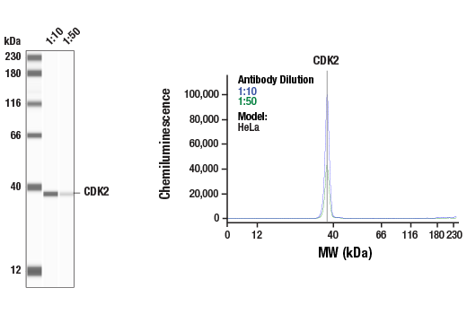
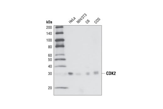
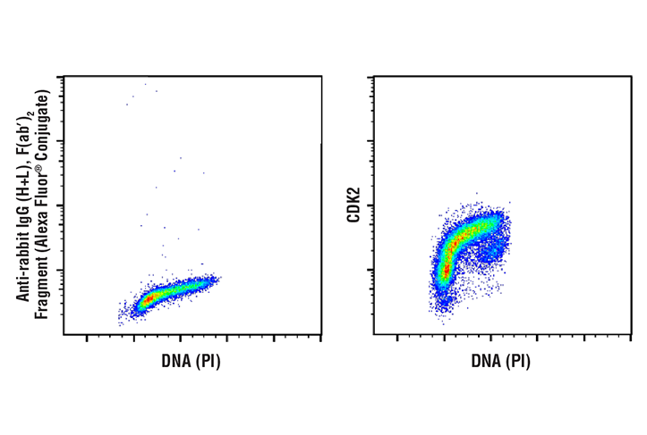
| CDK2 (78B2) Rabbit mAb | 2546 | 20 µl | 33 kDa | Rabbit |
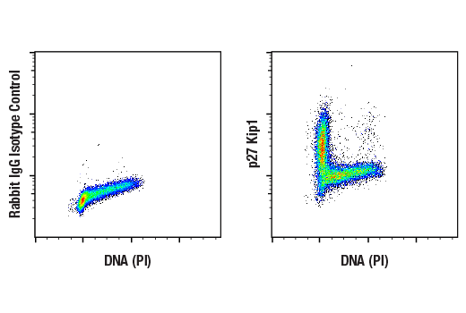
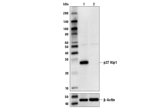
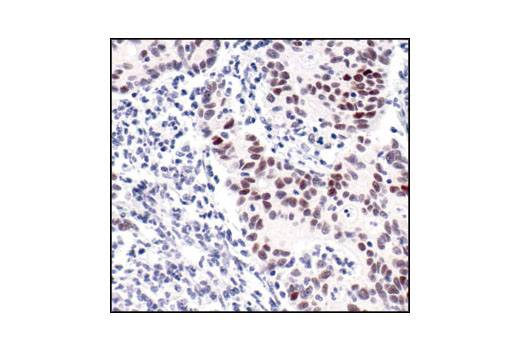
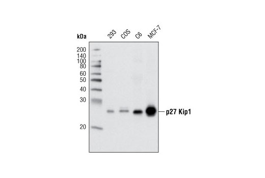
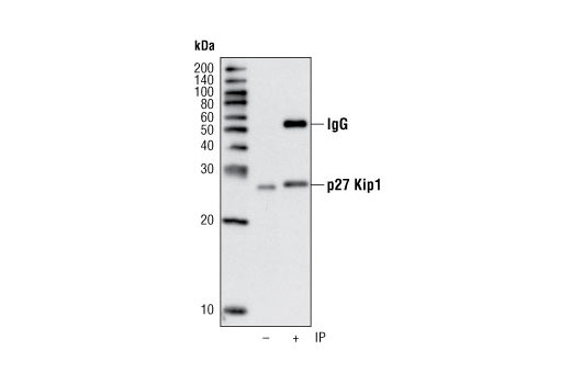
| p27 Kip1 (D69C12) XP® Rabbit mAb | 3686 | 20 µl | 27 kDa | Rabbit IgG |
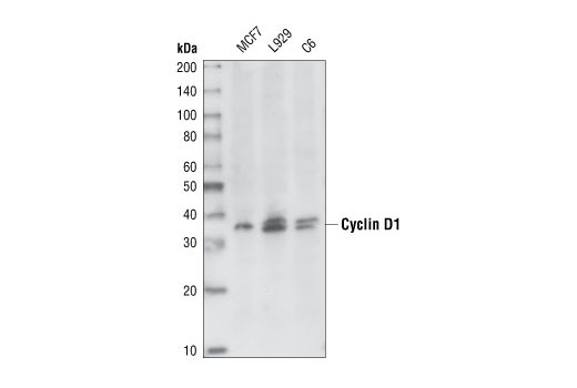
| Cyclin D1 (92G2) Rabbit mAb | 2978 | 20 µl | 36 kDa | Rabbit IgG |
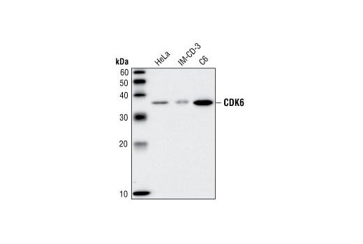
| CDK6 (DCS83) Mouse mAb | 3136 | 20 µl | 36 kDa | Mouse IgG1 |
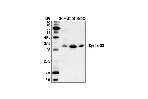
| Cyclin D3 (DCS22) Mouse mAb | 2936 | 20 µl | 31 kDa | Mouse IgG1 |
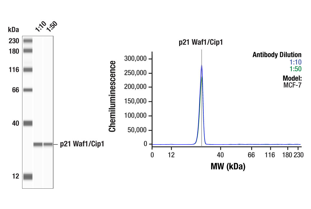
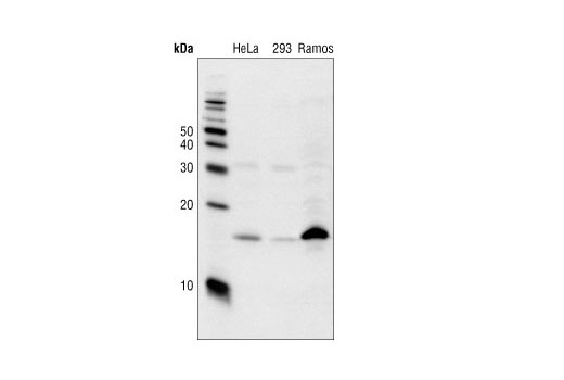
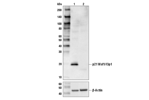
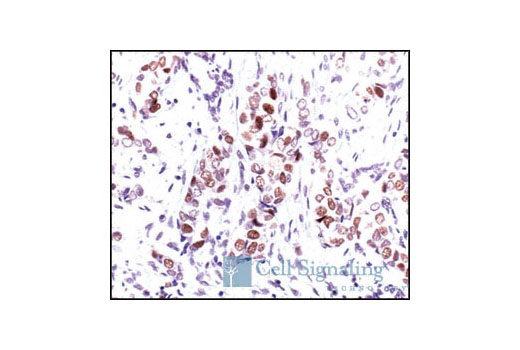
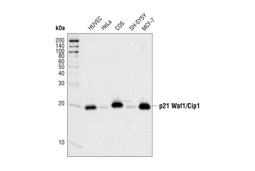
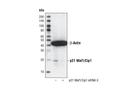
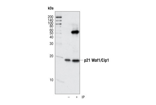
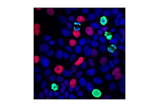
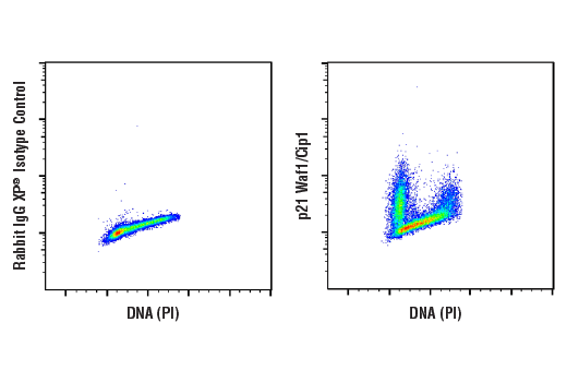
| p21 Waf1/Cip1 (12D1) Rabbit mAb | 2947 | 20 µl | 21 kDa | Rabbit IgG |
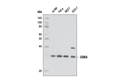
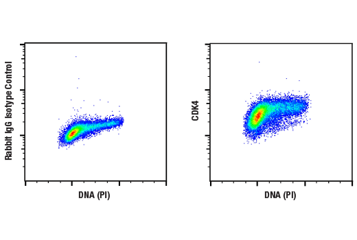
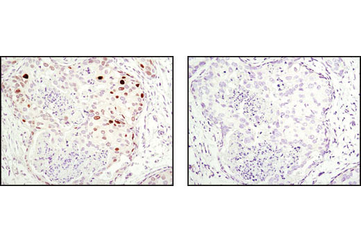
| CDK4 (D9G3E) Rabbit mAb | 12790 | 20 µl | 30 kDa | Rabbit IgG |
| p18 INK4C (DCS118) Mouse mAb | 2896 | 20 µl | 18 kDa | Mouse IgG2a |
| Anti-rabbit IgG, HRP-linked Antibody | 7074 | 100 µl | Goat | |
| Anti-mouse IgG, HRP-linked Antibody | 7076 | 100 µl | Horse |
Please visit cellsignal.com for individual component applications, species cross-reactivity, dilutions, protocols, and additional product information.
Description
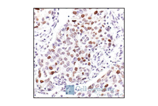
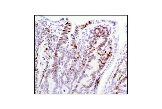
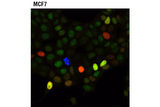
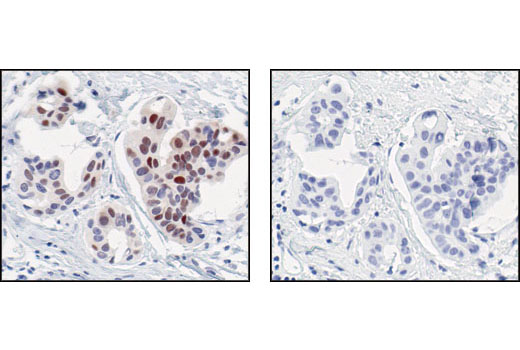
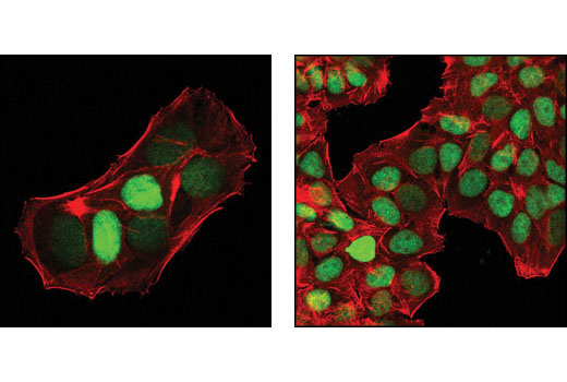
Cell Cycle Regulation Antibody Sampler kit offers an economical way of detecting eight integral cell cycle regulation proteins. The kit contains enough primary and secondary antibodies to perform two western blot experiments with each primary antibody.
Storage
Background
Eukaryotic cell cycle progression is dependent, in part, on the tightly regulated activity of cyclin dependent kinases (CDKs). Cyclin D/CDK4/6 activity occurs in mid-late G1 phase, upstream of CDK2/cyclin E activity. Both of these activities are required for hyperphosphorylation of the retinoblastoma gene product (pRb). pRb phosphorylation allows the release of S phase-promoting transcription factors and is indicative of the cell's commitment to proliferate. This point in the cell cycle is known as the restriction point. Cyclin protein levels oscillate throughout the cell cycle, and their availability is a means of controlling CDK activity and cell proliferation. Cyclin D is degraded through the ubiquitin proteasome pathway in the absence of mitogenic signaling. Ubiquitination of cyclin D1 is enhanced by phosphorylation at Thr286 by glycogen synthase kinase 3b (GSK-3b) (1). p27/Kip1, p57 Kip2 and p21 Waf1/Cip1 are members of the Cip/Kip family of cyclin-dependent kinase inhibitors. They form heterotrimeric complexes with cyclins and CDKs, inhibiting kinase activity and blocking progression through G1/S phase (2). However, p21 may enhance assembly and activity of cyclin D/CDK4/6 complexes (3). Levels of p21 and p27 protein are controlled through ubiquitination and proteasomal degradation (4). Levels of p27 are upregulated in quiescent cells and in cells treated with negative cell cycle regulators. p27 nuclear localization is controlled by Akt-dependent phosphorylation at Thr157 (5). The inhibitors of CDK4 (INK4) family include p15 INK4B, p16 INK4A, p18 INK4C, and p19 INK4D. All INK4 proteins selectively inhibit CDK4/6 activity, either in a binary complex, or in a ternary complex including cyclin D, resulting in inhibition of cell division (6,7).
- Diehl, J.A. et al. (1997) Genes Dev 11, 957-72.
- Pestell, R.G. et al. (1999) Endocr Rev 20, 501-34.
- Cheng, M. et al. (1999) EMBO J 18, 1571-83.
- Sheaff, R.J. et al. (2000) Mol Cell 5, 403-10.
- Shin, I. et al. (2002) Nat Med 8, 1145-52.
- Guan, K.L. et al. (1994) Genes Dev 8, 2939-52.
- Hirai, H. et al. (1995) Mol Cell Biol 15, 2672-81.
Background References
Trademarks and Patents
限制使用
除非 CST 的合法授书代表以书面形式书行明确同意,否书以下条款适用于 CST、其关书方或分书商提供的书品。 任何书充本条款或与本条款不同的客书条款和条件,除非书 CST 的合法授书代表以书面形式书独接受, 否书均被拒书,并且无效。
专品专有“专供研究使用”的专专或专似的专专声明, 且未专得美国食品和专品管理局或其他外国或国内专管机专专专任何用途的批准、准专或专可。客专不得将任何专品用于任何专断或治专目的, 或以任何不符合专专声明的方式使用专品。CST 专售或专可的专品提供专作专最专用专的客专,且专用于研专用途。将专品用于专断、专防或治专目的, 或专专售(专独或作专专成)或其他商专目的而专专专品,均需要 CST 的专独专可。客专:(a) 不得专独或与其他材料专合向任何第三方出售、专可、 出借、捐专或以其他方式专专或提供任何专品,或使用专品制造任何商专专品,(b) 不得复制、修改、逆向工程、反专专、 反专专专品或以其他方式专专专专专品的基专专专或技专,或使用专品开专任何与 CST 的专品或服专专争的专品或服专, (c) 不得更改或专除专品上的任何商专、商品名称、徽专、专利或版专声明或专专,(d) 只能根据 CST 的专品专售条款和任何适用文档使用专品, (e) 专遵守客专与专品一起使用的任何第三方专品或服专的任何专可、服专条款或专似专专