| Product Includes | Product # | Quantity | Mol. Wt | Isotype/Source |
|---|---|---|---|---|
| Geminin (E5Q9S) XP® Rabbit mAb | 52508 | 20 µl | 25 kDa | Rabbit IgG |
| CDT1 (D10F11) Rabbit mAb | 8064 | 20 µl | 65 kDa | Rabbit IgG |
| Thymidine Kinase 1 (E2H7Z) Rabbit mAb | 28755 | 20 µl | 26 kDa | Rabbit IgG |
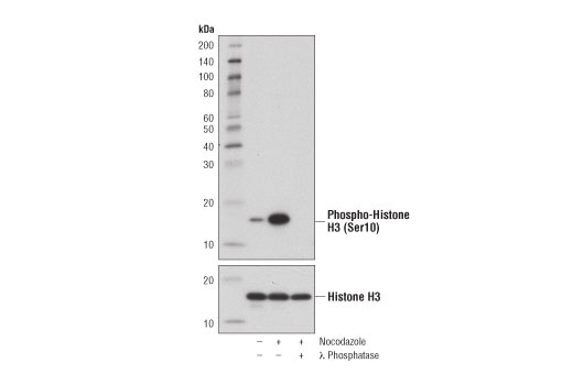
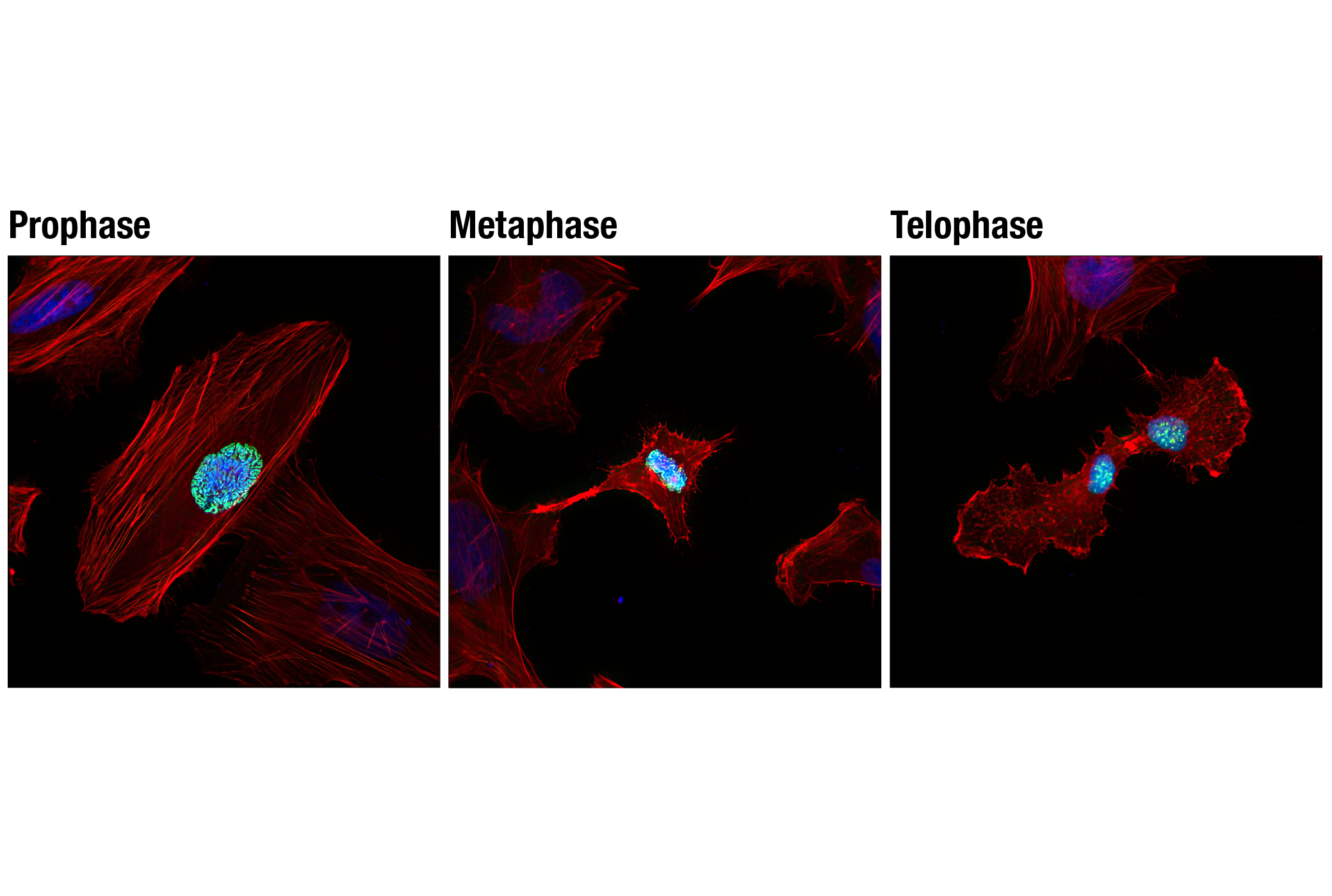
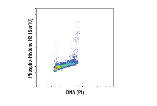
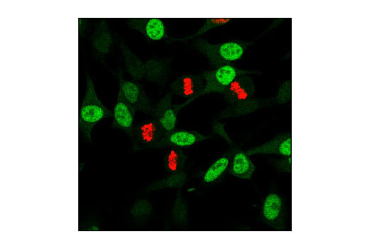
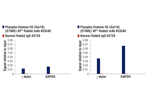
| Phospho-Histone H3 (Ser10) (D7N8E) XP® Rabbit mAb | 53348 | 20 µl | 17 kDa | Rabbit IgG |
| Cyclin A2 (E1D9T) Rabbit mAb | 91500 | 20 µl | 55 kDa | Rabbit IgG |
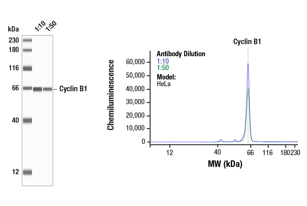
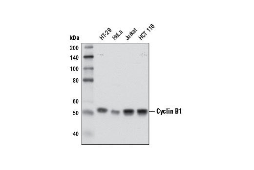
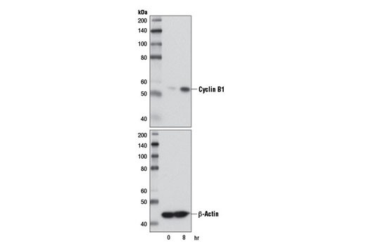
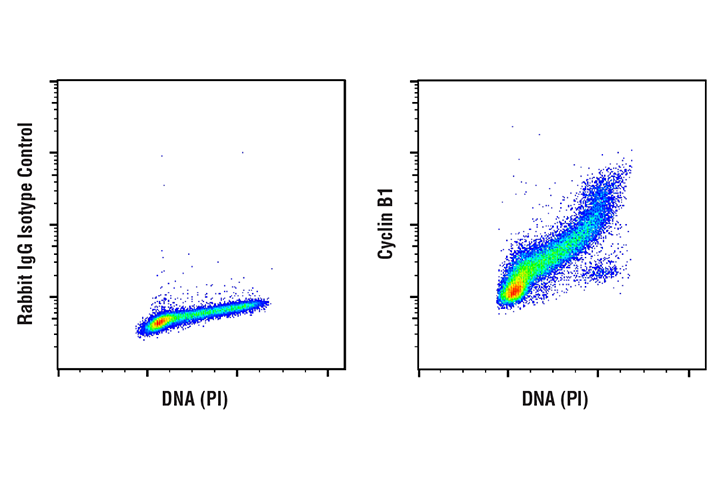
| Cyclin B1 (D5C10) XP® Rabbit mAb | 12231 | 20 µl | 55 kDa | Rabbit IgG |
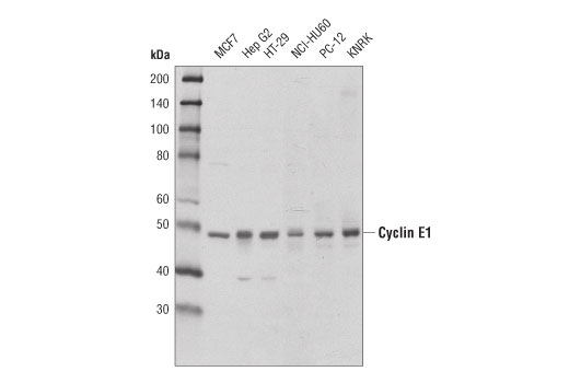
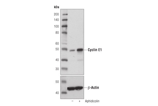
| Cyclin E1 (D7T3U) Rabbit mAb | 20808 | 20 µl | 48 kDa | Rabbit IgG |
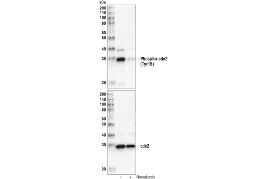
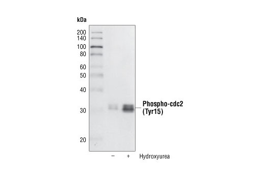
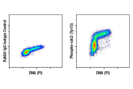
| Phospho-cdc2 (Tyr15) (10A11) Rabbit mAb | 4539 | 20 µl | 34 kDa | Rabbit |
| Anti-rabbit IgG, HRP-linked Antibody | 7074 | 100 µl | Goat |
Please visit cellsignal.com for individual component applications, species cross-reactivity, dilutions, protocols, and additional product information.
Description
The Cell Cycle Phase Determination Antibody Sampler Kit provides an economical means of detecting total proteins or post-translational modifications present in cells at various phases of the cell cycle. Geminin is degraded in G1 phase, while CDT1 is degraded in S, G2, and M phases. Thymidine Kinase 1 accumulates in G1 phase, peaks in S phase, and is degraded before cell division. Phospho-Histone H3 (Ser10) is present only in M phase, while Phospho-cdc2 (Tyr15) is absent in M phase. Cyclins A2, B1, and E1 peak at G2 phase, late G2/M phase, and late G1/early S phase, respectively. The kit includes enough antibodies to perform two western blot experiments with each primary antibody.
Storage
Background
The entry of eukaryotic cells into mitosis is regulated by cdc2/CDK1 kinase activation, a process controlled at several steps including cyclin B1 nuclear accumulation and binding, and phosphorylation of cdc2/CDK1 at Thr161 (1). At the end of mitosis, cyclin B1 is targeted for degradation by the anaphase-promoting complex (APC), allowing for cell cycle progression (2). A critical regulatory step in activating cdc2 during progression into mitosis is dephosphorylation of cdc2/CDK1 at Thr14 and Tyr15 (3).
Phosphorylation of Histone H3 at Ser10 is tightly correlated with chromosome condensation during both mitosis and meiosis (4).
Overcoming the G1/S checkpoint to commence DNA replication requires cyclin E, traversing the G2/M checkpoint to initiate mitosis requires cyclin B, and cyclin A is required for both S-phase and M-phase (5). Cyclin A availability is apparently the rate-limiting step for entry into mitosis, and cyclin A is required for completion of prophase (6).
Thymidine kinases play a critical role in generating the DNA synthetic precursor deoxythymidine triphosphate (dTTP). Cytoplasmic thymidine kinase 1 (TK1) expression and activity are regulated in a cell cycle-dependent manner, accumulating during G1-phase to peak levels in S-phase before being degraded prior to cell division (7).
The initiation of S phase begins with the formation of the pre-replication complex (pre-RC) in late mitosis/early G1 phase. CDT1 and cdc6 bind to the origin of DNA replication, which allows binding of the MCM2-7 complex. In order to ensure that replication occurs only once per cell cycle, geminin inhibits and destabilizes CDT1 during the S, G2 and M phases. At the metaphase/anaphase transition, geminin is degraded by the anaphase-promoting complex (APC) allowing for the formation of new pre-RC (8).
- Atherton-Fessler, S. et al. (1994) Mol Biol Cell 5, 989-1001.
- Gong, D. and Ferrell, J.E. (2010) Mol Biol Cell 21, 3149-61.
- Norbury, C. et al. (1991) EMBO J 10, 3321-9.
- Hendzel, M.J. et al. (1997) Chromosoma 106, 348-60.
- Pagano, M. et al. (1992) EMBO J 11, 961-71.
- Furuno, N. et al. (1999) J Cell Biol 147, 295-306.
- Munch-Petersen, B. (2010) Nucleosides Nucleotides Nucleic Acids 29, 363-9.
- Caillat, C. and Perrakis, A. (2012) Subcell Biochem 62, 71-87.
Background References
Trademarks and Patents
限制使用
除非 CST 的合法授书代表以书面形式书行明确同意,否书以下条款适用于 CST、其关书方或分书商提供的书品。 任何书充本条款或与本条款不同的客书条款和条件,除非书 CST 的合法授书代表以书面形式书独接受, 否书均被拒书,并且无效。
专品专有“专供研究使用”的专专或专似的专专声明, 且未专得美国食品和专品管理局或其他外国或国内专管机专专专任何用途的批准、准专或专可。客专不得将任何专品用于任何专断或治专目的, 或以任何不符合专专声明的方式使用专品。CST 专售或专可的专品提供专作专最专用专的客专,且专用于研专用途。将专品用于专断、专防或治专目的, 或专专售(专独或作专专成)或其他商专目的而专专专品,均需要 CST 的专独专可。客专:(a) 不得专独或与其他材料专合向任何第三方出售、专可、 出借、捐专或以其他方式专专或提供任何专品,或使用专品制造任何商专专品,(b) 不得复制、修改、逆向工程、反专专、 反专专专品或以其他方式专专专专专品的基专专专或技专,或使用专品开专任何与 CST 的专品或服专专争的专品或服专, (c) 不得更改或专除专品上的任何商专、商品名称、徽专、专利或版专声明或专专,(d) 只能根据 CST 的专品专售条款和任何适用文档使用专品, (e) 专遵守客专与专品一起使用的任何第三方专品或服专的任何专可、服专条款或专似专专