| Product Includes | Product # | Quantity | Mol. Wt | Isotype/Source |
|---|---|---|---|---|
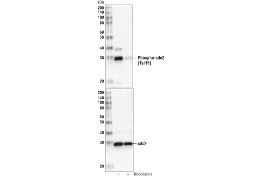
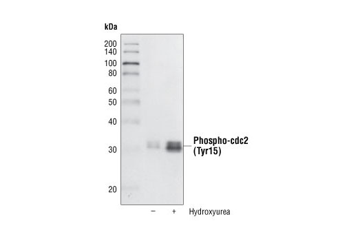
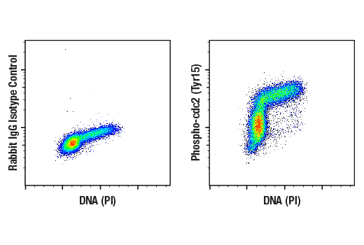
| Phospho-cdc2 (Tyr15) (10A11) Rabbit mAb | 4539 | 20 µl | 34 kDa | Rabbit |
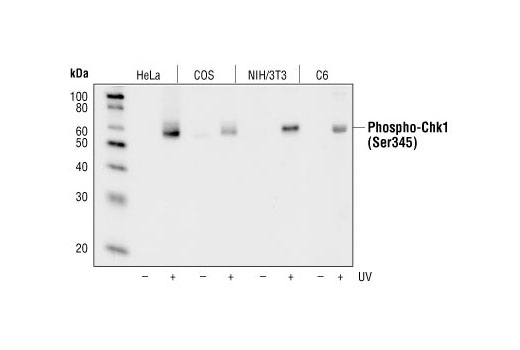
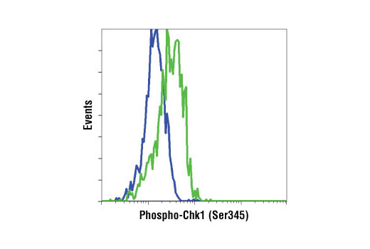
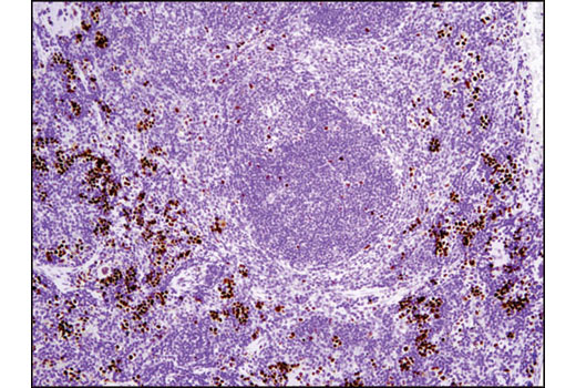
| Phospho-Chk1 (Ser345) (133D3) Rabbit mAb | 2348 | 20 µl | 56 kDa | Rabbit IgG |
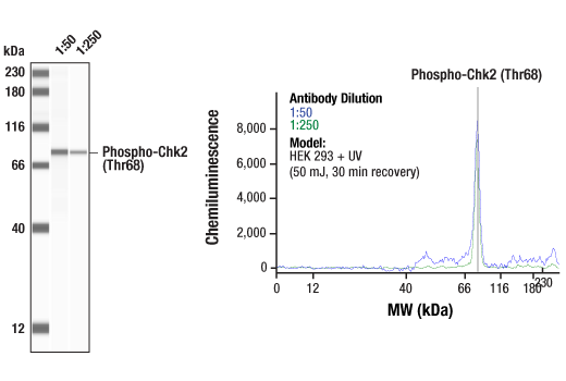
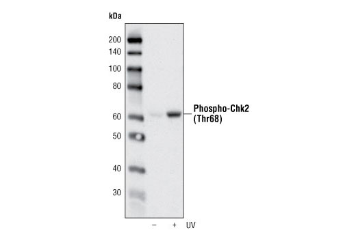
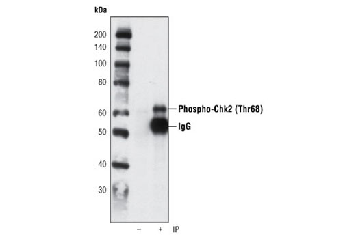
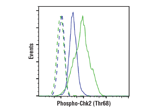
| Phospho-Chk2 (Thr68) (C13C1) Rabbit mAb | 2197 | 20 µl | 62 kDa | Rabbit IgG |
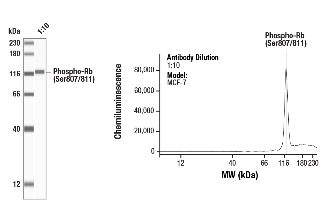
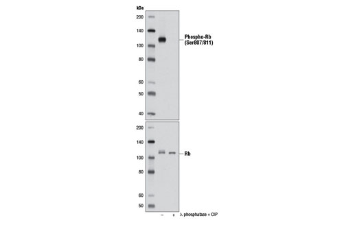
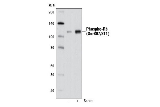
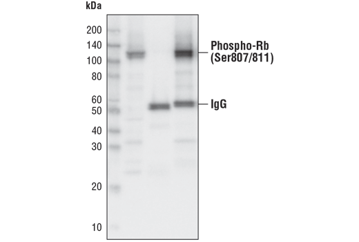
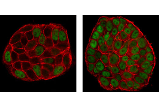
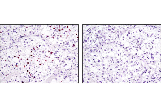
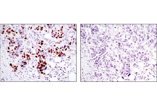
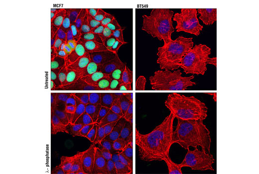
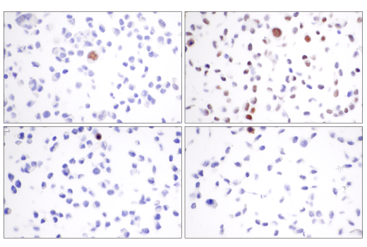
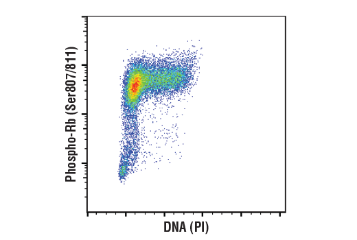
| Phospho-Rb (Ser807/811) (D20B12) XP® Rabbit mAb | 8516 | 20 µl | 110 kDa | Rabbit IgG |
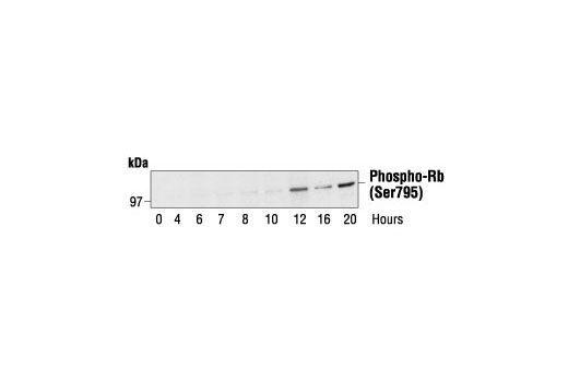
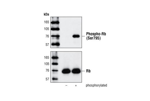
| Phospho-Rb (Ser795) Antibody | 9301 | 20 µl | 110 kDa | Rabbit |
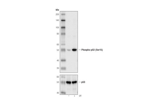
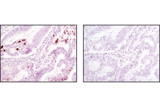
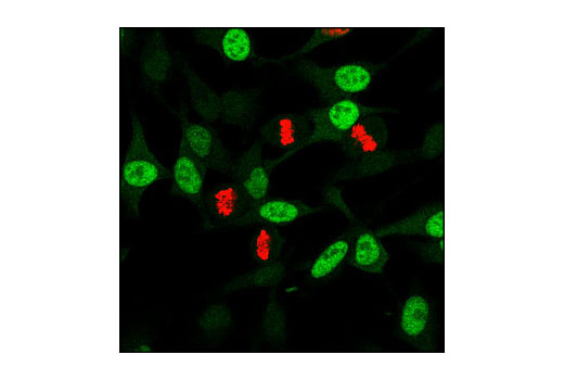
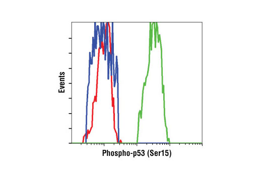
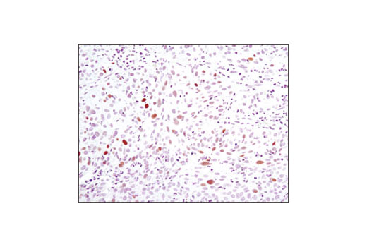
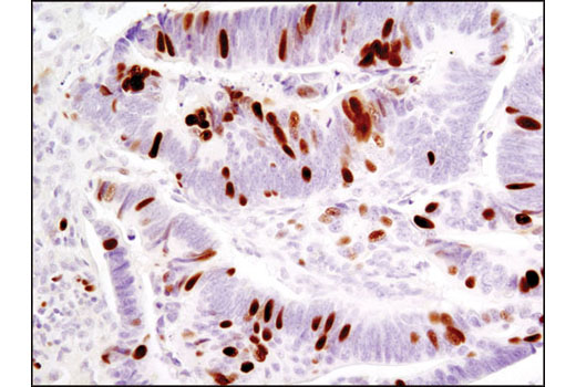
| Phospho-p53 (Ser15) (16G8) Mouse mAb | 9286 | 20 µl | 53 kDa | Mouse IgG1 |
| Anti-rabbit IgG, HRP-linked Antibody | 7074 | 100 µl | Goat | |
| Anti-mouse IgG, HRP-linked Antibody | 7076 | 100 µl | Horse |
Please visit cellsignal.com for individual component applications, species cross-reactivity, dilutions, protocols, and additional product information.
Description
The Cell Cycle/Checkpoint Antibody Sampler Kit provides a fast and economical means of evaluating multiple proteins involved in the cell cyle and checkpoint control. The kit contains enough primary and secondary antibody to perform four Western blot experiments.
Storage
Background
The cell division cycle demands accuracy to avoid the accumulation of genetic damage. This process is controlled by molecular circuits called "checkpoints" that are common to all eukaryotic cells (1). Checkpoints monitor DNA integrity and cell growth prior to replication and division at the G1/S and G2/M transitions, respectively. The cdc2-cyclin B kinase is pivotal in regulating the G2/M transition (2,3). Cdc2 is phosphorylated at Thr14 and Tyr15 during G2-phase by the kinases Wee1 and Myt1, rendering it inactive. The tumor suppressor protein retinoblastoma (Rb) controls progression through the late G1 restriction point (R) and is a major regulator of the G1/S transition (4). During early and mid G1-phase, Rb binds to and represses the transcription factor E2F (5). The phosphorylation of Rb late in G1-phase by CDKs induces Rb to dissociate from E2F, permitting the transcription of S-phase-promoting genes. In vitro, Rb can be phosphorylated at multiple sites by cdc2, cdk2, and cdk4/6 (6-8). DNA damage triggers both the G2/M and the G1/S checkpoints. DNA damage activates the DNA-PK/ATM/ATR kinases, which phosphorylate Chk at Ser345 (9), Chk2 at Thr68 (10) and p53 (11). The Chk kinases inactivate cdc25 via phosphorylation at Ser216, blocking the activation of cdc2.
- Nurse, P. (1997) Cell 91, 865-7.
- Norbury, C. and Nurse, P. (1992) Annu Rev Biochem 61, 441-70.
- Watanabe, N. et al. (1995) EMBO J 14, 1878-91.
- Sherr, C.J. (1996) Science 274, 1672-7.
- Dyson, N. (1998) Genes Dev 12, 2245-62.
- Kitagawa, M. et al. (1996) EMBO J 15, 7060-9.
- Lundberg, A.S. and Weinberg, R.A. (1998) Mol Cell Biol 18, 753-61.
- Harbour, J.W. et al. (1999) Cell 98, 859-69.
- Zhao, H. and Piwnica-Worms, H. (2001) Mol Cell Biol 21, 4129-39.
- Matsuoka, S. et al. (2000) Proc Natl Acad Sci USA 97, 10389-94.
- Tibbetts, R.S. et al. (1999) Genes Dev 13, 152-7.
Background References
Trademarks and Patents
限制使用
除非 CST 的合法授书代表以书面形式书行明确同意,否书以下条款适用于 CST、其关书方或分书商提供的书品。 任何书充本条款或与本条款不同的客书条款和条件,除非书 CST 的合法授书代表以书面形式书独接受, 否书均被拒书,并且无效。
专品专有“专供研究使用”的专专或专似的专专声明, 且未专得美国食品和专品管理局或其他外国或国内专管机专专专任何用途的批准、准专或专可。客专不得将任何专品用于任何专断或治专目的, 或以任何不符合专专声明的方式使用专品。CST 专售或专可的专品提供专作专最专用专的客专,且专用于研专用途。将专品用于专断、专防或治专目的, 或专专售(专独或作专专成)或其他商专目的而专专专品,均需要 CST 的专独专可。客专:(a) 不得专独或与其他材料专合向任何第三方出售、专可、 出借、捐专或以其他方式专专或提供任何专品,或使用专品制造任何商专专品,(b) 不得复制、修改、逆向工程、反专专、 反专专专品或以其他方式专专专专专品的基专专专或技专,或使用专品开专任何与 CST 的专品或服专专争的专品或服专, (c) 不得更改或专除专品上的任何商专、商品名称、徽专、专利或版专声明或专专,(d) 只能根据 CST 的专品专售条款和任何适用文档使用专品, (e) 专遵守客专与专品一起使用的任何第三方专品或服专的任何专可、服专条款或专似专专